Urethra
The urethra is a fibromuscular tube transporting urine from the urinary bladder to the exterior of the body.
The urethra is the sole urologic organ that shows any significant anatomic difference between males and females[1], due to the dual role of the male urethra in transporting both urine and semen[1]. In both sexes, the urethra begins inferior and central to the two ureteral openings at the three points of a triangular-shaped area at the base of the bladder called the trigone[1]. From this origin, the urethra tracks posterior and inferior to the pubic symphysis[1]. The proximal urethra is lined by transitional epithelium, whereas the terminal portion is a nonkeratinized, stratified squamous epithelium[1]. In the male, pseudostratified columnar epithelium lines the urethra between these two cell types[1]. Voiding is regulated by an involuntary autonomic nervous system-controlled internal urinary sphincter, consisting of smooth muscle, and a voluntary skeletal muscle that forms the external urinary sphincter below it[1]. Mucous glands are found along much of the length of the urethra and protect the urethra from extremes of urine pH[1].
DefinitionSphincter control
The internal urethral sphincter is involuntary smooth muscle, while the external urethral sphincter is voluntary skeletal muscle[1].
The male urethra averages 20 cm in length and passes through the prostate gland immediately inferior to the bladder[1]. It is divided into four regions: the preprostatic urethra, the prostatic urethra, the membranous urethra, and the spongy or penile urethra[1]. The preprostatic urethra is very short and incorporated into the bladder wall[1]. The prostatic urethra passes through the prostate gland[1], where it receives sperm via the ejaculatory ducts and secretions from the seminal vesicles[1]. Paired Cowper’s glands (bulbourethral glands) produce and secrete mucus into the urethra to buffer urethral pH during sexual stimulation[1]. The membranous urethra passes through the deep muscles of the perineum, where it is invested by the overlying urethral sphincters[1]. The spongy urethra exits at the tip, known as the external urethral orifice, of the penis after passing through the corpus spongiosum[1].
| Region | Location and Relations | Associated Secretions |
|---|---|---|
| Preprostatic | Incorporated into the bladder wall | None |
| Prostatic | Passes through the prostate gland | Seminal and prostatic secretions |
| Membranous | Passes through deep muscles of the perineum | Bulbourethral mucus |
| Spongy | Passes through the corpus spongiosum to the exterior | Pre-ejaculate |
The shorter female urethra contributes to the higher incidence of bladder infections in females[1]. Innervation of the urethra is the same in both males and females[1]. Voluntary control of micturition requires a mature and intact sacral micturition center[1].
Urethral regions in the male
- Preprostatic urethraIncorporated into the bladder wall
- Prostatic urethraPasses through the prostate gland
- Membranous urethraPasses through deep perineal muscles
- Spongy urethraPasses through corpus spongiosum to the external orifice
Figure 1Gray1142
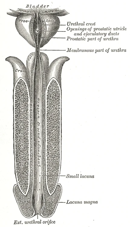
Long description
The male urethra laid open on its anterior (upper) surface.

Figure 1: The male urethra laid open on its anterior (upper) surface.
The female urethra is a short tube, measuring about 4 cm in length. It begins at the internal urethral orifice in the urinary bladder and courses inferiorly and anteriorly through the pelvic floor to terminate at the external urethral orifice. The external urethral orifice is embedded in the anterior vaginal wall inferior to the clitoris, superior to the vaginal opening, and medial to the labia minora. Because of its short length, the female urethra is less of a barrier to fecal bacteria than the longer male urethra, which explains the greater incidence of urinary tract infections in females. [1]
DefinitionMicturition
Micturition is the medical term for urination or voiding, regulated by a coordinated interplay between autonomic and somatic pathways.
The male urethra is considerably longer and serves as a shared passage for both urine and semen. It is divided into prostatic, membranous, and spongy parts. Fluid from the ejaculatory ducts enters the prostatic urethra, where secretions from the prostate are added to form semen. The shaft of the penis surrounds the spongy urethra within the corpus spongiosum.
| Sphincter | Muscle type | Innervation | Function |
|---|---|---|---|
| Internal urethral sphincter | Smooth muscle | Autonomic parasympathetic/sympathetic | Involuntary control of bladder emptying |
| External urethral sphincter | Skeletal muscle | Pudendal nerve (S2 to S4) | Voluntary control of urinary continence |
Voluntary control of the external urethral sphincter is a function of the pudendal nerve, which arises in the sacral region of the spinal cord via the S2 to S4 nerves of the sacral plexus. As bladder volume reaches about 150 mL, an urge to void is sensed but is easily overridden by conscious contraction of the external sphincter. As the bladder fills further, urges become harder to ignore, and voluntary constraint eventually fails as bladder volume approaches 300 to 400 mL, resulting in incontinence. [1]
Micturition Reflex Pathway
- Bladder fillingStretch receptors in the bladder wall transmit nerve impulses to the sacral spinal cord.
- Question: Bladder volume reaches threshold?
Yes
- Parasympathetic outflowCauses detrusor contraction and relaxation of the internal urethral sphincter.
- Somatic inhibitionInhibits motor neurons to relax the external urethral sphincter for voiding.
No
- Continence maintainedVoluntary somatic control keeps the external urethral sphincter contracted.
The urethra is a fibromuscular tube that conveys urine from the bladder to the exterior. Its anatomical course and relations differ significantly between sexes, reflecting the dual role of the male urethra in both urinary and reproductive functions. In both sexes, the urethra begins at the internal urethral orifice of the bladder.
DefinitionInternal Urethral Orifice
The opening at the base of the bladder where the urethra begins, located at the trigone.
In the male, the urethra is divided into four segments: prostatic, membranous, bulbar, and penile (spongy). The prostatic urethra passes through the prostate gland. Anteriorly, it is related to the pubic symphysis via the puboprostatic ligaments. Posteriorly, it is related to the seminal vesicles and the ampulla of the ductus deferens. The ejaculatory ducts open into the prostatic urethra on the seminal colliculus. The membranous urethra is the shortest and narrowest part, passing through the urogenital diaphragm. It is surrounded by the external urethral sphincter, a voluntary skeletal muscle. Posteriorly, it is related to the rectum. The bulbar urethra passes through the bulb of the penis. It is covered by the bulbospongiosus muscle. The penile urethra runs through the corpus spongiosum of the penis. It ends at the external urethral orifice on the glans penis.
NoteClinical Pearl
The male urethra is approximately 20 cm long, with the prostatic and membranous parts being fixed, while the penile part is mobile.
In the female, the urethra is shorter, approximately 4 cm long. It passes anterior to the vagina and posterior to the pubic symphysis. It is embedded in the anterior vaginal wall. The external urethral orifice is located between the clitoris and the vaginal opening. The urethra is supported by the pubourethral ligaments and the levator ani muscle.
| Segment | Anterior Relation | Posterior Relation | Lateral Relation |
|---|---|---|---|
| Prostatic | Pubic symphysis (via ligaments) | Seminal vesicles, Ampulla of ductus deferens | Prostatic venous plexus |
| Membranous | Pubic arch | Rectum | External urethral sphincter |
| Bulbar | Bulbospongiosus muscle | Corpus spongiosum | Crura of penis |
| Penile | Skin of penis | Corpus spongiosum | Corpora cavernosa |
The female urethra is closely related to the vagina. Anteriorly, it is related to the pubic symphysis. Posteriorly, it is related to the anterior vaginal wall. Laterally, it is related to the levator ani muscle. The urethra is supported by the endopelvic fascia.
Figure 2Gray1155 a
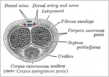
Long description
Cross-section of a human penis, illustration from Henry Gray's anatomy book. The labelling of the urethral cavernous body has been supplemented with a synonym from a more recent source: Pschyrembel online.

Figure 2: Cross-section of a human penis, illustration from Henry Gray's anatomy book.
The seminal vesicles are posterior to the bladder base. The rectum is posterior to the prostate. The pubic symphysis is anterior to the bladder.
Exam trapCommon Mistake
Do not confuse the internal urethral sphincter (smooth muscle, involuntary) with the external urethral sphincter (skeletal muscle, voluntary).
The blood supply to the urethra comes from branches of the internal iliac artery. The prostatic urethra is supplied by the inferior vesical artery. The membranous urethra is supplied by the artery to the bulb. The penile urethra is supplied by the dorsal artery of the penis. Venous drainage follows the arteries. Lymphatic drainage from the prostatic urethra goes to the internal iliac nodes. The membranous and penile urethra drain to the external iliac nodes. The female urethra drains to the internal iliac nodes.
The urethra serves as the terminal conduit for urine expulsion in both sexes and semen in males. Its internal architecture is defined by distinct epithelial linings and muscular arrangements that facilitate continence and voiding. The lining transitions from the bladder neck downwards, reflecting functional changes from storage to expulsion.
The verumontanum is a raised area on the posterior wall of the prostatic urethra where the ejaculatory ducts open. The spongy urethra runs through the corpus spongiosum of the penis, ending at the navicular fossa near the glans.
The epithelial lining varies along the course. The proximal portion, continuous with the bladder, is lined by transitional epithelium. This tissue is structurally suited for large volume fluctuations, resembling columnar epithelia when empty and transitioning to a squamous appearance when stretched. As the urethra descends, the lining changes. The prostatic urethra retains transitional epithelium. The membranous urethra is lined by stratified or pseudostratified columnar epithelium. The spongy urethra, particularly in the navicular fossa, is lined by stratified squamous epithelium, providing protection against friction and potential pathogens. [1]
Muscular control is critical for urinary continence. The internal urethral sphincter, located at the bladder neck, consists of smooth muscle fibres from the detrusor muscle. The detrusor muscle is comprised of irregular crisscrossing bands of smooth muscle. Contraction of the detrusor facilitates voiding, while relaxation allows storage. The external urethral sphincter is a voluntary skeletal muscle located at the membranous urethra. It provides conscious control over micturition. [1]
In males, the urethra also transports semen. During ejaculation, sperm exit the tail of the epididymis and are pushed by smooth muscle contraction to the ductus deferens. The fluid, now containing both sperm and seminal vesicle secretions, moves into the ejaculatory duct. The paired ejaculatory ducts transport this seminal fluid into the prostate gland. The prostatic urethra receives these secretions, mixing them with prostatic fluid before expulsion through the urethral lumen. This dual function necessitates coordinated muscular action to prevent retrograde ejaculation and ensure efficient expulsion. [2]
The arterial supply of the urethra is derived from branches of the internal iliac artery, specifically the internal pudendal artery. This vessel enters the perineum through the urogenital diaphragm. It then courses along the lateral aspect of the ischiorectal fossa within the pudendal canal. The internal pudendal artery gives off several branches that contribute to the vascularisation of the pelvic organs. One significant branch is the artery of the bulb, which supplies the bulb of the penis and the proximal urethra. Another branch is the deep artery of the penis, which runs along the dorsum of the corpus spongiosum. These vessels anastomose extensively to ensure adequate blood flow.
The deep artery of the penis runs superiorly along the corpus spongiosum. The urethra lies centrally, surrounded by the spongy tissue. The corpora cavernosa are positioned dorsally. The arterial branches penetrate the tunica albuginea.
The physiological basis of erection involves vasocongestion. Both sexual arousal and REM sleep can induce an erection. Penile erections result from the engorgement of tissues due to increased arterial inflow exceeding venous outflow. During sexual arousal, nitric oxide is released from nerve endings near blood vessels within the corpora cavernosa and spongiosum. This release activates a signaling pathway. The pathway results in the relaxation of smooth muscles surrounding the penile arteries. This relaxation causes the arteries to dilate. The dilation increases the amount of blood entering the penis. It also induces endothelial cells in the penile arterial walls to secrete nitric oxide. This secretion perpetuates the vasodilation. [2]
The urethra itself receives blood from the internal pudendal artery via its branches. The prostatic urethra is supplied by branches from the inferior vesical artery. The membranous urethra receives blood from the artery of the bulb. The spongy urethra is supplied by the dorsal artery and the artery of the bulb. These vessels form a rich plexus within the submucosa. This ensures that the urethral mucosa remains well-vascularised. The venous drainage follows the arterial supply. The blood drains into the internal pudendal veins. These veins accompany the arteries and drain into the internal iliac veins.
DefinitionVasocongestion
Engorgement of tissues due to more arterial blood flowing into the penis than is leaving in the veins. [2]
| Segment | Artery | Source |
|---|---|---|
| Prostatic | Inferior vesical artery | Internal iliac artery |
| Membranous | Artery of the bulb | Internal pudendal artery |
| Spongy | Dorsal artery of penis | Internal pudendal artery |
| Spongy | Artery of the bulb | Internal pudendal artery |
The clinical significance of this supply is evident in erectile dysfunction. Impairment of nitric oxide release can prevent smooth muscle relaxation. This prevents adequate arterial dilation. Consequently, vasocongestion does not occur. The penis remains flaccid. Understanding the anatomical course of these arteries is crucial for surgical procedures. Injury to the internal pudendal artery can lead to significant haemorrhage. Knowledge of the branching pattern helps in localising the source of bleeding. The extensive anastomoses provide collateral circulation. This protects the urethra from ischaemia during minor vascular insults.
The venous drainage of the urethra is intimately linked to the haemodynamics of the erectile tissues that surround it. Understanding this pathway requires a grasp of how blood flow is regulated during sexual arousal, specifically the mechanism that maintains rigidity. The primary structures involved are the deep dorsal vein of the penis and the associated plexuses that drain the corpora cavernosa and the spongy urethra.
At the centre lies the spongy urethra, surrounded by the corpus spongiosum. Flanking this are the two corpora cavernosa.
The physiological basis for this drainage pattern is critical for understanding erectile function. During sexual stimulation, arterial inflow increases significantly. This leads to a rapid increase in blood volume within the erectile chambers. As these chambers fill, the internal pressure rises sharply. This increased pressure compresses the thin-walled penile venules against the tough tunica albuginea of the corpora cavernosa. This compression effectively traps blood within the erectile tissue, preventing venous drainage. The result is the maintenance of erection. The balance between arterial inflow and restricted venous outflow is what sustains rigidity.
The average length of an erect penis measures approximately 15 cm, though this can vary depending on the flaccid dimensions of the individual. This variation highlights the importance of understanding the underlying vascular mechanics rather than relying on static measurements. The urethra itself does not have a dedicated, isolated venous system separate from the erectile tissues. Instead, it shares drainage pathways with the corpus spongiosum. The emissary veins from the urethral mucosa drain into the submucosal plexus, which then communicates with the veins of the corpus spongiosum. These veins eventually join the deep dorsal vein or drain directly into the prostatic plexus. [2]
Clinical pearlKey Concept
Erection is maintained not by arterial inflow alone, but by the compression of venules that prevents venous outflow.
Disruption of this drainage mechanism can lead to erectile dysfunction. If the venules fail to compress adequately, blood drains too quickly, and rigidity is lost. Conversely, if arterial inflow is insufficient, the chambers do not fill enough to generate the necessary pressure. The urethra, being part of the corpus spongiosum, remains relatively compliant to allow for ejaculation, but its venous drainage is still subject to the same haemodynamic principles. The prostatic venous plexus serves as a major collecting point for blood from the prostate, bladder, and the proximal urethra. This plexus is valveless, allowing for retrograde flow in certain pathological conditions, such as benign prostatic hyperplasia.
| Structure | Primary Drainage | Termination |
|---|---|---|
| Deep Dorsal Vein | Circumflex Veins | Prostatic Plexus |
| Corpus Spongiosum | Submucosal Plexus | Prostatic Plexus |
| Prostatic Plexus | Internal Iliac Veins | Common Iliac Vein |
The clinical relevance of this anatomy is seen in conditions affecting the venous plexuses. For instance, trauma to the perineum can damage the prostatic plexus, leading to significant haematuria or erectile dysfunction. Understanding the precise course of these veins is essential for surgical interventions in the pelvic region. The deep dorsal vein is a key landmark during radical prostatectomy, where careful ligation is required to minimise blood loss and preserve erectile function. The relationship between the urethra and these veins underscores the integrated nature of the male reproductive and urinary systems.
DefinitionAnatomical watershed
The lymphatic drainage of the urethra follows a strict anatomical division based on its sphincteric and fascial relations.
The anterior part of the male urethra and the female urethra drain into the superficial inguinal lymph nodes. This pathway is clinically significant because infections or malignancies in the distal urethra often present with palpable inguinal lymphadenopathy. The lymphatic vessels from the penile urethra and the bulbous urethra travel alongside the dorsal neurovascular structures before entering the superficial inguinal nodes located in the femoral triangle. [1]
The posterior part of the male urethra and the prostate drain into the internal iliac, external iliac, and sacral lymph nodes. This deep drainage pattern reflects the embryological origin of the prostatic and membranous urethra from the urogenital sinus. Pathology in these regions, such as prostate cancer or posterior urethral strictures, may lead to metastasis or inflammation in the pelvic sidewall nodes rather than the groin. [1]
This illustration provides an anterior view of the pelvis and perineum, oriented facing the patient, displaying the deep and superficial pathways of lymphatic vessels ascending from the tubular urethra toward the pelvic sidewalls and inguinal regions. The diagram highlights the transition point at the urogenital diaphragm, where the drainage shifts from the superficial inguinal group to the internal iliac group. Key labelled structures include the superficial inguinal nodes, the internal iliac nodes, and the lymphatic trunks connecting the urethral segments to these nodal basins. [1]
| Urethral Segment | Primary Lymph Node Group | Secondary Drainage Destination |
|---|---|---|
| Anterior urethra / Female urethra | Superficial inguinal lymph nodes | Deep inguinal and external iliac nodes |
| Posterior urethra / Prostate | Internal iliac lymph nodes | Common iliac and lumbar nodes |
Urethral Lymphatic Flow
- Identify urethral segmentAnterior versus posterior urethra.
- Question: Is it anterior or female urethra?
Yes
- Drain to superficial inguinal lymph nodes
No
- Drain to internal iliac and external iliac lymph nodes
The innervation of the urethra and the micturition reflex arc involves a complex interplay between autonomic and somatic nervous systems. Understanding this requires tracing the pathways from the spinal cord to the peripheral effectors, ensuring that voluntary control and involuntary reflexes are coordinated correctly.
From this center, the pelvic nerves emerge, carrying parasympathetic efferent fibres. These fibres travel alongside the hypogastric nerves, which convey sympathetic input.
The autonomic nervous system regulates the involuntary aspects of urination. Sympathetic activity is mediated by the hypogastric nerves. These nerves suppress contraction of the detrusor muscle during the filling phase of the bladder. This suppression allows the bladder to expand without increasing pressure significantly. Simultaneously, sympathetic tone helps maintain closure of the internal urethral sphincter.
Parasympathetic control is executed via the pelvic nerves. As the bladder fills, stretch receptors send afferent signals over these sacral pelvic nerves. These signals activate parasympathetic neurons in the sacral micturition center. The resulting efferent response releases acetylcholine at the neuromuscular junctions of the detrusor muscle. This chemical release produces detrusor contraction, leading to bladder emptying. Voluntary micturition requires an intact spinal cord to transmit these signals effectively. [1]
Somatic control is provided by the pudendal nerve. This nerve arises from the sacral micturition center and supplies the external urinary sphincter. Since this sphincter consists of voluntary skeletal muscle, it is under conscious control. Actions by cholinergic neurons maintain contraction of this muscle during bladder filling. This sustained contraction ensures continence. When the decision to void is made, voluntary inhibition of the pudendal nerve allows the external sphincter to relax. This relaxation, combined with detrusor contraction, permits urine flow. [1]
The coordination between these systems is critical. The hypogastric, pelvic, and pudendal nerves work in concert to control urination. Disruption of any part of this pathway, such as damage to the pudendal nerve or the spinal cord, can lead to urinary dysfunction. The somatic component allows for the voluntary initiation and cessation of micturition, while the autonomic components manage the storage and release phases.
DefinitionSacral Micturition Center
A group of neurons in the sacral spinal cord (S2-S4) that coordinates the reflex arc for bladder emptying.
| Nerve | Type | Primary Action |
|---|---|---|
| Hypogastric | Sympathetic | Suppresses detrusor contraction; maintains internal sphincter tone. |
| Pelvic | Parasympathetic | Stimulates detrusor contraction; promotes bladder emptying. |
| Pudendal | Somatic | Controls voluntary external sphincter; maintains continence. |
The urethra serves as the terminal conduit for the urinary and reproductive systems, exhibiting distinct histological features that reflect its dual functional roles in males and females. Understanding the epithelial transitions is fundamental to grasping its physiological resilience and susceptibility to pathology.
In the male urethra, the epithelium undergoes significant transformation along its course. The prostatic urethra, which traverses the prostate gland, is lined by transitional epithelium (urothelium). This stratified, multilayered tissue is designed to withstand the distension and pressure changes associated with urine flow from the bladder. As the urethra extends into the membranous segment, the lining remains transitional but becomes thinner. The most substantial histological shift occurs in the spongy (penile) urethra. Here, the epithelium transitions to non-keratinised stratified squamous epithelium in the distal portions, providing robust protection against friction and potential irritants. This region also contains numerous mucous glands, known as Littre’s glands, which secrete mucus to lubricate the urethral canal, facilitating the passage of semen during ejaculation and urine during micturition.
The female urethra is considerably shorter, measuring approximately 4 cm in length, and is primarily lined by stratified squamous epithelium, particularly in the distal third near the external urethral orifice. This epithelial type offers enhanced protection against mechanical stress and microbial invasion, which is critical given the urethra’s proximity to the anus and vagina. The proximal portion may exhibit transitional epithelium, similar to the male prostatic urethra, reflecting its embryological origin from the urogenital sinus.
DefinitionUrothelium
A specialized stratified epithelium capable of stretching, found in the urinary tract, including the prostatic urethra.
The vascular supply and innervation support these histological structures, ensuring adequate nutrient delivery and sensory feedback. The submucosa contains a rich network of blood vessels and lymphatics, which are essential for immune surveillance and tissue repair.
For a more detailed exploration of these histological layers, including microscopic diagrams and specific staining characteristics, please refer to the dedicated histology page. This resource provides comprehensive visual aids that complement the textual description of epithelial transitions and glandular distributions.
Understanding these histological nuances is crucial for medical students, as variations in epithelial type can influence the presentation and management of urethral disorders. For instance, the presence of transitional epithelium in the prostatic urethra makes it susceptible to conditions affecting the bladder, such as cystitis, which may extend proximally. Conversely, the stratified squamous epithelium in the distal male and female urethra is more resistant to such extensions but may be affected by local trauma or infection.
Clinical pearlClinical Correlation
The transition from transitional to squamous epithelium in the male urethra is a key landmark for understanding the spread of infections and the site of potential strictures.
The histological architecture of the urethra is not merely a passive conduit but an active participant in urinary and reproductive health. The interplay between epithelial type, glandular secretion, and vascular supply ensures the urethra’s functionality while highlighting areas of vulnerability. Medical students must appreciate these details to accurately diagnose and treat urethral pathologies, ensuring optimal patient outcomes.
The formation of the urethra is deeply intertwined with the hormonal environment established during embryogenesis. To understand the distinct anatomical differences between male and female urethral development, one must first appreciate the critical role of androgens. The continued presence of testosterone is necessary to keep the male reproductive system working properly, and Leydig cells produce approximately 6 to 7 mg of testosterone per day. This hormonal signal drives the differentiation of the genital tubercle and urogenital sinus into male-specific structures, including the prostatic and membranous urethra. Without this sustained androgenic influence, the default developmental pathway leads to the formation of the shorter, purely spongy female urethra. [2]
Testicular steroidogenesis (the manufacture of androgens, including testosterone) results in testosterone concentrations that are 100 times higher in the testes than in the circulation. This massive local concentration is vital not only for internal genitalia development but also for the proper fusion of the urogenital folds in males, which enclose the penile urethra. In females, the ovaries secrete small amounts of testosterone, although most is converted to estradiol. Consequently, the lack of significant local testosterone prevents the fusion of the urogenital folds, leaving the urethral opening separate from the vaginal introitus. [2]
Maintaining these normal concentrations of testosterone promotes spermatogenesis, whereas low levels of testosterone can lead to infertility. While this statement primarily addresses adult function, the developmental origins of this system rely on the initial surge of testosterone during the first trimester. In addition to intratesticular secretion, testosterone is also released into the systemic circulation and plays an important role in muscle development, bone growth, the development of secondary sex characteristics, and maintaining libido (sex drive) in both males and females. The systemic effects highlight the broad impact of androgens, but the local embryological effects are specific to the urogenital sinus derivatives. [2]
DefinitionTestosterone Role
The continued presence of testosterone is necessary to keep the male reproductive system working properly, and Leydig cells produce approximately 6 to 7 mg of testosterone per day.
The divergence in urethral length and course is a direct result of these early hormonal instructions. The male urethra traverses the prostate, the urogenital diaphragm, and the entire length of the penis. In contrast, the female urethra is much shorter, running from the bladder neck to the vestibule. Understanding this developmental split is essential for clinical practice, as anomalies in testosterone production or reception can lead to disorders of sexual development affecting urethral anatomy.
Radiological imaging of the male and female lower urinary tract demonstrates the distinct segments of the urethra using contrast agents.
The bladder lies posterior to the pubic bone and anterior to the rectum in males, with the prostate positioned inferior to the bladder. In females, the anatomy is similar, minus the prostate, and with the addition of the uterus positioned superior and posterior to the bladder, while the bladder itself lies anterior to the uterus. During late pregnancy, its capacity is reduced due to compression bycompensatory mechanisms and the enlarging uterus, resulting in increased frequency of urination. The bladder functions as a retroperitoneal organ whose dome distends superiorly when the bladder is filling with urine. [1]
Below the prostatic urethra lies the short membranous urethra, traversing the urogenital diaphragm. Continuing distally, the spongy urethra runs through the length of the penis within the corpus spongiosum, terminating at the external urethral meatus.
NoteImaging modality
Retrograde urethrography outlines the urethral lumen clearly by introducing water-soluble contrast media through the external urethral meatus.
| Segment | Location | Surrounding Structure |
|---|---|---|
| Prostatic urethra | Below bladder neck | Prostate gland |
| Membranous urethra | Urogenital diaphragm | Sphincter urethrae |
| Spongy urethra | Length of penis | Corpus spongiosum |
The urethra is the tubular structure responsible for conveying urine from the bladder to the exterior of the body. In males, it also serves as the terminal conduit for semen during ejaculation. This dual function necessitates a complex anatomical arrangement involving distinct segments and sphincteric mechanisms. The male urethra is significantly longer than the female counterpart, measuring approximately 18 to 20 centimetres in length. It is divided into two main parts: the posterior urethra and the anterior urethra. The posterior urethra comprises the prostatic and membranous sections, while the anterior urethra includes the bulbar and penile (or spongy) segments. [1]
The prostatic urethra is the widest and most dilatable part of the male urethra. It passes directly through the prostate gland, lying posterior to the pubic symphysis and anterior to the rectum. A key feature is the urethral crest, a longitudinal ridge on the posterior wall. On this crest lies the colliculus seminalis, or verumontanum, which is a raised area where the ejaculatory ducts and prostatic utricle open. This landmark is crucial during transurethral surgery to avoid damaging adjacent structures.
The membranous urethra is the shortest and narrowest segment. It traverses the urogenital diaphragm, passing through the deep perineal pouch. It is surrounded by the external urethral sphincter, which provides voluntary control over micturition. This segment lies inferior to the prostate and anterior to the perineal membrane. Its fixed position makes it susceptible to injury during pelvic fractures, particularly those involving the pubic rami.
The bulbar urethra is the first part of the anterior urethra. It lies within the bulb of the penis, supported by the bulbospongiosus muscle. The penile urethra extends along the ventral aspect of the corpus spongiosum, ending at the external urethral orifice on the glans penis. The navicular fossa is a slight dilation at the distal end.
The caption indicates this is a sagittal section showing the relationship of the urethra to the bladder neck, prostate, and pelvic floor muscles. Recognising these landmarks helps in identifying the site of urethral rupture. For instance, a straddle injury typically affects the bulbar urethra, while pelvic fractures often disrupt the membranous urethra. Understanding these spatial relationships is essential for accurate diagnosis and management of urethral trauma.
The male urethra is a dual-purpose conduit, serving both the urinary and reproductive systems. It is anatomically divided into two distinct segments: the anterior urethra and the posterior urethra. This division is critical for understanding the clinical presentation of urethral injuries and strictures.
The anterior urethra is the longer segment, extending from the external urethral orifice to the membranous urethra. It is further subdivided into the bulbar and penile (pendulous) urethra. The penile urethra runs along the ventral aspect of the corpus spongiosum. It is fixed to the corpora cavernosa by the Buck’s fascia. The bulbar urethra is the most common site for urethral stricture formation following trauma. It lies within the bulb of the penis. The navicular fossa is a slight dilation at the tip of the glans penis. It is the widest part of the anterior urethra.
The posterior urethra is shorter and lies within the pelvic cavity. It is divided into the prostatic and membranous urethra. The prostatic urethra passes through the prostate gland. It receives the ejaculatory ducts and the prostatic ducts. The verumontanum (seminal colliculus) is a prominent elevation on the posterior wall. It is the landmark for the opening of the ejaculatory ducts. The membranous urethra is the shortest and narrowest part of the entire urethra. It passes through the urogenital diaphragm. It is surrounded by the external urethral sphincter, which is under voluntary control. This segment is vulnerable to fracture during pelvic bone injuries.
Urethral injuries are classified based on their location. Anterior urethral injuries are often associated with straddle injuries. Posterior urethral injuries are typically linked to pelvic fractures. A high-riding prostate on digital rectal examination suggests a posterior urethral disruption. Blood at the meatus is a cardinal sign of urethral injury. Retrograde urethrography is the diagnostic test of choice when injury is suspected. Catheterisation should be avoided until injury is ruled out.
Exam trapCommon Trap
Do not confuse the external urethral orifice with the prostatic urethra. The external orifice is the terminal opening, not the internal sphincter.
Clinical pearlKey Fact
The bulbous urethra is the most common site for anterior urethral strictures due to its fixation to the pubic arch.
| Segment | Location | Key Feature |
|---|---|---|
| Prostatic | Within prostate | Receives ejaculatory ducts |
| Membranous | Urogenital diaphragm | Narrowest part |
| Bulbar | Bulb of penis | Common site of stricture |
| Penile | Corpus spongiosum | Longest segment |
Students frequently confuse the anatomical course of the ureter with that of the urethra, leading to errors in localising pathology. The ureters are retroperitoneal structures that lead from the renal pelvis of the kidney to the trigone area at the base of the bladder. In contrast, the urethra is the terminal conduit for urine excretion from the bladder. Another frequent error involves the mechanism of urine transport. Students often assume gravity assists the flow of urine from the kidneys to the bladder, but peristaltic contractions move urine without the aid of gravity. [1]
A significant pitfall relates to the clinical significance of the ureteral insertion. Students may overlook the importance of the oblique course of the ureter through the bladder wall. Children born lacking this oblique course are susceptible to “vesicoureteral reflux,” which dramatically increases their risk of serious UTI. Pregnancy also increases the likelihood of reflux and UTI due to physiological changes. When considering bladder emptying, students might attribute all voiding force to the detrusor muscle. While the detrusor muscle contracts with significant force in the young, the bladder’s strength diminishes with age. Voluntary contractions of abdominal skeletal muscles can increase intra-abdominal pressure to promote more forceful bladder emptying in older adults. [1]
Regarding the male reproductive system, a common mistake is confusing the origin of semen components. Students often believe sperm constitute the majority of ejaculate volume. In reality, sperm make up only 5 percent of the final volume of semen. The bulk of semen is produced by three critical accessory glands: the seminal vesicles, the prostate, and the bulbourethral glands. Another error involves the blood-testis barrier. Students may think all testicular cells are exposed to the bloodstream. [2]
Finally, students often misidentify the location of spermatogenesis. It occurs specifically within the seminiferous tubules that make up most of the testis. The testes are located in a skin-covered, highly pigmented, muscular sack called the scrotum that extends from the body behind the penis. This location is crucial because sperm production proceeds more efficiently when the testes are kept 2 to 4°C below core body temperature. Loss of control of micturition is called incontinence, resulting in voiding when the bladder contains about 250 mL urin [1][2]
At the bedside, understanding the urethra and associated structures is vital for managing urinary retention and incontinence. Clinicians assess bladder volume to determine if voluntary abdominal contraction is needed for emptying in elderly patients. The presence of incontinence, defined as voiding when the bladder contains about 250 mL urine, suggests a loss of control requiring neurological evaluation. In males, benign prostatic hyperplasia is a common cause of obstruction. Mild to moderate symptoms are treated with medication, whereas severe enlargement of the prostate is treated by surgery in which a portion of the prostate tissue is removed. Knowledge of the prostate’s position relative to the urethra is essential for interpreting symptoms of urinary flow obstruction. [2]
In the operating theatre, anatomical precision is required during procedures involving the lower urinary tract. Surgeons must identify the trigone area at the base of the bladder, where the ureters lead from the renal pelvis. During cystoscopy or urethral catheterisation, awareness of the urethral length and course prevents iatrogenic injury. For male reproductive surgery, such as vasectomy or hernia repair, the ductus deferens must be located. From each epididymis, each ductus deferens extends superiorly into the abdominal cavity through the inguinal canal in the abdominal wall. It continues posteriorly to the pelvic cavity, ending posterior to the bladder where it dilates in a region called the ampulla. Misidentification here can lead to significant bleeding or damage to adjacent structures. [2]
On imaging, the retroperitoneal course of the ureters is a key landmark. The ureters are approximately 30 cm long. Radiologists look for the characteristic peristaltic movement or dilation indicative of obstruction. The loose adventitial layer composed of collagen and fat anchors the ureters between the parietal peritoneum and the posterior abdominal wall. In paediatric imaging, the absence of the oblique ureteral insertion is a critical finding. This anatomical defect predisposes children to vesicoureteral reflux. Recognising this on voiding cystourethrograms allows for early intervention to prevent serious UTI. [1]
In examinations, students must accurately describe the histological and anatomical features of the reproductive tract. The inner mucosa of the ureters is lined with transitional epithelium and scattered goblet cells that secrete protective mucus. For the testes, students must distinguish between the seminiferous tubules and the rete testes. The tightly coiled seminiferous tubules form the bulk of each testis. Sperm move from the lumens of the seminiferous tubules into the straight tubules, then into the rete testes. They leave the testis through the 15 to 20 efferent ductules that cross the tunica albuginea. Understanding the blood-testis barrier is also frequently tested. Sertoli cells extend physically around the germ cells from the peripheral basement membrane of the seminiferous tubules to the lumen. This structural arrangement is fundamental to maintaining spermatogenesis. [1] [2]
Urethral and Bladder Assessment
- Patient presents with urinary symptomsAssess volume and control
- Question: Bladder volume > 250 mL with incontinence?
Yes
- Evaluate for incontinenceCheck neurological control
No
- Assess obstructionCheck prostate and urethra
- Determine managementMedication or surgery for prostate
The ureters are retroperitoneal and lead from the renal pelvis to the trigone area at the base of the bladder. [1] Urine transport relies on peristaltic contractions, not gravity, moving urine without the aid of gravity. [1] The ureters are approximately 30 cm long and anchored by a loose adventitial layer. Loss of control of micturition is called incontinence and results in voiding when the bladder contains about 250 mL urine. [1] Sperm make up only 5 percent of the final volume of semen. The testes are kept 2 to 4°C below core body temperature for efficient sperm production. [2] Regarding the male reproductive system, a common mistake is confusing the origin of semen components. Students often believe sperm constitute the majority of ejaculate volume. In reality, sperm make up only 5 percent of the final volume of semen. The bulk of semen is produced by three critical accessory glands: the seminal vesicles, the prostate, and the bulbourethral glands. Another error involves the blood-testis barrier. Tight junctions between these sustentacular cells create the blood–testis barrier, which keeps bloodborne substances from reaching the germ cells [2] From each epididymis, each ductus deferens extends superiorly into the abdominal cavity through the inguinal canal.
Answer each one in your head first, then open the answer.
What is the approximate length of the ureters?
Show answerHide answer
30 cmWhat percentage of semen volume is composed of sperm?
Show answerHide answer
5 percentWhat is the temperature difference required for efficient sperm production?
Show answerHide answer
2 to 4°C below core body temperatureWhere does the ductus deferens end posterior to the bladder?
Show answerHide answer
AmpullaWhat condition results from lacking the oblique course of the ureter?
Show answerHide answer
Vesicoureteral reflux
- Betts JG, Young KA, Wise JA, et al. Gross Anatomy of Urine Transport. OpenStax, Anatomy and Physiology 2e (CC BY-NC-SA 4.0). Accessed .
- Betts JG, Young KA, Wise JA, et al. Anatomy and Physiology of the Testicular Reproductive System. OpenStax, Anatomy and Physiology 2e (CC BY-NC-SA 4.0). Accessed .
Figure credits
Educational content for medical students and doctors. It is not a substitute for clinical judgement or local guidelines.