Rectum and anal canal
Anatomical structures and physiological mechanisms of the rectum and anal canal.
The rectum is the final segment of the large intestine, receiving food residue from the sigmoid colon near the third sacral vertebra[1]. It extends anterior to the sacrum and coccyx, measuring approximately 20.3 cm (8 in) in length[1]. Although the term rectum implies straightness, the organ follows the curved contour of the sacrum, creating three lateral bends[1]. These bends form internal transverse folds known as rectal valves, which help separate feces from gas to prevent simultaneous passage.
Figure 1Gray1082
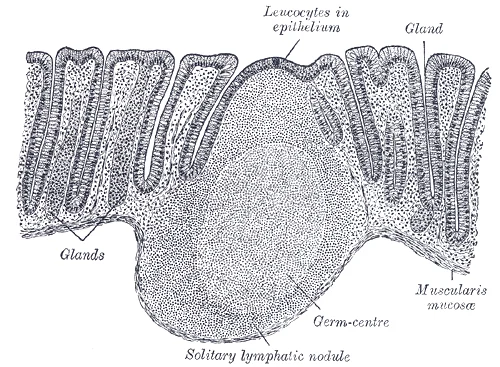
Long description
Section of mucous membrane of human rectum. (Sobotta.) X 60.

Figure 1: Section of mucous membrane of human rectum (Sobotta.) X 60.
The anal canal is the last 3.8–5 cm (1.5–2 in) of the alimentary canal, located in the perineum outside the abdominopelvic cavity[1]. It opens to the exterior at the anus and contains two sphincters[1]. The internal anal sphincter consists of smooth muscle under involuntary control, while the external anal sphincter is skeletal muscle under voluntary control[1]. Except when defecating, both usually remain closed[1].
The pectinate line, or dentate line, is a horizontal, jagged band running circumferentially just below the anal sinuses[1]. This line represents the junction between the hindgut and external skin[1]. The mucosa above this line is fairly insensitive, whereas the area below is very sensitive[1]. This difference in pain threshold arises because the upper region is innervated by visceral sensory fibers, and the lower region by somatic sensory fibers[1].
Haemorrhoids involve the venous plexuses within the anal columns[1]. Anal fissures typically occur in the sensitive lower region below the pectinate line due to somatic innervation[1]. Continence is maintained by the tonic contraction of both sphincters[1]. The process of defecation begins when mass movements force feces from the colon into the rectum, stretching the rectal wall and provoking the defecation reflex[1].
DefinitionPectinate Line
A jagged band representing the junction between the hindgut and external skin, marking a significant change in innervation and blood supply.
| Structure | Muscle Type | Control |
|---|---|---|
| Internal Anal Sphincter | Smooth | Involuntary |
| External Anal Sphincter | Skeletal | Voluntary |
Figure 2Gray1079
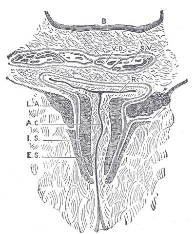
Long description
Coronal section through the anal canal. (Symington.) B. Cavity of urinary bladder. V.D. Ductus deferens. S.V. Seminal vesicle. R. Second part of rectum. A.C. Anal canal. L.A. Levator ani. I.S. Sphincter ani internus. E.S. Sphincter ani externus.

Figure 2: Coronal section through the anal canal ((symington.) b).
The rectum and anal canal represent the terminal portion of the alimentary canal. The rectum follows the curve of the sacrum and coccyx, while the anal canal passes inferiorly and posteriorly through the pelvic floor to terminate at the anus. The mouth and anus are both open to the external environment, meaning food and wastes within the alimentary canal are technically considered to be outside the body. Only through the process of absorption do the nutrients in food enter into and nourish the body's inner space. [2]
Although the rectum and anal canal lack teniae coli and haustra, they possess well-developed layers of muscularis. In the rest of the large intestine, three bands of smooth muscle called teniae coli make up the longitudinal muscle layer of the muscularis, except at its terminal end. Tonic contractions of the teniae coli bunch up the colon into a succession of pouches called haustra, which are responsible for the wrinkled appearance of the colon, and small fat-filled sacs of visceral peritoneum called epiploic appendages are attached to the teniae coli. By contrast, the rectum and anal canal lack both teniae coli and haustra, yet they feature well-developed layers of muscularis that create the strong contractions needed for defecation. The entry of feces into the rectum activates the defecation reflex. [1]
External hemorrhoids are dilated vascular structures located outside the anal verge, originating from the inferior hemorrhoidal plexus and covered by sensitive stratified squamous epithelium. The wall of the rectum and anal canal shares the general four-layer tissue plan of the alimentary canal, comprising mucosa, submucosa, muscularis, and serosa. Intrinsic innervation is supplied by the enteric nervous system, which runs from the esophagus to the anus and contains approximately 100 million motor, sensory, and interneurons, a feature unique to this system compared to all other parts of the peripheral nervous system. These enteric neurons are grouped into two plexuses, with the myenteric plexus or plexus of Auerbach lying in the muscularis layer to govern motility, rhythm, and force of contractions, and the submucosal plexus or plexus of Meissner lying in the submucosal layer to regulate digestive secretions. Blood supply to these terminal regions is delivered by arteries branching from the abdominal aorta, specifically via the inferior mesenteric artery. The lamina propria houses numerous blood and lymphatic vessels along with clusters of lymphocytes that make up mucosa-associated lymphoid tissue to defend against foodborne bacteria and other pathogens. [2]
Figure 3Gray1083
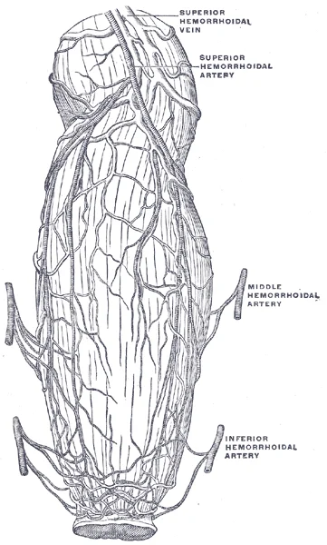
Long description
The bloodvessels of the rectum and anus, showing the distribution and anastomosis on the posterior surface near the termination of the gut. (Poirier and Charpy)

Figure 3: The bloodvessels of the rectum and anus, showing the distribution and anastomosis on the posterior surface near the termination of the gut (Poirier and Charpy).
Figure 4Gray1081
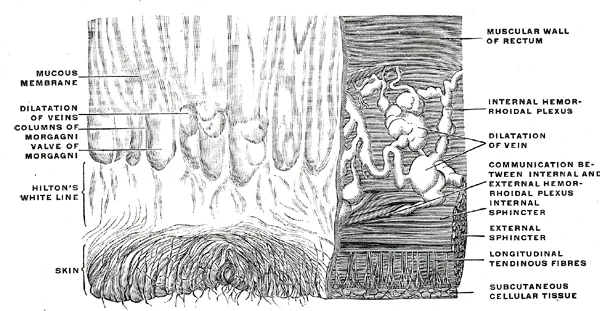
Long description
Inner wall of the lower end of the rectum and anus. On the right the mucous membrane has been removed to show the dilatation of the veins and how they pass through the muscular wall to anastomose with the external hemorrhoidal plexus. (Luschka.)

Figure 4: Inner wall of the lower end of the rectum and anus.
Figure 5Gray1080
Long description
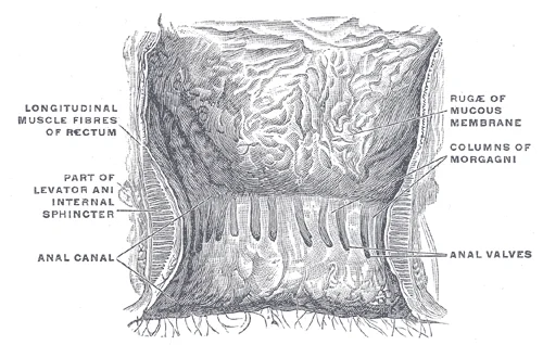
The interior of the anal cami and lower part of the rectum, showing the columns of Morgagni and the anal valves between their lower ends. The columns were more numerous in the specimen than usual. (Cunningham.)

Figure 5: The interior of the anal cami and lower part of the rectum, showing the columns of Morgagni and the anal valves between their lower ends.
The digestive organs within the abdominal cavity are held in place by the peritoneum, a broad serous membranous sac made up of squamous epithelial tissue surrounded by connective tissue. During fetal development, portions of the large intestine, including the rectum, remain completely or partially posterior to the peritoneum. Thus, the location of these structures is described as retroperitoneal. In this sagittal view, the rectum follows the sacrococcygeal curve, lying anterior to the sacrum and coccyx. The upper third of the rectum is covered by peritoneum on its anterior and lateral surfaces, the middle third has peritoneum only on its anterior surface, and the lower third lies completely below the peritoneal reflection. [2] [1]
Figure 6Gray1077
Long description
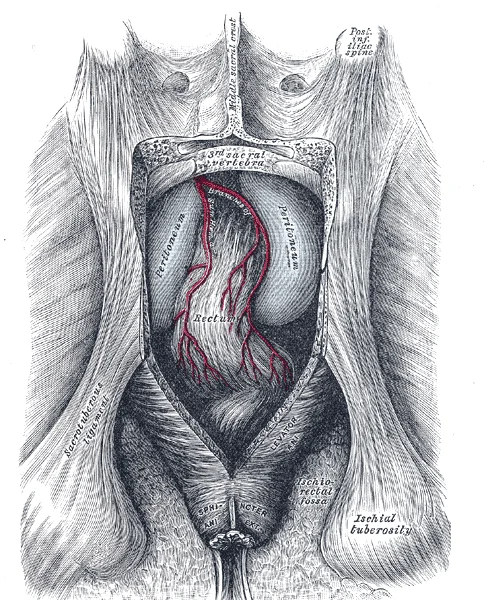
The posterior aspect of the rectum exposed by removing the lower part of the sacrum and the coccyx.

Figure 6: The posterior aspect of the rectum exposed by removing the lower part of the sacrum and the coccyx.
| Structure | Position and Relations |
|---|---|
| Rectum | Lies anterior to the sacrum, showing lateral and anteroposterior curvatures |
| Anal canal | Passes inferiorly through the pelvic floor to the exterior |
| Pectinate line | Marks the transition from internal endoderm to external ectoderm |
| Sphincters | Internal smooth muscle and external skeletal muscle rings |
The rectum exhibits three lateral flexures and an anteroposterior curve that follows the sacrococcygeal contour. Inferiorly, the rectum narrows into the anal canal, which is surrounded by the internal and external anal sphincters. Together with the puborectalis muscle, these sphincters form the anorectal ring at the anorectal junction. The pectinate line divides the upper visceral zone from the lower somatic zone of the anal canal. Pathological downward displacement of the anal cushions leads to internal haemorrhoids above this line, while external haemorrhoids occur below it. An anal fissure represents a tear in the anal mucosa distal to the pectinate line.
DefinitionRetroperitoneal relations
During fetal development, portions of the large intestine, including the rectum, remain completely or partially posterior to the peritoneum.
Anorectal Anatomy
- RectumFollows sacrococcygeal curve
- Question: Anal Canal Region?
Above Pectinate Line
- Internal haemorrhoids and columnar epithelium
Below Pectinate Line
- External haemorrhoids and stratified squamous epithelium
The large intestine represents the terminal segment of the alimentary canal. Its primary physiological roles are to complete the absorption of nutrients and water, synthesize specific vitamins, form faeces, and eliminate waste from the body. Anatomically, the large intestine is subdivided into four distinct regions: the cecum, the colon, the rectum, and the anus. The transition from the small intestine is controlled by the ileocecal valve, which regulates the flow of chyme into the large intestine. [1]
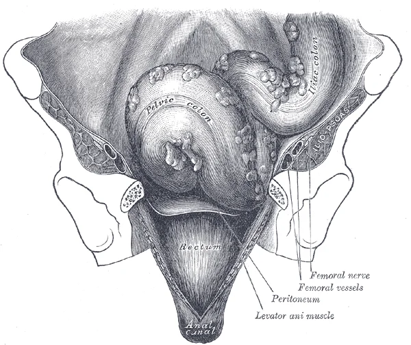
The rectum is positioned in the retroperitoneum. This anatomical placement is consistent with the ascending and descending colon, whereas the transverse and sigmoid colon are tethered to the posterior abdominal wall by the mesocolon. The rectum lies directly anterior to the sacrum, following the curvature of the pelvic cavity. In this view, the rectum is seen descending from the sigmoid colon, resting against the sacral promontory and the sacral canal. The sacral promontory forms the anterior lip of the superior base of the sacrum. Lateral to this, the auricular surface joins the ilium to form the sacroiliac joints. The rectum continues inferiorly, passing through the pelvic diaphragm to become the anal canal. [1] [3]
Figure 7Gray1076
Long description
Iliac colon, sigmoid or pelvic colon, and rectum seen from the front, after removal of pubic bones and bladder.

Figure 7: Iliac colon, sigmoid or pelvic colon, and rectum seen from the front, after removal of pubic bones and bladder.
Within the anal canal, the internal hemorrhoids are located in the submucosa of the upper anal canal, specifically above the pectinate line. These structures are vascular cushions composed of dilated venous plexuses. They are covered by insensate columnar epithelium, distinguishing them from the somatic innervation below the pectinate line. Externally, they are related to the external anal sphincter, while internally they lie adjacent to the anal columns of Morgagni.
DefinitionInternal hemorrhoids
Varicosities of the submucosal venous plexuses above the pectinate line, covered by insensate columnar epithelium.
| Feature | Location | Function / Feature |
|---|---|---|
| Rectum | Retroperitoneum | Terminal storage of faeces |
| Anal canal | Inferior to rectum | Elimination of faeces |
| Internal hemorrhoids | Submucosa above pectinate line | Vascular cushions |
The superior rectal artery represents the direct continuation of the inferior mesenteric artery, crossing the left common iliac vessels to descend into the sigmoid mesocolon before dividing into right and left branches that supply the upper rectum. The middle rectal artery arises from the anterior division of the internal iliac artery, running medially on the pelvic floor to supply the muscular wall of the middle and lower rectum and anastomosing freely with both the superior rectal and inferior rectal arteries. The inferior rectal artery originates from the internal pudendal artery within the pudendal canal (Alcock canal), traversing the ischioanal fossa horizontally to pierce the anal sphincter mechanism and supply the anal canal below the pectinate line.
Figure 8Gray1078
Long description
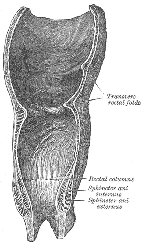
Coronal section of rectum and anal canal.

Figure 8: Coronal section of rectum and anal canal.
| Artery | Origin | Distribution |
|---|---|---|
| Superior rectal artery | Inferior mesenteric artery | Upper rectum |
| Middle rectal artery | Internal iliac artery | Middle and lower rectum |
| Inferior rectal artery | Internal pudendal artery | Anal canal |
Clinical pearlClinical anastomoses
The rich anastomoses between the superior, middle, and inferior rectal arteries create a continuous collateral network linking the systemic circulation with the portal venous system via the inferior mesenteric venous return.
While the superior mesenteric artery is the main arterial supply for the small intestine, [1][2]
The venous drainage of the rectum and anal canal is a critical anatomical feature because it creates a portosystemic anastomosis, linking the portal circulation with the systemic circulation. This connection is clinically significant as it allows for the bypass of the liver in conditions such as portal hypertension. To understand this drainage, one must first appreciate the arterial supply, which dictates the venous return. The rectum receives its blood supply primarily from the superior rectal artery, a continuation of the inferior mesenteric artery. The lower part of the anal canal is supplied by the inferior rectal arteries, branches of the internal pudendal arteries.
The middle rectal veins drain the middle portion of the rectum and anastomose with the superior rectal veins, draining into the internal iliac veins. The inferior rectal veins drain the lower anal canal and anastomose with the middle rectal veins, draining into the internal pudendal veins and then the internal iliac veins. The pectinate line serves as the anatomical landmark separating the upper two-thirds from the lower one-third of the anal canal, corresponding to the transition between the superior and middle/inferior rectal venous drainage systems.
The superior rectal vein is the primary venous drainage for the rectum. It ascends within the mesorectum and drains into the inferior mesenteric vein. The inferior mesenteric vein then joins the splenic vein, which merges with the superior mesenteric vein to form the hepatic portal vein. This pathway directs blood to the liver for processing. In contrast, the middle and inferior rectal veins drain into the systemic circulation. The middle rectal veins drain into the internal iliac veins, while the inferior rectal veins drain into the internal pudendal veins. Both of these eventually join the internal iliac veins and then the common iliac veins, leading to the inferior vena cava.
This dual drainage system allows for collateral circulation. If portal pressure increases, blood can flow from the portal system to the systemic system through these anastomoses. This can lead to the formation of varices, particularly in the lower oesophagus and the anal canal. The anal canal varices are known as haemorrhoids. Understanding the precise location and connections of these veins is essential for diagnosing and treating conditions related to portal hypertension and local vascular disorders.
The lymphatic drainage of the rectum and anal canal is defined by the anatomical boundary known as the pectinate (dentate) line. This landmark is critical because it separates the embryological hindgut from the proctodeum, resulting in distinct vascular, lymphatic, and neural pathways above and below this level.
The mucosa above the pectinate line drains into the internal iliac nodes and the inferior mesenteric nodes. Specifically, the upper rectum drains primarily to the inferior mesenteric lymph nodes, which follow the inferior mesenteric artery. The lower rectum and upper anal canal drain into the internal iliac lymph nodes. These nodes are situated along the internal iliac vessels within the pelvis. From here, efferent vessels ascend to the common iliac nodes and then to the para-aortic nodes. This pathway is significant because malignant cells from adenocarcinomas of the rectum typically spread via this route. The lymphatic vessels run in the submucosa and muscularis propria, converging towards the mesorectum. [1]
The skin of the anal canal below the pectinate line drains into the superficial inguinal lymph nodes. This drainage pathway is distinct from the pelvic nodes above. The superficial inguinal nodes are located in the upper medial thigh, just below the inguinal ligament. They receive lymph from the lower anal canal, the perianal skin, and the lower vagina in females. Metastasis from squamous cell carcinomas of the anal canal often involves these nodes. Understanding this separation is vital for staging and surgical planning.
The autonomic innervation also respects the pectinate line. Above the line, the rectum receives parasympathetic innervation from the pelvic splanchnic nerves (S2–S4) and sympathetic innervation from the superior hypogastric plexus. These nerves are visceral and insensitive to pain, though they mediate stretch and distension. Below the line, the anal canal is innervated by the inferior rectal nerves, branches of the pudendal nerve (S2–S4). These are somatic nerves, providing conscious sensation to pain, temperature, and touch. This explains why procedures above the line are often painless under local anaesthesia, while those below are not.
The pectinate line is marked as the dividing boundary.
DefinitionPectinate Line
The pectinate line is the anatomical landmark separating the hindgut-derived upper anal canal from the proctodeum-derived lower anal canal, dictating distinct lymphatic and neural pathways.
| Region | Boundary | Primary Nodes | Clinical Relevance |
|---|---|---|---|
| Upper Rectum | Above pectinate line | Inferior mesenteric | Adenocarcinoma spread |
| Lower Rectum | Above pectinate line | Internal iliac | Adenocarcinoma spread |
| Anal Canal | Below pectinate line | Superficial inguinal | Squamous cell carcinoma spread |
The innervation of the rectum and anal canal is complex, involving both autonomic and somatic pathways. This dual supply explains the distinct sensory experiences and reflexes associated with these structures. The autonomic nervous system provides involuntary control, while the somatic nervous system allows for conscious perception and voluntary control.
The mucosa here is insensitive to pain but sensitive to stretch.
The somatic innervation is crucial for the lower anal canal. The inferior anal nerve, a branch of the pudendal nerve (S2-S4), supplies the skin of the anal verge and the lower one-third of the anal canal. This area is richly innervated by somatic sensory fibres. Consequently, it is highly sensitive to pain, temperature, and touch. This contrasts with the upper rectum, where visceral afferents travel with sympathetic fibres to T11-L2, causing referred pain to the lower back and abdomen.
The external anal sphincter is a skeletal muscle under voluntary control. It is innervated by the inferior anal nerve and branches of the perineal nerve. The internal anal sphincter is smooth muscle, controlled by the autonomic nervous system. Sympathetic stimulation causes contraction, while parasympathetic stimulation causes relaxation.
DefinitionReferred Pain
Pain perceived in a location other than the site of the painful stimulus, due to convergence of visceral and somatic afferents in the spinal cord.
The transition zone, or pectinate line, marks the boundary between the two embryological origins and their respective nerve supplies. Above this line, pain is poorly localised. Below this line, pain is sharp and well-localised. This distinction is vital for clinical examination and diagnosis.
| Structure | Nerve Supply | Type | Sensation |
|---|---|---|---|
| Upper 2/3 Anal Canal | Superior Rectal Plexus (Autonomic) | Visceral | Stretch, not pain |
| Lower 1/3 Anal Canal | Inferior Anal Nerve (Somatic) | Somatic | Pain, Touch, Temperature |
| External Anal Sphincter | Inferior Anal Nerve | Somatic | Voluntary Control |
| Internal Anal Sphincter | Autonomic Plexus | Autonomic | Involuntary Control |
Understanding this anatomy helps explain why haemorrhoids above the pectinate line are painless, while those below are extremely painful. It also guides the technique for regional anaesthesia during anal surgery.
The rectum and anal canal represent the terminal segments of the large intestine, transitioning from abdominal viscera to the perineum. Understanding their histology requires appreciating the shift from simple columnar epithelium to stratified squamous epithelium, a change that reflects their distinct functional roles in storage versus voluntary expulsion.
DefinitionRectal Mucosa
The rectum is lined by simple columnar epithelium with abundant goblet cells, facilitating lubrication for faecal passage.
Unlike the rest of the colon, the rectum lacks taeniae coli and epiploic appendages. The mucosa contains simple columnar epithelium with numerous goblet cells, which secrete mucus to reduce friction. The lamina propria contai [1]
Exam trapAnal Canal Transition
Do not confuse the anal canal's epithelial transition with the rectal mucosa; the pectinate line marks a crucial embryological and histological boundary.
The anal canal is divided into upper and lower parts by the pectinate (dentate) line. This line is a zig-zag ridge formed by the anal valves. Above the pectinate line, the epithelium is columnar, transitioning to non-keratinised stratified squamous epithelium. This region is derived from the hindgut and is innervated by visceral afferents, meaning it is insensitive to pain but sensitive to stretch. This area is innervated by somatic afferents, making it highly sensitive to pain, temperature, and touch. The vascular supply also changes: above the line, blood is drained by the superior rectal vein into the portal system; below the line, it is drained by the inferior rectal vein into the systemic circulation. Lymphatic drainage follows the same pattern: above the line drains to internal iliac nodes, while below the line drains to superficial inguinal nodes.
Clinical pearlClinical Correlation
Haemorrhoids above the pectinate line are internal and painless; those below are external and painful due to somatic innervation.
The muscularis externa of the anal canal is unique. The inner circular layer thickens to form the internal anal sphincter, which is smooth muscle and under involuntary control. The outer longitudinal layer continues into the anal canal. External to this is the external anal sphincter, composed of skeletal muscle under voluntary control. It has three parts: subcutaneous, superficial, and deep. The puborectalis muscle, part of the levator ani, forms a sling around the anorectal junction, maintaining the anorectal angle which is crucial for continence.
| Muscle | Type | Control | Innervation |
|---|---|---|---|
| Internal Anal Sphincter | Smooth | Involuntary | Autonomic (Sympathetic & Parasympathetic) |
| External Anal Sphincter | Skeletal | Voluntary | Inferior Rectal Nerve (Branch of Pudendal Nerve) |
| Puborectalis | Skeletal | Voluntary | Direct branches from S3, S4 |
The histological distinction at the pectinate line is critical for clinical practice. Diseases such as fissures and fistulae are classified based on their position relative to this line. The transition from columnar to squamous epithelium also affects the susceptibility to certain pathologies, with squamous cell carcinoma being more common below the line, while adenocarcinoma typically arises above it. The rich vascular network in the submucosa of the upper anal canal is prone to dilation, leading to haemorrhoids. Understanding these histological features provides the foundation for diagnosing and managing anorectal disorders.
The rectum and anal canal arise from distinct embryological origins, a fact critical for understanding their vascular, lymphatic, and epithelial characteristics. The hindgut gives rise to the upper two-thirds of the anal canal and the rectum. This region is lined by simple columnar epithelium, similar to the rest of the gastrointestinal tract, and is supplied by the superior rectal artery, a continuation of the inferior mesenteric artery. The lower one-third of the anal canal, known as the anal verge or pectinate line region, derives from the ectoderm of the proctodeum. This ectodermal origin means it is lined by stratified squamous epithelium and is innervated by somatic nerves, making it sensitive to pain, temperature, and touch, unlike the visceral innervation of the upper region.
The pectinate (dentate) line marks the junction between the hindgut and the proctodeum. It is a crucial anatomical landmark. Below this line, drainage is to the superficial inguinal nodes. This distinction is vital in oncology, as it determines the pattern of metastasis for carcinomas arising in these regions. The vascular supply also changes at this line. The superior rectal artery supplies the area above, while the middle and inferior rectal arteries, branches of the internal iliac and internal pudendal arteries respectively, supply the area below.
The muscular wall of the rectum consists of the inner circular and outer longitudinal layers of smooth muscle. The internal anal sphincter, derived from the circular muscle layer, is under involuntary control. The puborectalis muscle, part of the levator ani, forms a sling around the anorectal junction, maintaining the anorectal angle which is essential for fecal continence.
DefinitionPectinate Line
The pectinate line is the embryological junction between the hindgut and proctodeum, marking a change in epithelium, innervation, vascular supply, and lymphatic drainage.
The development of the vertebral column also influences pelvic anatomy. During fetal development, the body is flexed anteriorly into the fetal position, giving the entire vertebral column a single curvature that is concave anteriorly. Each of these is thus called a primary curve because they are retained from the original fetal curvature of the vertebral column. This sacrococcygeal curve provides the posterior boundary of the pelvic cavity, housing the sacrum and coccyx, which support the pelvic viscera including the rectum. [3]
| Muscle | Origin | Insertion | Action | Nerve Supply |
|---|---|---|---|---|
| Internal Anal Sphincter | Circular smooth muscle of rectum | Skin around anal verge | Constricts anal canal (involuntary) | Autonomic (sympathetic/parasympathetic) |
| External Anal Sphincter | Anococcygeal ligament | Perineal body | Constricts anal canal (voluntary) | Inferior rectal nerve (branch of pudendal) |
| Puborectalis | Pubic bone | Anorectal junction | Maintains anorectal angle | Branches of S3, S4 |
Radiological imaging of the lower gastrointestinal tract requires a precise understanding of cross-sectional planes to delineate the pelvic viscera from surrounding fibroadipose tissues and muscular boundaries.
Below this line, the mesorectal fascia appears as a fine, smooth hyperintense border enclosing the perirectal fat and lymph nodes around the rectum.
DefinitionMesorectal fascia
The circumferential envelope of loose connective tissue enclosing the rectum, perirectal fat, and lymphovascular structures.
| Muscle | Origin | Insertion | Action | Nerve Supply |
|---|---|---|---|---|
| Puborectalis | Posterior surface of pubic bone | Loops behind anorectal junction | Maintains anorectal angle | S4 nerve roots |
| Internal anal sphincter | Continuation of circular rectal smooth muscle | Intersphincteric groove | Involuntary closure of anal canal | Pelvic splanchnic nerves |
| External anal sphincter | Anococcygeal body and perineal body | Perineal body and skin | Voluntary closure of anal canal | Pudendal nerve |
The rectum forms the terminal part of the large intestine, beginning at the rectosigmoid junction in front of the third sacral vertebra. It follows the anterior curve of the sacrum and coccyx, presenting three lateral flexures (superior, intermediate, and inferior) that correspond to internal mucosal folds known as the valves of Houston.
Relations of the rectum differ between its upper intraperitoneal third, middle extraperitoneal third, and lower extraperitoneal third. Anteriorly, the upper part relates to the rectouterine pouch in females or rectovesical pouch in males, while the lower two-thirds relate to the posterior wall of the vagina or the prostate gland, seminal vesicles, and base of the bladder. Posteriorly, the rectum rests against the sacrum, coccyx, median sacral vessels, and sympathetic trunk.
DefinitionAnorectal ring
The palpable muscular ring formed by the fusion of the puborectalis muscle, internal sphincter, and external sphincter at the anorectal junction, essential for maintaining faecal continence.
The interior of the upper half of the anal canal displays six to ten vertical mucosal folds called rectal columns of Morgagni, which are united inferiorly by small crescentic folds termed anal valves. Above these valves lie the anal sinuses, which receive the ducts of anal glands.
| Muscle | Origin | Insertion | Action | Nerve Supply |
|---|---|---|---|---|
| Puborectalis | Back of pubic bone | Loops behind anorectal junction | Maintains anorectal angle | S4 and perineal branch of pudendal nerve |
| Internal anal sphincter | Thickened inner circular smooth muscle | Continuous with smooth muscle of colon | Involuntary closure of anal canal | Pelvic splanchnic nerves (parasympathetic) |
| External anal sphincter | Tip of coccyx and perineal body | Perineal body and skin | Voluntary closure of anal canal | Inferior rectal branch of pudendal nerve |
Above the dentate line, the submucosal venous plexus gives rise to internal haemorrhoids, whereas the external venous plexus below the line gives rise to external haemorrhoids.
- The pectinate line is a watershed: above it derives from hindgut (visceral innervation, columnar epithelium, portal venous drainage, internal iliac lymphatics); below it derives from proctodeum (somatic innervation, stratified squamous epithelium, systemic venous drainage, inguinal lymphatics).
- Internal hemorrhoids originate above the pectinate line, are covered by insensate columnar epithelium, and bleed painlessly; external hemorrhoids arise below the line, are covered by sensitive squamous epithelium, and thrombose painfully.
- The internal anal sphincter provides 85% of resting tone (involuntary); the external anal sphincter and puborectalis provide voluntary squeeze and maintain the anorectal angle (~90°).
- Peritoneal coverage grades caudally: upper third of rectum is intraperitoneal (anterior and lateral), middle third is covered anteriorly only, lower third and anal canal are entirely extraperitoneal.
- The rectoanal inhibitory reflex (sampling reflex) causes transient internal sphincter relaxation upon rectal distension, allowing mucosal discrimination of contents before voluntary defecation.
- Anal fissures occur predominantly in the posterior midline (90%) below the pectinate line, where somatic innervation makes them exquisitely painful and sphincter spasm impairs healing.
- Venous drainage above the pectinate line enters the portal system (superior rectal → inferior mesenteric → portal vein); below the line it enters the systemic circulation (inferior rectal → pudendal → internal iliac → IVC), creating a portosystemic anastomosis.
Exam trapVertebral count confusion
Students often confuse the fusion timeline of the sacrum, which is formed by the fusion of five sacral vertebrae, a process that does not begin until after the age of 20. [3]
Medical students frequently mistake the structural classification of the coccyx, remembering that the coccyx, or tailbone, is derived from the fusion of four very small coccygeal vertebrae. Another frequent error is misidentifying weight-bearing regions, forgetting that the sacrum is a triangular-shaped bone that is thick and wide across its superior base where it is weight bearing and then tapers down to an inferior, non-weight bearing apex. [3]
Learners often mislocate the primary rotation pivot of the cervical spine, confusing the axis with other levels, whereas the second cervical (C2) vertebra is called the axis, because it serves as the axis for rotation when turning the head toward the right or left. [3]
A further error involves the identification of cranial sutures and landmarks on the lateral skull. Students routinely misjudge the exact location of the weakest part of the skull, which is the pterion, located approximately two finger widths above the zygomatic arch and a thumb’s width posterior to the upward portion of the zygomatic bone.
Misunderstanding nasal anatomy is also common. When looking into the nasal cavity from the front of the skull, two bony plates are seen projecting from each lateral wall, where the larger of these is the inferior nasal concha, an independent bone of the skull. [4]
Students frequently misinterpret skull base anatomy, failing to recognize that the sphenoid bone is a single, complex bone of the central skull that serves as a keystone bone because it joins with almost every other bone of the skull. [4]
Finally, learners confuse the boundaries of the cranial fossae, overlooking that the posterior cranial fossa is the most posterior and deepest portion of the cranial cavity containing the cerebellum of the brain. [4]
Clinical pearlClinical landmarks
Recognising surface landmarks such as the median sacral crest, a bumpy ridge that is the remnant of the fused spinous processes, helps clinicians orient themselves during spinal procedures. [3]
In the operating theatre, surgeons must be acutely aware of regional skeletal boundaries and weak points. Knowledge of the lateral skull is crucial during neurosurgical approaches and trauma management, where clinicians recall that the pterion is the weakest part of the skull formed at the intersection of four bones.
On imaging, radiologists interpret complex craniofacial structures by identifying key landmarks such as the sella turcica, located at the midline of the middle cranial fossa, named for its resemblance to the horse saddles used by the Ottoman Turks. [4]
In examinations, students are routinely tested on structural relationships, such as identifying the dens, a bony projection that extends upward from the vertebral body of the axis to join with the inner aspect of the anterior arch of the atlas, held in place by the transverse ligament. [3]
Bedside examinations of the back require an understanding of spinal ligaments, such as the supraspinous ligament located on the posterior side of the vertebral column, where it interconnects the spinous processes of the thoracic and lumbar vertebrae. [3]
When examining the nasal airway, clinicians visualise the nasal septum, where the upper portion of the nasal septum is formed by the perpendicular plate of the ethmoid bone and the lower portion is the vomer bone. [4]
Finally, interpretation of skull radiographs requires mastery of the cranial fossae, noting that the anterior cranial fossa is the most anterior and the shallowest of the three cranial fossae, overying the orbits and containing the frontal lobes of the brain. [4]
Anatomical Identification Pathway
- Examine vertebral or cranial specimenIdentify regional features
- Question: Is the structure part of the vertebral column?
Yes
- Check for fusion patterns like the sacrum formed by five sacral vertebrae
No
- Assess cranial fossae, e.g. posterior cranial fossa containing the cerebellum
- The vertebral column consists of a sequence of vertebrae separated and united by intervertebral discs.
- The second cervical vertebra is the axis, featuring the dens which permits head rotation.
- The sacrum is formed by the fusion of five sacral vertebrae starting after age 20.
- The coccyx is derived from four very small coccygeal vertebrae.
- The pterion marks the weakest part of the lateral skull.
- The sphenoid bone acts as a keystone bone in the central skull base.
- The posterior cranial fossa houses the cerebellum and is the deepest cranial fossa.
Answer each one in your head first, then open the answer.
Which vertebra acts as the axis for head rotation?
Show answerHide answer
The second cervical (C2) vertebraHow many sacral vertebrae fuse to form the sacrum?
Show answerHide answer
Five sacral vertebraeWhat is the anatomical name for the tailbone?
Show answerHide answer
CoccyxWhich cranial landmark is considered the weakest part of the skull?
Show answerHide answer
PterionWhich bone serves as a keystone bone joining almost every other bone of the skull?
Show answerHide answer
Sphenoid bone
- Betts JG, Young KA, Wise JA, et al. The Small and Large Intestines. OpenStax, Anatomy and Physiology 2e (CC BY-NC-SA 4.0). Accessed .
- Betts JG, Young KA, Wise JA, et al. Overview of the Digestive System. OpenStax, Anatomy and Physiology 2e (CC BY-NC-SA 4.0). Accessed .
- Betts JG, Young KA, Wise JA, et al. The Vertebral Column. OpenStax, Anatomy and Physiology 2e (CC BY-NC-SA 4.0). Accessed .
- Betts JG, Young KA, Wise JA, et al. The Skull. OpenStax, Anatomy and Physiology 2e (CC BY-NC-SA 4.0). Accessed .
Figure credits
- Figure 1: Henry Vandyke Carter, via Wikimedia Commons, Public domain, Public domain.
- Figure 2: Henry Vandyke Carter, via Wikimedia Commons, Public domain, Public domain.
- Figure 3: Henry Vandyke Carter, via Wikimedia Commons, Public domain, Public domain.
- Figure 4: Henry Vandyke Carter, via Wikimedia Commons, Public domain, Public domain.
- Figure 5: Henry Vandyke Carter, via Wikimedia Commons, Public domain, Public domain.
- Figure 6: Henry Vandyke Carter, via Wikimedia Commons, Public domain, Public domain.
- Figure 7: Henry Vandyke Carter, via Wikimedia Commons, Public domain, Public domain.
- Figure 8: Henry Vandyke Carter, via Wikimedia Commons, Public domain, Public domain.
Educational content for medical students and doctors. It is not a substitute for clinical judgement or local guidelines.