Male reproductive organs
Anatomy of the male reproductive tract including testes scrotum epididymis and spermatic cord structures.
The male reproductive system is unique for its role in producing sperm and transferring them to the female reproductive tract[1]. The paired testes are crucial components that produce both sperm and androgens, with testosterone being the most important androgen in male humans[1]. These organs are housed in a skin-covered, highly pigmented, muscular sack called the scrotum that extends from the body behind the penis[1]. This external location is vital because sperm production proceeds more efficiently when the testes are kept 2 to 4°C below core body temperature[1].
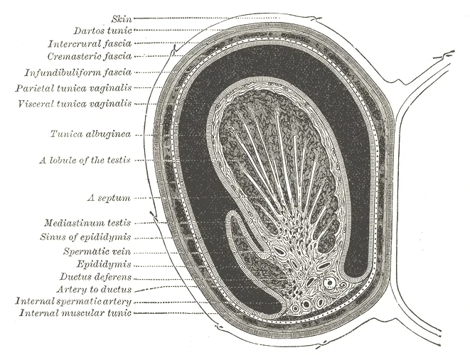
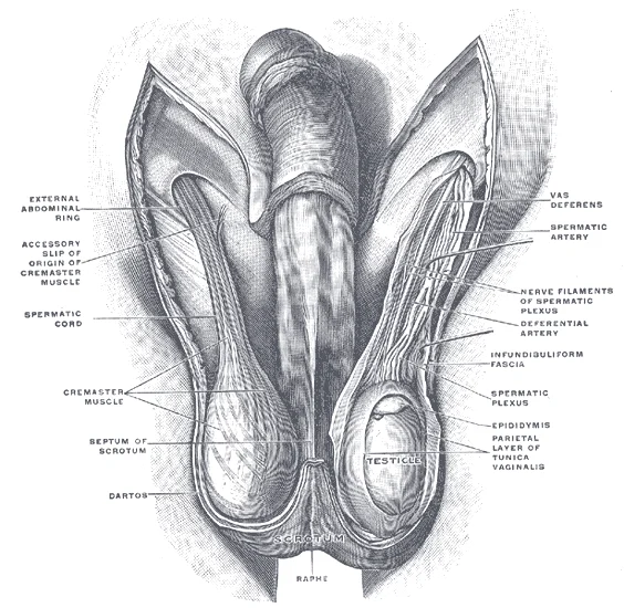
The scrotal wall consists of several layers. The dartos muscle makes up the subcutaneous muscle layer of the scrotum and continues internally to form the scrotal septum, a wall that divides the scrotum into two compartments, each housing one testis[1]. Descending from the internal oblique muscle of the abdominal wall are the two cremaster muscles, which cover each testis like a muscular net[1]. By contracting simultaneously, the dartos and cremaster muscles can elevate the testes in cold weather or water, moving the testes closer to the body and decreasing the surface area of the scrotum to retain heat[1]. Conversely, as the environmental temperature increases, the scrotum relaxes, moving the testes farther from the body core and increasing scrotal surface area to promote heat loss[1]. Externally, the scrotum has a raised medial thickening on the surface called the raphae[1].
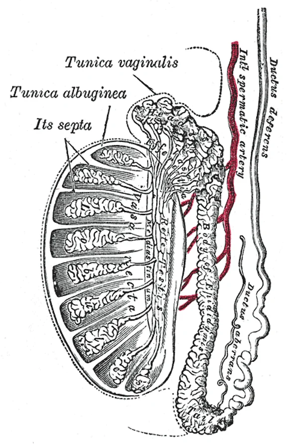
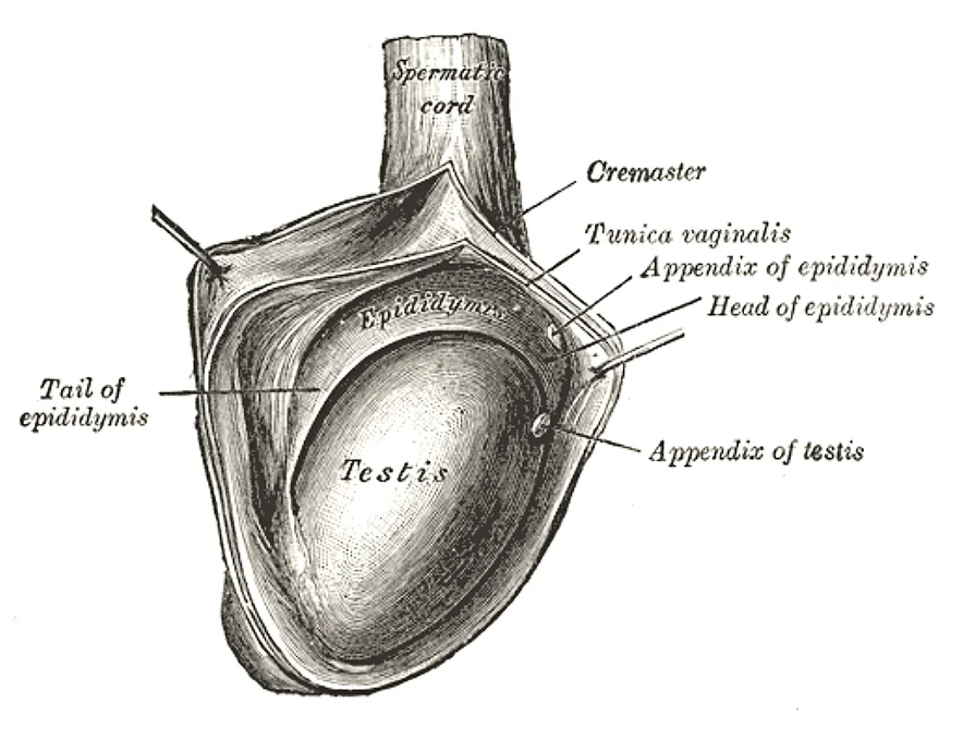
Adult testes are paired ovals, each approximately 4 to 5 cm in length[1]. They are surrounded by two distinct layers of protective connective tissue[1]. The outer tunica vaginalis is a serous membrane that has both a parietal and a thin visceral layer[1]. Beneath the tunica vaginalis is the tunica albuginea, a tough, white, dense connective tissue layer covering the testis itself[1]. The tunica albuginea invaginates to form septa that divide the testis into 300 to 400 structures called lobules[1]. Within the lobules, sperm develop in structures called seminiferous tubules[1].
Figure 1Gray1149
Long description
Vertical section of the testis, to show the arrangement of the ducts.

Figure 1: Vertical section of the testis, to show the arrangement of the ducts.
Figure 2Gray1148
Long description
The right testis, exposed by laying open the tunica vaginalis.

Figure 2: The right testis, exposed by laying open the tunica vaginalis.
Figure 3Gray1145
Long description
Transverse section through the left side of the scrotum and the left testis. The sac of the tunica vaginalis is represented in a distended condition. (Diagrammatic.) (Delépine.)

Figure 3: Transverse section through the left side of the scrotum and the left testis.
Figure 4Gray1144
Long description
The scrotum. The penis has been turned upward, and the anterior wall of the scrotum has been removed. On the right side, the spermatic cord, the infundibuliform fascia, and the Cremaster muscle are displayed; on the left side, the infundibuliform fascia has been divided by a longitudinal incision pa

Figure 4: The scrotum.
Figure 5Gray1143
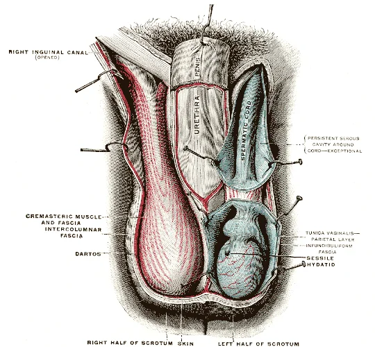
Long description
The scrotum. On the left side the cavity of the tunica vaginalis has been opened; on the right side only the layers superficial to the Cremaster have been removed. (Testut.)

Figure 5: The scrotum.
During the seventh month of the developmental period of a male fetus, each testis moves through the abdominal musculature to descend into the scrotal cavity[1]. This process is called the descent of the testis[1]. Cryptorchidism is the clinical term used when one or both of the testes fail to descend into the scrotum prior to birth[1]. This condition requires attention as undescended testes may not function properly for spermatogenesis.
DefinitionTesticular Descent
The descent of the testis occurs during the seventh month of fetal development, moving through the abdominal musculature into the scrotal cavity[1].
The regulation of testosterone concentrations throughout the body is critical for male reproductive function[1]. The intricate interplay between the endocrine system and the reproductive system ensures that the testes maintain the necessary hormonal environment for sperm production[1]. If testosterone is not present during embryonic development, female sexual organs will develop instead. A gene on the Y chromosome called SRY is critical in stimulating a cascade of events that simultaneously stimulate testis development and repress the development of female structures. Testosterone produced by Leydig cells in the embryonic testis stimulates the development of male sexual organs.
| Muscle | Origin | Action | Nerve Supply |
|---|---|---|---|
| Dartos | Subcutaneous tissue | Elevates scrotum in cold | Autonomic |
| Cremaster | Internal oblique | Elevates testis in cold | Genital branch of genitofemoral |
In this section, the typical reproductive anatomy of XY individuals is discussed, where terms like "male" refer to biological sex. The penis functions as the male organ of copulation, or sexual intercourse. It remains flaccid during non-sexual actions such as urination, and becomes turgid and rod-like with sexual arousal. This stiffness during erection allows the organ to penetrate the vagina and deposit semen into the female reproductive tract. The bulk of semen is produced by three critical accessory glands: the seminal vesicles, the prostate, and the bulbourethral glands. Several accessory organs and ducts aid sperm maturation and transport seminal components to the penis, which may deliver sperm to the female reproductive tract [P2 [1]
DefinitionBiological Sex
In this context, male refers to biological sex as determined by X/Y chromosomes, hormones, sex organs, and physical characteristics.
The penis is positioned centrally inferior to the pubic symphysis, featuring a root, a body or shaft, and a distal expanded tip called the glans penis. Flanking the penis inferiorly is the scrotum, a cutaneous pouch housing the testes. Cells that can become either the glans penis or the glans clitoris form the glans penis when exposed to testosterone, whereas they differentiate into the clitoris without testosterone[1]. Testosterone is a steroid hormone produced by Leydig cells, also termed interstitial cells due to their location between the seminiferous tubules in the testes. Leydig cells secrete testosterone by the seventh week of embryonic development, reaching peak concentrations in the second trimester. [2]
| Structure or Hormone | Source or Cell Type | Primary Function |
|---|---|---|
| Testosterone | Leydig cells (interstitial cells) | Anatomical differentiation and spermatogenesis activation |
| Sustentacular cells | Testicular tissue | Trigger degradation of the female Müllerian duct[1] [2] |
| Accessory Glands | Seminal vesicles, prostate, bulbourethral glands | Produce the bulk of semen volume |
Embryonic Duct Differentiation
- Embryonic Ducts PresentBoth Wolffian and Müllerian duct systems exist initially.
- Question: Testosterone present?
Yes
- Wolffian duct growsForms epididymis, ductus deferens, and seminal vesicles.
No
- Wolffian duct degradesMale internal structures regress.
Figure 6EB1911 Reproductive System, in Anatomy - pelvis section showing bladder and prostate
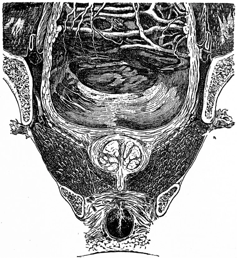
Long description
Coronal section through the pelvis of a human male, showing the relations of the bladder above, prostate and bulb below.

Figure 6: Coronal section through the pelvis of a human male, showing the relations of the bladder above, prostate and bulb below.
DefinitionPeritoneal and visceral relations
The male pelvic viscera exhibit precise anatomical relationships with the peritoneum superiorly and adjacent pelvic structures anteriorly, posteriorly, and laterally.
The urinary bladder lies most anteriorly behind the pubic bones, covered superiorly by the peritoneum which reflects onto the anterior abdominal wall. Directly inferior to the urinary bladder sits the prostate gland, which surrounds the prostatic urethra.
This enlargement does not usually cause problems; however, abnormal growth of the prostate, or benign prostatic hyperplasia (BPH), can cause constriction of the urethra as it passes through the middle of the prostate gland, leading to a number of lower urinary tract symptoms, such as a frequent and intense urge to urinate, a weak stream, and a sensation that the bladder has not emptied completely. By age 60, approximately 40 percent of males have some degree of BPH. By age 80, the number of affected individuals has jumped to as many as 80 percent. Treatments for BPH attempt to relieve the pressure on the urethra so that urine can flow more normally. [1]
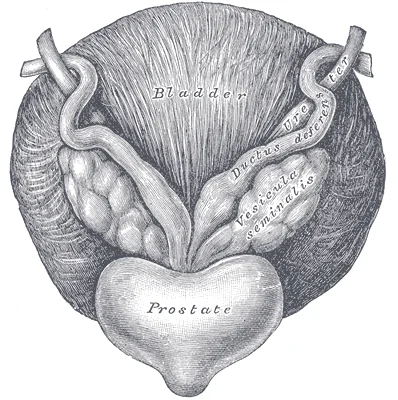
The paired seminal vesicles are glands that contribute approximately 60 percent of the semen volume. Seminal vesicle fluid contains large amounts of fructose. As sperm pass through the ampulla of the ductus deferens at ejaculation, they mix with fluid from the associated seminal vesicle. [1]
| Structure | Anterior Relations | Posterior Relations | Superior Relations |
|---|---|---|---|
| Urinary bladder | Pubic symphysis | Rectum, seminal vesicles | Peritoneum, loops of ileum |
| Prostate gland | Pubic bones, retropubic space | Rectum | Urinary bladder neck |
| Seminal vesicles | Urinary bladder base | Rectum | Peritoneum of rectovesical pouch |
Pelvic Viscera Arrangement
- Anterior: Urinary BladderRests behind pubic symphysis, inferiorly related to prostate.
- Question: Midline Structures
Prostate
- Surrounds prostatic urethra, inferior to bladder neck.
Seminal Vesicles
- Posterior to bladder base, superior to prostate.
- Posterior: RectumSeparated from prostate and seminal vesicles by rectovesical pouch.
Figure 7EB1911 Reproductive System, in Anatomy - testis and epididymis
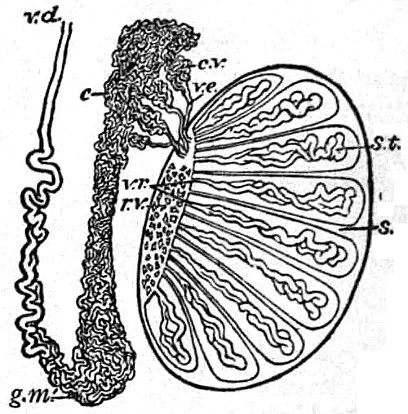
Long description
Diagram to illustrate the structure of the testis and epididymis (components of the reproductive system of the human male).

Figure 7: Diagram to illustrate the structure of the testis and epididymis (components of the reproductive system of the human male).
The interior of the male reproductive system features the testes, which form the bulk of the male gonadal tissue. Spermatogenesis occurs in the seminiferous tubules that form the bulk of each testis. The process begins at puberty, after which time sperm are produced constantly throughout a male's life. One production cycle, from spermatogonia through formed sperm, takes approximately 64 days. A new cycle starts approximately every 16 days, although this timing is not synchronous across the seminiferous tubules. [1]
Spermatogenesis begins with mitotic division of spermatogonia (stem cells) to produce primary spermatocytes that undergo the two divisions of meiosis to become secondary spermatocytes, then the haploid spermatids. During spermiogenesis, spermatids are transformed into spermatozoa (formed sperm). Early spermatids look very similar to cells in the earlier stages of spermatogenesis, with a round shape, central nucleus, and large amount of cytoplasm. A process called spermiogenesis transforms these early spermatids, reducing the cytoplasm, and beginning the formation of the parts of a true sperm. The fifth stage of germ cell formation—spermatozoa, or formed sperm—is the end result of this process, which occurs in the portion of the tubule nearest the lumen. Eventually, the sperm are released into the lumen and are moved along a series of ducts in the testis toward a structure called the epididymis for the next step of sperm maturation. [1]
The seminiferous tubules converge centrally within each testis lobule. From the tubules, sperm enter the straight tubules and the rete testis, located in the mediastinum testis posteriorly. Efferent ductules then carry the sperm out of the testis superiorly into the head of the epididymis. The epididymis lies applied to the posterior and superior border of the testis, running as a comma-shaped structure divided into a head superiorly, a body centrally, and a tail inferiorly. The more mature sperm are then stored in the tail of the epididymis (the final section) until ejaculation occurs. [1]
DefinitionLeydig cell function
Leydig cells produce approximately 6 to 7 mg of testosterone per day. [1]
Testicular steroidogenesis (the manufacture of androgens, including testosterone) results in testosterone concentrations that are 100 times higher in the testes than in the circulation. Maintaining these normal concentrations of testosterone promotes spermatogenesis, whereas low levels of testosterone can lead to infertility. [1]
| Parameter | Value |
|---|---|
| Duration of one spermatogenic cycle | Approximately 64 days |
| Timing of new cycle initiation | Approximately every 16 days |
| Daily testosterone production | Approximately 6 to 7 mg per day |
| Intratesticular vs circulating testosterone | 100 times higher in the testes |
The central structure is the prostate gland, situated anterior to the rectum at the base of the bladder, surrounding the prostatic urethra. The arterial supply to the testes originates from the testicular arteries, which arise directly from the abdominal aorta. These vessels descend through the inguinal canal to reach the testes. The prostate gland receives its blood supply primarily from the inferior vesical arteries, branches of the internal iliac artery. The bulbourethral glands, located posterolateral to the membranous urethra, are supplied by branches of the internal pudendal artery. The penis is supplied by the internal pudendal artery, which gives rise to the deep artery of the penis and the dorsal artery of the penis. The corpora cavernosa and corpus spongiosum are engorged during erection due to increased arterial inflow.
Figure 8Gray1158
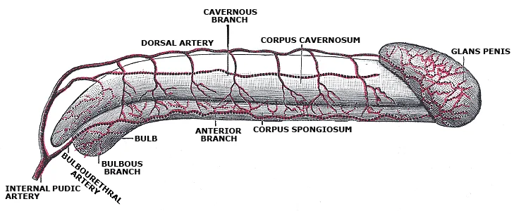
Long description
Diagram of the arteries of the penis. (Testut.)

Figure 8: Diagram of the arteries of the penis (Testut.).
Figure 9Gray1156
Long description
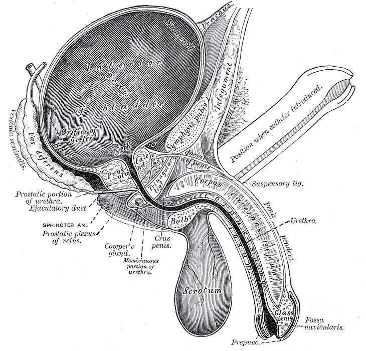
Vertical section of bladder, penis, and urethra.

Figure 9: Vertical section of bladder, penis, and urethra.
Figure 10Gray1155
Long description
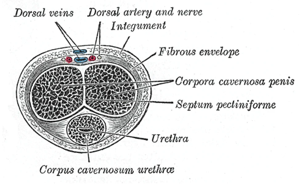
Transverse section of the penis. Labelled illustration from Wikimedia Commons, used under its open licence.

Figure 10: Transverse section of the penis.
Figure 11Gray1154
Long description
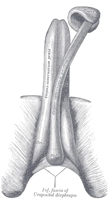
The constituent cavernous cylinders of the penis. The glans and anterior part of the corpus cavernosum urethræ are detached from the corpora cavernosa penis and turned to one side.

Figure 11: The constituent cavernous cylinders of the penis.
Figure 12Gray1152
Long description
Fundus of the bladder with the vesiculæ seminales.

Figure 12: Fundus of the bladder with the vesiculæ seminales.
The arterial supply to the male reproductive organs is complex and region-specific. The testes are supplied by the testicular arteries, which are unique in arising directly from the abdominal aorta. The prostate gland is supplied by the inferior vesical arteries, which branch from the internal iliac artery. These vessels penetrate the prostate capsule to supply the glandular and muscular tissues. The bulbourethral glands receive blood from the internal pudendal artery, specifically from branches that supply the urogenital diaphragm. The penis is supplied by the internal pudendal artery, which enters the perineum and gives rise to the deep artery of the penis. This artery runs within the corpus cavernosum and supplies the erectile tissue. The corpus spongiosum is supplied by the artery of the bulb, a branch of the internal pudendal artery.
The regulation of blood flow to the erectile tissues is critical for sexual function. During sexual arousal, nitric oxide is released from nerve endings near blood vessels within the corpora cavernosa and spongiosum. This nitric oxide activates a signaling pathway that results in relaxation of the smooth muscles surrounding the penile arteries. This dilation increases the amount of blood that can enter the penis. The endothelial cells in the penile arterial walls also secrete nitric oxide, perpetuating the vasodilation. This process leads to vasocongestion, or engorgement of the tissues because of more arterial blood flowing into the penis than is leaving in the veins. The increased pressure within the corpora cavernosa compresses the emissary veins against the tunica albuginea, reducing venous outflow and maintaining the erection. [1]
DefinitionVasocongestion
Engorgement of tissues due to increased arterial inflow exceeding venous outflow, leading to erection.
The prostate gland sits anterior to the rectum at the base of the bladder surrounding the prostatic urethra. It is formed of both muscular and glandular tissues. The glandular tissue secretes an alkaline, milky fluid that is critical to first coagulate and then decoagulate the semen following ejaculation. The temporary thickening of semen helps retain it within the female reproductive tract.
| Structure | Arterial Supply | Origin |
|---|---|---|
| Testes | Testicular arteries | Abdominal aorta |
| Prostate | Inferior vesical arteries | Internal iliac artery |
| Bulbourethral glands | Internal pudendal artery branches | Internal iliac artery |
| Penis | Internal pudendal artery | Internal iliac artery |
Blood supply to the male reproductive organs, including the testes and penis, is critical for both baseline metabolic function and physiological responses such as erection. During an erection, a rapid increase in blood volume fills the erectile chambers, and the increased pressure of the filled chambers compresses the thin-walled penile venules, preventing venous drainage of the penis. The result of this increased blood flow to the penis and reduced blood return from the penis is erection. Depending on the flaccid dimensions of a penis, it can increase in size slightly or greatly during erection, with the average length of an erect penis measuring approximately 15 cm. [1]
Figure 13Gray1157
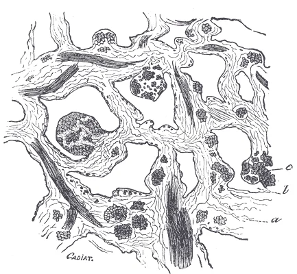
Long description
Section of corpus cavernosum penis in a non-distended condition. (Cadiat.) a. Trabeculæ of connective tissue, with many elastic fibers and bundles of plain muscular tissue, some of which are cut across ( c ). b. Blood sinuses.

Figure 13: Section of corpus cavernosum penis in a non-distended condition (Cadiat.) a.
Flanking the deep dorsal vein are the paired dorsal arteries and nerves. The prostatic venous plexus surrounds the inferolateral surfaces of the prostate gland and communicates closely with the vesical plexus of the urinary bladder. From this interconnected network, blood flows into the paired internal iliac veins, which lie posteromedial to the internal iliac arteries on the lateral pelvic wall. The internal iliac veins then ascend to join the common iliac veins, ultimately returning blood to the inferior vena cava.
Figure 14Gray1147
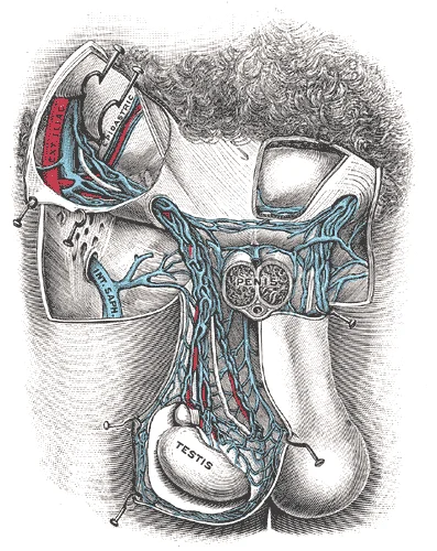
Long description
Spermatic veins. (Testut.). Labelled illustration from Wikimedia Commons, used under its open licence.

Figure 14: Spermatic veins (Testut.).
Figure 15Gray1159
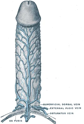
Long description
Veins of the penis. (Testut.). Labelled illustration from Wikimedia Commons, used under its open licence.

Figure 15: Veins of the penis (Testut.).
DefinitionVenous Occlusion
The physiological mechanism of penile erection relies entirely on the compression of thin-walled venules by expanding sinusoidal chambers, effectively halting venous outflow.
Figure 16Gray1136
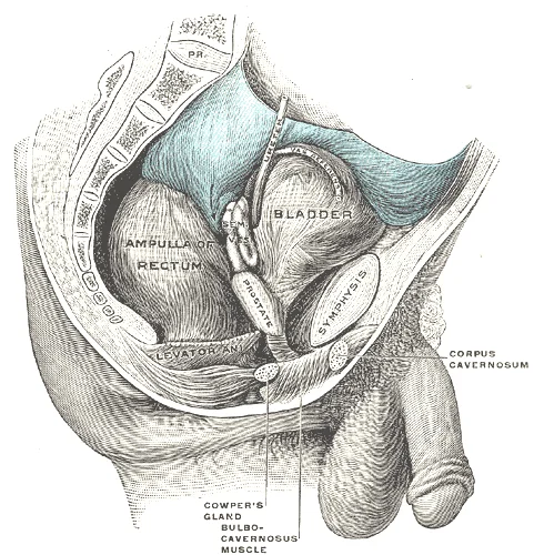
Long description
Male pelvic organs seen from right side. Bladder and rectum distended; relations of peritoneum to the bladder and rectum shown in blue. The arrow points to the rectovesical pouch.

Figure 16: Male pelvic organs seen from right side.
The lymphatic architecture of the male internal genitalia reflects its embryological ascent from the posterior abdominal wall. Testicular lymphatics ascend within the spermatic cord as part of the testicular vascular pedicle. They cross the pelvic brim to reach the posterior abdominal wall.
DefinitionRetroperitoneal destination
Testicular lymph drains primarily into the lumbar or para-aortic nodes, mirroring the high posterior abdominal origin of the gonad.
The testis lies within the scrotum, and its primary lymphatic vessels ascend directly through the inguinal canal inside the spermatic cord.
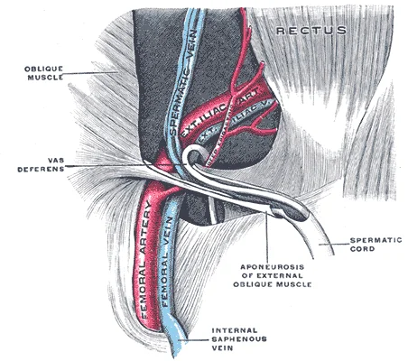
Figure 17Gray1146
Long description
The spermatic cord in the inguinal canal. (Poirier and Charpy.)

Figure 17: The spermatic cord in the inguinal canal (Poirier and Charpy.).
When evaluating pathological conditions, clinicians must remember that testicular cancer metastases bypass pelvic nodes and spread directly to retroperitoneal para-aortic nodes. Conversely, malignancies of the scrotum drain to superficial inguinal nodes. Low-resource settings rely heavily on careful physical examination of these nodal basins when advanced imaging is unavailable.
| Structure | Primary Drainage Node | Alternative Drainage Node |
|---|---|---|
| Testis | Para-aortic nodes | None |
| Scrotum | Superficial inguinal nodes | None |
Testicular Lymphatic Pathway
- TestisOrigin of lymphatic vessels within the organ.
- Spermatic cordAscends through the inguinal canal.
- Posterior abdominal wallCrosses over the psoas major muscle.
- Para-aortic nodesTermination site at the level of lumbar vertebra two.
Unlike the testis, the scrotum and distal penile skin drain differently. Their lymphatic channels track downward and laterally to empty into the superficial inguinal lymph nodes situated in the femoral triangle. Recognising this anatomical split is vital for staging testicular malignancies versus scrotal skin lesions. Additional considerations apply in older adults and children, where altered tissue planes or congenital malformations can occasionally shift expected lymphatic pathways.
===CLAims===
DefinitionAutonomic and Somatic Control
The male reproductive organs receive a dual nerve supply consisting of autonomic fibers for involuntary control and somatic fibers for voluntary movement and sensation.
These pathways merge within the pelvic plexuses to reach the target organs.
Somatic supply travels via the pudendal nerve. This nerve provides motor innervation to the perineal muscles and sensory innervation to the skin of the external genitalia.
Referred pain occurs because visceral afferents from the reproductive organs enter the same spinal cord segments as somatic afferents from overlying dermatomes. For example, pathology within deep pelvic structures can be perceived as pain radiating to the lower back or thighs.
Follow the cavernous nerves as they pierce the perineal membrane to enter the root of the penis, lying posterolateral to the membranous urethra. Identify the pudendal nerve exiting the greater sciatic foramen, crossing the ischial spine, and entering the Alcock canal on the lateral wall of the ischioanal fossa alongside the internal pudendal vessels.
| Component | Nerve Source | Main Function |
|---|---|---|
| Corpus cavernosum | Parasympathetic S2-S4 | Erection via vasodilation |
| Corpus spongiosum | Parasympathetic S2-S4 | Erection and urethral support |
| Glans penis | Pudendal nerve (S2-S4) | Cutaneous sensation |
| Prepuce | Pudendal nerve (S2-S4) | Protective sensory feedback |
link to the histology page.
DefinitionPubertal onset
Pubertal changes begin around the age of eight or nine when luteinizing hormone production becomes detectable. [2]
The first physiological changes of puberty are preceded years prior by nocturnal releases of luteinizing hormone during sleep. In pre-pubertal children, the hypothalamic-pituitary negative feedback system demonstrates very high sensitivity, meaning minimal concentrations of androgens or estrogens suppress gonadotropin-releasing hormone, luteinizing hormone, and follicle-stimulating hormone production. [2]
As individuals approach puberty, sensitivity in the hypothalamus and pituitary to negative feedback decreases, requiring progressively larger concentrations of sex steroid hormones to halt gonadotropin production. Simultaneously, the gonads increase in sensitivity to follicle-stimulating hormone and luteinizing hormone signals, becoming far more responsive than during childhood. These dual shifts drive slowly increasing gonadotropin levels, leading to gonadal enlargement, maturation, elevated sex hormone secretion, and the initiation of spermatogenesis. [1] [2]
Pubertal timing is influenced by genetics, environment, psychological stress, and nutrition. Historical United States data illustrate how improved nutrition lowered the average age of menarche from approximately 17 years in 1860 down to approximately 12.75 years by 1960. Additionally, stored body fat correlates with the secretion of leptin by adipose cells, playing a strong contributory role in triggering maturation across both sexes. Different sex hormone concentrations across the sexes also drive the development and function of secondary sexual characteristics. [1] [2]
| Factor | Mechanism or historical shift |
|---|---|
| Nutrition | Improved nutrition decreased menarche age from 17 years in 1860 to 12.75 years in 1960 |
| Adipose tissue | Stored body fat corresponds with leptin secretion by adipose cells to influence maturation |
| Feedback sensitivity | Hypothalamic and pituitary sensitivity to negative feedback decreases as puberty approaches |
Endocrine cascade of puberty
- Age 8 to 9Detectable nocturnal LH release begins.
- Question: Feedback sensitivity?
High
- Pre-pubertal state: low GnRH, LH, and FSH.
Decreased
- Rising LH and FSH drive gonadal maturation and spermatogenesis.
Embryonic formation of the reproductive tract begins early in gestation, establishing the foundational architecture for future physiological function. For more information and a figure of differentiation of the gonads, seek additional content on fetal development. This early phase is critical, as errors in differentiation can lead to significant congenital anomalies. Understanding this binary decision-making process is essential for diagnosing disorders of sex development. [2]
Within the testes, gametogenesis takes place inside specialized compartments that maintain a unique microenvironment. The sustentacular cells, also known as Sertoli cells, play a pivotal role in nurturing the developing spermatozoa and forming the blood-testis barrier. This barrier is crucial for preventing an autoimmune response against the haploid germ cells, which express novel antigens not recognized by the immune system during fetal life. [1]
Germ cell development progresses from the basement membrane—at the perimeter of the tubule—toward the lumen. This directional migration is a hallmark of spermatogenesis. Immature germ cells, such as spermatogonia, reside near the basement membrane, where they undergo mitotic division to maintain the stem cell pool. As they differentiate into spermatocytes and then spermatids, they move progressively inward. Let’s look more closely at these cell types. The final stage involves the release of mature spermatozoa into the lumen, a process known as spermiation. This organized progression ensures that only morphologically normal sperm are released into the reproductive tract. [1]
Clinical pearlTubular arrangement
The structural organization of the seminiferous tubule ensures that immature germ cells reside peripherally while mature spermatozoa are released into the central lumen.
This spatial arrangement allows for efficient nutrient exchange and waste removal. The basement membrane provides structural support and acts as a selective filter. Any disruption in this gradient can lead to impaired spermatogenesis and infertility. In clinical practice, understanding this histological layout helps in interpreting biopsy results. For instance, a lack of germ cells near the lumen may indicate a blockage or maturation arrest. Conversely, an accumulation of cells at the basement membrane might suggest a defect in differentiation.
| Cell Type | Location | Primary Function |
|---|---|---|
| Sustentacular Cells | Adjacent to basement membrane | Support, nourishment, blood-testis barrier |
| Spermatogonia | Basement membrane | Stem cell reservoir, mitosis |
| Primary Spermatocytes | Near basement membrane | Meiosis I |
| Secondary Spermatocytes | Middle layers | Meiosis II |
| Spermatids | Near lumen | Spermiogenesis |
| Spermatozoa | Lumen | Motility, fertilization |
This table summarizes the hierarchical organization within the tubule. Each cell type has a distinct role in the production of viable sperm. The transition from one stage to the next is tightly regulated by hormonal signals, particularly follicle-stimulating hormone and testosterone. Disruptions in these signals can halt development at any stage, leading to azoospermia or oligospermia. Recognizing these patterns is vital for diagnosing male infertility. [1]
Radiological imaging of the male reproductive organs relies on cross-sectional modalities, particularly magnetic resonance imaging, which offers superior soft-tissue contrast resolution compared to computed tomography.
DefinitionSagittal Pelvic Plane
A midline sagittal slice reveals the spatial relationships between the urinary bladder, prostate gland, rectum, and external genitalia in a single continuous plane.
Immediately inferior to the base of the urinary bladder lies the prostate gland, which displays a zonal architecture on high-resolution T2-weighted magnetic resonance sequences.
| Structure | T2 Signal Characteristics | Anatomical Relation |
|---|---|---|
| Urinary bladder | High fluid signal | Superior to the prostate gland |
| Prostatic peripheral zone | High signal intensity | Posterior and lateral aspect of the prostate |
| Prostatic transition zone | Low to intermediate signal | Surrounds the proximal prostatic urethra |
| Seminal vesicles | High signal intensity fluid | Posterior to the urinary bladder base |
Pelvic Organ Identification
- Locate urinary bladderIdentify the fluid-filled anterior structure.
- Question: Structure inferior to bladder?
Yes
- Identify prostate gland and evaluate zonal architecture
No
- Scan posteriorly for seminal vesicles
Posterior to the prostate and seminal vesicles, the rectum descends in the curve of the sacrum and coccyx, separated from the reproductive structures by the rectovesical fascia. Familiarity with these radiological landmarks enables accurate staging of pelvic pathology and precise identification of anatomical boundaries during surgical and interventional procedures.
The external genitalia comprise the scrotum and the penis. The scrotum is a cutaneous pouch divided into two lateral compartments by a midline septum, housing the testes and lower spermatic cords. The penis consists of three cylindrical bodies of cavernous erectile tissue enclosed by deep fascia, termed Buck fascia, and skin. Two dorsal corpora cavernosa and one ventral corpus spongiosum, which expands distally to form the glans penis, make up the erectile framework.
The testis is an ovoid reproductive organ suspended within the scrotum by the spermatic cord. Each testis is surrounded by a tough fibrous capsule, the tunica albuginea. Septa extend from this capsule to divide the interior into about 250 conical lobules, each containing one to four seminiferous tubules where spermatogenesis occurs.
DefinitionTunica albuginea
A dense fibrous capsule that envelops the testis and sends inward septa to compartmentalize the glandular parenchyma into lobules.
The skin forms the outermost covering, deep to which lies the dartos muscle blended with superficial fascia. Successively deeper are the external spermatic fascia, cremasteric muscle and fascia, and internal spermatic fascia derived from the abdominal wall layers. Deep to the internal spermatic fascia is the parietal layer of the tunica vaginalis, separated by a potential cavity containing serous fluid from the visceral layer adherent to the tunica albuginea. The tunica albuginea encases the testicular parenchyma, which converges posteriorly at the mediastinum testis. Within the testis, convoluted seminiferous tubules drain via straight tubules into the rete testis, which exits through efferent ductules into the epididymis. The epididymis lies posterolateral to the testis, consisting of a superior head, intermediate body, and inferior tail continuous with the ductus deferens. Accompanying vessels include the testicular artery and the pampiniform plexus, embedded within the spermatic cord structures. [1]
Clinical pearlSpermatogonia mitosis
Two identical diploid cells result from spermatogonia mitosis. One of these cells remains a spermatogonium, while the other becomes a primary spermatocyte, representing the next stage in the process of spermatogenesis. [1]
Clinical pearlMeiosis I chromosome separation
This process results in two distinct cells, called secondary spermatocytes, each carrying only half the number of chromosomes. [1]
Clinical pearlMeiosis II division
A second round of cell division, known as meiosis II, occurs in both of the secondary spermatocytes. [1]
Clinical pearlSpermatid formation
This second meiotic division results in a total of four cells possessing only half of the original number of chromosomes, and each of these new cells is designated as a spermatid. [1]
Clinical pearlMitochondrial energy supply
Adenosine triphosphate produced by the mitochondria powers the flagellum, which extends continuously from the neck and the mid-piece through the tail of the sperm, thereby enabling the entire sperm cell to move. [1]
Exam trapMeiosis chromosome trap
Medical students often confuse the stages of division; remember that meiosis I separates the 23 pairs of chromosomes, whereas meiosis II divides each of the 23 already replicated chromosomes.
Exam trapSpermatid yield trap
A common misconception is that a single secondary spermatocyte produces four cells; in reality, the complete sequence starting from one primary spermatocyte through meiosis I and meiosis II yields a total of four spermatids.
Students frequently confuse the anatomical continuity of the male and female reproductive tracts, leading to errors in embryology and clinical examination. A primary error is assuming the labia minora and clitoris are homologous to the scrotum and penis rather than the glans penis and prepuce. The superior, anterior portions of the labia minora come together to encircle the clitoris (or glans clitoris), an organ that originates from the same cells as the glans penis and has abundant nerves that make it important in sexual sensation and orgasm. This homology explains why conditions affecting one structure often have parallels in the other, such as cysts or infections. [3]
Another frequent mistake involves the permanence of sterilisation procedures. Students may underestimate the psychological and surgical finality of a vasectomy. Because the ductus deferens is physically accessible within the scrotum, surgical sterilization to interrupt sperm delivery can be performed by cutting and sealing a small section of the ductus (vas) deferens. Although it may be possible to reverse a vasectomy, clinicians consider the procedure permanent, and advise people to undergo it only if they are certain they no longer wish to have children. This distinction is vital for pre-operative counselling. [1]
Misunderstanding the menstrual cycle phases often leads to incorrect interpretations of hormonal feedback loops. A common error is believing that the corpus luteum produces estrogen primarily during the luteal phase. Instead, the surge of LH also stimulates a change in the granulosa and theca cells that remain in the follicle after the oocyte has been ovulated. This change is called luteinization, and it transforms the collapsed follicle into a new endocrine structure called the corpus luteum, a term meaning “yellowish body”. Instead of estrogen, the luteinized granulosa and theca cells of the corpus luteum begin to produce large amounts of the sex steroid hormone progesterone, a hormone that critical for the establishment and maintenance of pregnancy. [3]
Students also often confuse the layers of the endometrium and their shedding patterns. It is incorrect to assume the entire endometrium sheds during menstruation. This inner functional layer provides the proper site of implantation for the fertilized egg, and—should fertilization not occur—it is only the stratum functionalis layer of the endometrium that sheds during menstruation. The stratum basalis remains intact to regenerate the functionalis. [3]
Finally, learners frequently misidentify the site of fertilisation within the uterine tubes. While the infundibulum captures the oocyte, fertilisation rarely occurs there. The wide distal infundibulum flares out with slender, finger-like projections called fimbriae. The middle region of the tube, called the ampulla, is where fertilization often occurs. Recognising this anatomy is crucial for understanding ectopic pregnancy locations. [3]
In the operating theatre, knowledge of the spermatic cord is essential for hernia repairs and vasectomies. During a vasectomy, the surgeon isolates the ductus deferens within the spermatic cord. The ductus deferens is a thick, muscular tube that is bundled together inside the scrotum with connective tissue, blood vessels, and nerves into a structure called the spermatic cord. Identifying the vas deferens among the other cord structures prevents injury to the testicular artery and pampiniform plexus. [1]
On imaging, particularly ultrasound, understanding the uterine tube anatomy helps in diagnosing hydrosalpinx or ectopic pregnancy. The uterine tubes (also called fallopian tubes or oviducts) serve as the conduit of the oocyte from the ovary to the uterus. The isthmus is the narrow medial end of each uterine tube that is connected to the uterus. The wide distal infundibulum flares out with slender, finger-like projections called fimbriae. The middle region of the tube, called the ampulla, is where fertilization often occurs. Radiologists look for dilation in the ampulla or fluid in the fimbriae. [3]
At the bedside, clinical examination of the scrotum relies on knowing the contents of the spermatic cord. The ductus deferens is physically accessible within the scrotum, allowing for palpation during a vasectomy or hernia assessment. During ejaculation, sperm exit the tail of the epididymis and are pushed by smooth muscle contraction to the ductus deferens. Palpating the thick, muscular tube helps confirm patency or identify abnormalities.
In examinations, questions often test the hormonal regulation of the ovarian cycle to predict clinical outcomes. During the follicular phase of the ovarian cycle, the tertiary follicles are growing and secreting estrogen. At the same time, the stratum functionalis of the endometrium is thickening to prepare for a potential implantation. The post-ovulatory increase in progesterone, which characterizes the luteal phase, is key for maintaining a thick stratum functionalis. Understanding this sequence allows students to answer questions about amenorrhoea or abnormal bleeding. [3]
Furthermore, knowledge of the hymen is relevant in gynaecological history taking. The hymen is a thin membrane that sometimes partially covers the entrance to the vagina. An intact hymen cannot be used as an indication of “virginity”; even at birth, this is only a partial membrane, as menstrual fluid and other secretions must be able to exit the body, regardless of penile–vaginal intercourse. This fact is critical for sensitive patient communication and avoiding diagnostic bias. [3]
The menses phase of the menstrual cycle is the phase during which the lining is shed; that is, the days that the person menstruates. Although it averages approximately five days, the menses phase can last from 2 to 7 days, or longer. This range is important for distinguishing normal variation from pathological menorrhagia. [3]
DefinitionVasectomy
Surgical sterilization involving cutting and sealing the ductus deferens, considered permanent despite potential reversibility.
| Region | Description | Clinical Significance |
|---|---|---|
| Isthmus | Narrow medial end connected to uterus | Site of tubal ligation sometimes |
| Ampulla | Middle region, wider than isthmus | Most common site of fertilization |
| Infundibulum | Wide distal end with fimbriae | Captures oocyte from ovary |
Ovarian Cycle Hormonal Control
- Follicular PhaseTertiary follicles grow, secrete estrogen
- Question: Estrogen high enough?
Yes
- Positive FeedbackTriggers LH surge from anterior pituitary
No
- Continue GrowthNegative feedback maintains cycle
- LH SurgeInduces ovulation and luteinization
- Question: Ovulation occurs?
Yes
- Corpus Luteum FormationGranulosa/theca cells produce progesterone
No
- Cycle DisruptionAnovulation or luteinized unruptured follicle
- Luteal PhaseProgesterone maintains stratum functionalis
- Question: Implantation?
Yes
- Maintain Corpus LuteumhCG signals continued progesterone production
No
- Corpus Luteum DegradesProgesterone drops, menstruation begins
The ductus deferens is bundled in the spermatic cord with vessels and nerves. Vasectomy is considered permanent despite potential reversibility. The clitoris is homologous to the glans penis and has abundant nerves. The hymen is a partial membrane and does not indicate virginity. The ampulla of the uterine tube is the common site of fertilization. Only the stratum functionalis of the endometrium sheds during menstruation. The corpus luteum produces progesterone, not estrogen, after ovulation. Menstruation lasts 2 to 7 days, averaging five days.
Answer each one in your head first, then open the answer.
What structure is bundled with the ductus deferens in the spermatic cord?
Show answerHide answer
Connective tissue, blood vessels, and nervesWhich part of the uterine tube is the narrow medial end connected to the uterus?
Show answerHide answer
The isthmusWhat hormone does the corpus luteum primarily produce?
Show answerHide answer
ProgesteroneWhere does fertilization most commonly occur in the uterine tube?
Show answerHide answer
The ampullaWhat is the typical duration of the menses phase?
Show answerHide answer
2 to 7 days, averaging five days
- Betts JG, Young KA, Wise JA, et al. Anatomy and Physiology of the Testicular Reproductive System. OpenStax, Anatomy and Physiology 2e (CC BY-NC-SA 4.0). Accessed .
- Betts JG, Young KA, Wise JA, et al. Development of the Male and Female Reproductive Systems. OpenStax, Anatomy and Physiology 2e (CC BY-NC-SA 4.0). Accessed .
- Betts JG, Young KA, Wise JA, et al. Anatomy and Physiology of the Ovarian Reproductive System. OpenStax, Anatomy and Physiology 2e (CC BY-NC-SA 4.0). Accessed .
Figure credits
- Figure 1: Henry Vandyke Carter, via Wikimedia Commons, Public domain, Public domain.
- Figure 2: Henry Vandyke Carter, via Wikimedia Commons, Public domain, Public domain.
- Figure 3: Henry Vandyke Carter, via Wikimedia Commons, Public domain, Public domain.
- Figure 4: Henry Gray (1918) Anatomy of the Human Body (See "Book" section below) Bartleby.com: Gray's Anatomy, Plate 1144, via Wikimedia Commons, Public domain, Public domain.
- Figure 5: Henry Vandyke Carter, via Wikimedia Commons, Public domain, Public domain.
- Figure 6: From C. S. Wallace's Prostatic Enlargement, via Wikimedia Commons, Public domain, Public domain.
- Figure 7: From A. F. Dixon, Cunningham's Text-book of Anatomy., via Wikimedia Commons, Public domain, Public domain.
- Figure 8: Henry Vandyke Carter, via Wikimedia Commons, Public domain, Public domain.
- Figure 9: Henry Vandyke Carter, via Wikimedia Commons, Public domain, Public domain.
- Figure 10: Henry Vandyke Carter, via Wikimedia Commons, Public domain, Public domain.
- Figure 11: Henry Vandyke Carter, via Wikimedia Commons, Public domain, Public domain.
- Figure 12: Henry Vandyke Carter, via Wikimedia Commons, Public domain, Public domain.
- Figure 13: Henry Vandyke Carter, via Wikimedia Commons, Public domain, Public domain.
- Figure 14: Henry Vandyke Carter, via Wikimedia Commons, Public domain, Public domain.
- Figure 15: Henry Vandyke Carter, via Wikimedia Commons, Public domain, Public domain.
- Figure 16: Henry Vandyke Carter, via Wikimedia Commons, Public domain, Public domain.
- Figure 17: Henry Vandyke Carter, via Wikimedia Commons, Public domain, Public domain.
Educational content for medical students and doctors. It is not a substitute for clinical judgement or local guidelines.