Female reproductive organs
Anatomy of female reproductive organs including uterus, tubes, ovaries and supporting ligaments.
The more granulosa and theca cells a follicle has, the more estrogen it will produce in response to LH stimulation. [1]
DefinitionAdnexa
The term adnexa refers to the uterine tubes and ovaries, which are critical sites for ectopic pregnancy and adnexal torsion. [1]
The infundibulopelvic ligament suspends the ovary from the pelvic wall, containing the ovarian vessels. [1]
The uterus, also known as the Müllerian structure, develops from the Müllerian ducts. In typical female development, the Müllerian duct develops while the Wolffian duct degrades. This occurs in the absence of testosterone secretion from sustentacular cells. The internal reproductive structures, including the uterus and uterine tubes, form from these rudimentary duct systems. [2]
| Structure | Description |
|---|---|
| Infundibulopelvic | Suspends ovary to pelvic wall; contains ovarian vessels |
| Utero-ovarian | Connects ovary to uterus |
| Broad ligament | Peritoneal fold enclosing tubes and ovaries |
| Round ligament | Connects uterus to labia majora |
The lobes are surrounded by fat tissue, which determines the size of the breast; breast size differs between individuals and does not affect the amount of milk produced. [1]
NoteClinical Pearl
Adnexal torsion is a surgical emergency requiring prompt diagnosis to preserve ovarian function.
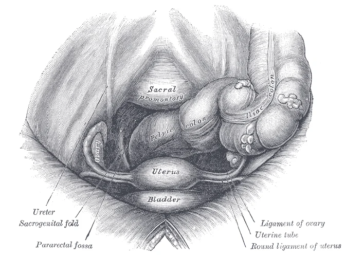
Surface marking of the uterus involves the pelvic cavity boundaries. The ovaries are located in the ovarian fossa on the lateral pelvic wall. The uterine tubes extend laterally from the uterus toward the ovaries. The nearby uterine tube receives the oocyte. [1]
The external female reproductive structures are referred to collectively as the vulva[1]. Anteriorly, the mons pubis is a pad of fat located over the pubic bone, which becomes covered in pubic hair after puberty[1]. Posterior to the mons pubis are the labia majora, which are folds of hair-covered skin[1].
Surface marking of the uterus involves the pelvic cavity boundaries. The ovaries are located in the ovarian fossa on the lateral pelvic wall. The uterine tubes extend laterally from the uterus toward the ovaries. The ampulla of the fallopian tubes is the site where fertilization typically occurs[1].
The uterine tubes have three layers: an outer serosa, a middle smooth muscle layer, and an inner mucosal layer[1].
In addition to its mucus-secreting cells, the inner mucosa contains ciliated cells that beat in the direction of the uterus, producing a current that is critical to move the oocyte[1]. Beating cilia help to transport the oocyte through the tube toward the uterus[1]. Fertilization occurs within the uterine tube, and the final stage of meiosis is completed[1]. If the oocyte is successfully fertilized, the resulting zygote will begin to divide into two cells, then four, and so on, as it makes its way through the
The ovary is suspended in the broad ligament, a double fold of peritoneum that extends laterally from the uterus to the pelvic wall. The ovary’s position is maintained by two key ligaments. The infundibulopelvic ligament, also known as the suspensory ligament of the ovary, connects the superior pole of the ovary to the pelvic wall. It contains the ovarian vessels, nerves, and lymphatics. The utero-ovarian ligament connects the inferior pole of the ovary to the lateral aspect of the uterus, just below the uterine tube. These ligaments anchor the ovary within the ovarian fossa on the lateral pelvic wall. [1]
The ovary itself comprises an outer covering of cuboidal epithelium called the ovarian surface epithelium. This layer is superficial to a dense connective tissue covering called the tunica albuginea. Beneath the tunica albuginea lies the cortex, or outer portion, of the organ. The cortex is composed of a tissue framework called the ovarian stroma that forms the bulk of the adult ovary. Oocytes develop within the outer layer of this stroma, each surrounded by supporting cells. This grouping of an [1]
Figure 1Gray1163
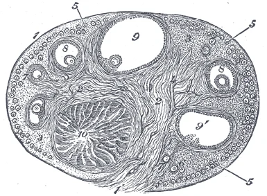
Long description
Section of the ovary. (After Schrön.) 1. Outer covering. 1’. Attached border. 2. Central stroma. 3. Peripheral stroma. 4. Bloodvessels. 5. Vesicular follicles in their earliest stage. 6, 7, 8. More advanced follicles. 9. An almost mature follicle. 9’. Follicle from which the ovum has escaped. 10. Co

Figure 1: Section of the ovary (After Schrön.) 1.
The broad ligament serves as a primary support for the uterus. It extends laterally from both sides of the uterus and attaches it to the pelvic wall. The round ligament attaches to the uterus near the uterine tubes, and extends to the labia majora. Finally, the uterosacral ligament stabilizes the uterus posteriorly by its connection from the cervix to the pelvic wall. These structures ensure the uterus remains in its anteverted position. The ovary lies posterior to the broad ligament, nestled between the ureter and the internal iliac artery. The infundibulopelvic ligament crosses over the external iliac vessels to reach the pelvic brim. This anatomical arrangement protects the ovarian vasculature while allowing mobility for follicular development. [1]
Figure 2Gray1161
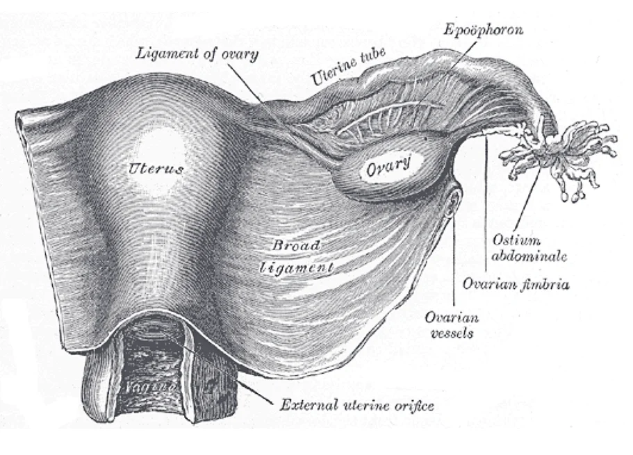
Long description
Uterus and right broad ligament, seen from behind. The broad ligament has been spread out and the ovary drawn downward.

Figure 2: Uterus and right broad ligament, seen from behind.
Figure 3Gray1162
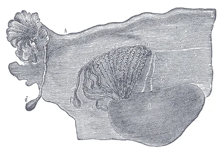
Long description
Adult ovary, epoöphoron, and uterine tube. (From Farre, after Kobelt.) a, a. Epoöphoron formed from the upper part of the Wolffian body. b. Remains of the uppermost tubes sometimes forming hydatids. c. Middle set of tubes. d. Some lower atrophied tubes. e. Atrophied remains of the Wolffian duct. f.

Figure 3: Adult ovary, epoöphoron, and uterine tube.
| Structure | Attachment | Function |
|---|---|---|
| Infundibulopelvic | Ovary to pelvic wall | Suspends ovary; contains vessels |
| Utero-ovarian | Ovary to uterus | Anchers inferior pole |
| Broad | Uterus to pelvic wall | Supports uterus; houses tubes |
| Round | Uterus to labia majora | Maintains anteversion |
The peritoneal cavity surrounds these organs. The parovarian cysts may arise from remnants in the broad ligament. The ovarian fossa is a shallow depression on the lateral pelvic wall where the ovary rests. It is bounded by the external iliac vessels anteriorly and the internal iliac vessels posteriorly. The ureter passes inferior to the uterine artery, a relationship known as 'water under the bridge'. This proximity is critical during surgical procedures to avoid injury. The ovarian ligament connects the uterus to the ovary, providing stability. The mesovarium, a portion of the broad ligament, supports the anterior border of the ovary and transmits vessels.
The uterine wall consists of three distinct histological layers, each with specific functional roles. The outermost layer is the perimetrium, a serous membrane that covers the exterior surface of the organ. This layer provides a smooth, protective covering for the uterus as it sits within the pelvic cavity. Beneath this is the myometrium, which constitutes the bulk of the uterine wall. This middle layer is composed of thick smooth muscle tissue responsible for generating uterine contractions. The muscle fibers within the myometrium are arranged in horizontal, vertical, and diagonal directions. This complex interwoven arrangement allows for powerful contractions during labor and less intense contractions, often experienced as cramps, which help expel menstrual blood. Additionally, anteriorly directed myometrial contractions occur near ovulation, potentially facilitating sperm transport through the reproductive tract. The innermost layer is the endometrium, which lines the uterine cavity and undergoes cyclical changes in preparation for potential implantation. [1]
The ovarian cycle involves predictable changes in oocytes and ovarian follicles over a roughly 28-day period. This cycle is interrelated with but distinct from the menstrual cycle. It encompasses two primary processes: oogenesis, the production of gametes, and folliculogenesis, the growth and development of ovarian follicles. During the reproductive years, these processes follow a consistent sequence. The larger amount of cytoplasm in the female gamete serves a critical function by supplying nutrients to the developing zygote between fertilization and implantation. Unlike sperm, which contribute only DNA at fertilization, the egg provides all cytoplasmic organelles, including mitochondria. Consequently, mitochondrial DNA is maternally inherited, allowing lineage tracing through female ancestors. Understanding this cellular contribution is vital for comprehending early embryonic development and genetic inheritance patterns. [1]
Puberty marks the stage where individuals achieve sexual maturity, driven by hormonal changes that are similar in mechanism but different in outcome between sexes. The process is initiated by a concerted release of hormones from the hypothalamus, anterior pituitary, and gonads. In females, the first visible change is typically the development of breast tissue, followed by the growth of axillary and pubic hair. A significant growth spurt usually begins between ages 9 and 11, potentially lasting two years or more. During this period, height can increase by approximately 3 inches per year. The final major event in female puberty is menarche, which signifies the start of menstruation. The sequence of these changes is predictable, although the exact timing varies between individuals. [2] [1]
Gametes are reproductive cells produced by gonads that combine to form offspring. The female gamete is significantly larger than the male gamete; the volume of a sperm cell is 85,000 times less than that of the female gamete. Sperm are produced at a rate of 100 to 300 million per day, whereas females typically ovulate only one oocyte per month. The sperm structure is specialized for its function, featuring a head, mid-piece, and tail. The head contains a compact haploid nucleus and is covered by [3]
The endometrium lines the central cavity. The ovaries are positioned laterally to the uterus, connected by the broad ligament. The fallopian tubes extend from the uterine horns to the fimbriated ends near the ovaries.
The arterial supply of the female reproductive organs relies primarily on the paired ovarian arteries and the uterine arteries, which form a major anastomotic network within the pelvis. It illustrates the abdominal aorta superiorly, giving rise laterally to the paired ovarian arteries which descend into the pelvis within the suspensory ligament of the ovary, crossing the pelvic brim to reach the hilum of the ovary. Medially and inferiorly, the internal iliac artery gives off the uterine artery, which runs horizontally within the base of the broad ligament toward the lateral margin of the uterus, crossing superior to the ureter. [1]
Figure 4Figure 28 02 01
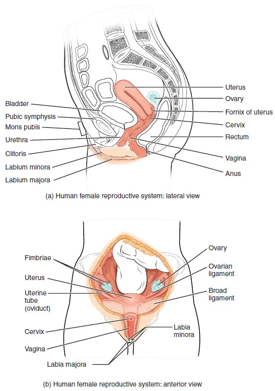
Long description
The major organs of the female reproductive system are located inside the pelvic cavity.

Figure 4: The major organs of the female reproductive system are located inside the pelvic cavity.
Figure 5Gray1165
Long description
Female pelvis and its contents, seen from above and in front.

Figure 5: Female pelvis and its contents, seen from above and in front.
DefinitionUterine and Ovarian Anastomosis
The uterine artery ascends along the lateral border of the uterus and anastomoses directly with the ovarian artery, providing dual collateral blood flow to both the uterus and the adnexa.
Within the myometrium, the uterine artery branches into arcuate arteries, radial arteries, and ultimately into the spiral arteries that supply the stratum functionalis of the endometrium. If no pregnancy occurs within approximately 10 to 12 days, or specifically 10 to 12 days, the corpus luteum degrades, leading to falling hormone levels and prostaglandin-mediated constriction of these spiral arteries. [1]
| Artery | Origin | Primary Destination | Clinical Relevance |
|---|---|---|---|
| Ovarian artery | Abdominal aorta | Ovary and uterine tube | Suspensory ligament contains vessels; site of adnexal torsion |
| Uterine artery | Internal iliac artery | Uterus and upper vagina | Crosses superior to ureter; supplies fibroids and gravid uterus |
| Spiral artery | Uterine radial artery | Endometrium (stratum functionalis) | Constricts and ruptures during menses; affected by prostaglandins |
Adnexal torsion involves the twisting of the ovary and uterine tube around the suspensory ligament and broad ligament, compromising the ovarian and uterine arterial inflow and leading to acute pelvic pain. Ectopic pregnancy most frequently implants within the uterine tube, where the local tubal branches of the ovarian and uterine vessels provide a precarious blood supply that can result in rupture as the trophoblast invades the wall. [1]
Pelvic Arterial Supply and Pathology
- Internal Iliac ArteryGives rise to the uterine artery running within the broad ligament.
- Question: Anastomosis intact?
Yes
- Dual supply to ovary and uterus via ovarian-uterine arcade.
No
- Ischaemia, adnexal torsion, or tubal rupture in ectopic pregnancy.
The venous drainage of the female reproductive organs follows a pattern that mirrors the arterial supply, ensuring efficient return of deoxygenated blood from the pelvic viscera to the systemic circulation. This system is critical for maintaining tissue health and facilitating the transport of metabolic waste products away from the reproductive structures.
The vagina, a muscular canal approximately 10 cm long, serves as the entrance to the reproductive tract and the exit during menses and childbirth. Its outer walls are formed into longitudinal columns or ridges, and the superior portion, known as the fornix, meets the protruding uterine cervix. The walls consist of an outer fibrous adventitia, a middle layer of smooth muscle, and an inner layer of stratified squamous epithelium resting on a lamina propria. The vaginal epithelium forms transverse [1]
The uterus is drained by the uterine venous plexus, which surrounds the cervix and lower uterine segment. This plexus drains into the internal iliac veins. The upper part of the uterus drains via the ovarian veins, which cross over the external iliac vessels. The uterine veins are valveless, allowing for bidirectional flow, which is clinically significant in the spread of infection or metastasis. The rich vascular network supports the thick muscular wall and the endometrial lining, which undergoes cyclic changes.
Early tertiary follicles with their fluid-filled antrum are stimulated by an increase in FSH, a gonadotropin produced by the anterior pituitary, to grow in the 28-day ovarian cycle. [1]
The lymphatic drainage of the female reproductive organs follows specific anatomical pathways that correspond to the embryological origin of each structure. Understanding these pathways is critical for predicting the spread of malignancies and infections.
The labia majora and minora drain to the superficial inguinal nodes. The clitoris and upper vagina drain to the internal iliac and sacral nodes. The ovaries and fallopian tubes drain to the para-aortic nodes. The uterus drains to the internal iliac, external iliac, and sacral nodes. The lower vagina drains to the superficial inguinal nodes. The upper vagina drains to the internal iliac nodes.
The labia majora and minora receive lymphatic drainage that flows primarily to the superficial inguinal lymph nodes. These nodes are located in the femoral triangle. They filter lymph from the lower abdominal wall and perineum. The clitoris also drains to the deep inguinal nodes. This pathway is significant because it bypasses the inguinal nodes in some cases.
The vagina has a dual lymphatic drainage pattern. The lower third of the vagina drains to the superficial inguinal lymph nodes. This is similar to the drainage of the labia. The middle third of the vagina drains to the internal iliac lymph nodes. The upper third of the vagina drains to the external iliac lymph nodes. This complex drainage reflects the embryological development of the urogenital sinus.
The uterus has extensive lymphatic connections. The fundus and body of the uterus drain to the external iliac and para-aortic lymph nodes. The cervix drains primarily to the internal iliac, external iliac, and sacral lymph nodes. This drainage pattern is crucial in the staging of cervical cancer.
The ovaries and fallopian tubes drain to the para-aortic lymph nodes. This pathway follows the gonadal vessels. The para-aortic nodes are located alongside the abdominal aorta. This drainage is distinct from the rest of the pelvis. It reflects the embryological descent of the gonads from the posterior abdominal wall.
The vulva, including the clitoris and labia, drains to the inguinal nodes. The superficial inguinal nodes are the first site of metastasis for many vulvar cancers. The deep inguinal nodes receive drainage from the clitoris and the vestibular bulbs.
DefinitionLymphatic Drainage
The flow of lymph from tissues to regional lymph nodes, determining the spread of disease.
| Structure | Primary Drainage |
|---|---|
| Labia majora/minora | Superficial inguinal nodes |
| Clitoris | Deep inguinal nodes |
| Lower vagina | Superficial inguinal nodes |
| Middle vagina | Internal iliac nodes |
| Upper vagina | External iliac nodes |
| Cervix | Internal/external iliac, sacral nodes |
| Ovaries/Fallopian tubes | Para-aortic nodes |
The nerves supplying the female reproductive organs include the pudendal nerve and the autonomic nervous system. The pudendal nerve provides somatic sensation to the vulva and lower vagina. The autonomic nerves regulate smooth muscle contraction and glandular secretion.
The superior and anterior portions of the labia minora come together to encircle the clitoris. The clitoris is an organ that originates from the same cells as the glans penis. It has abundant nerves that make it important in sexual sensation and orgasm. The hymen is a thin membrane that sometimes partially covers the entrance to the vagina. An intact hymen cannot be used as an indication of “virginity”. The vaginal opening is located between the opening of the urethra and the anus. It is flanked by outlets to the Bartholin’s glands. [1]
The innervation of the female reproductive organs is complex, involving both autonomic and somatic components that facilitate sensation, motor control, and vascular regulation. Understanding this supply is critical for interpreting referred pain patterns and surgical outcomes.
DefinitionAutonomic Innervation
The visceral components are primarily sympathetic and parasympathetic fibres that regulate smooth muscle contraction and glandular secretion.
Sympathetic fibres originate from the lower thoracic and upper lumbar spinal cord segments. These fibres travel via the sympathetic trunk to the superior and inferior hypogastric plexuses. From these plexuses, nerves extend to the uterus, fallopian tubes, and ovaries.
Parasympathetic fibres arise from the sacral spinal cord segments S2 to S4. These fibres travel through the pelvic splanchnic nerves to join the inferior hypogastric plexus. They provide parasympathetic innervation to the lower vagina and the clitoris, facilitating vasodilation and engorgement during sexual arousal.
Somatic innervation is provided by the pudendal nerve, which arises from the sacral plexus (S2-S4). This nerve supplies the external genitalia, including the labia majora, labia minora, and the lower third of the vagina. It also provides sensory innervation to the perineum. The clitoris, being homologous to the penis, receives dense somatic sensory innervation, making it highly sensitive to touch and pressure.
The superior hypogastric plexus lies anterior to the sacral promontory. Branches from this plexus descend to form the inferior hypogastric plexus, which is located on the lateral pelvic wall.
| Structure | Origin | Destination | Function |
|---|---|---|---|
| Pelvic Splanchnic Nerves | S2-S4 | Inferior Hypogastric Plexus | Parasympathetic to lower vagina/clitoris |
| Pudendal Nerve | S2-S4 | External Genitalia | Somatic sensation to labia/perineum |
| Sympathetic Trunk | T10-L2 | Uterus/Ovaries | Vasoconstriction, myometrial contraction |
Lesions to these nerves can result in significant dysfunction. Damage to the pudendal nerve during childbirth may lead to perineal numbness or dyspareunia. Autonomic damage during pelvic surgery can cause bladder dysfunction or sexual arousal difficulties.
Link to the histology page.
The female reproductive system undergoes significant histological and functional changes driven by hormonal feedback loops. Understanding these mechanisms is foundational for clinical practice.
DefinitionHormonal Feedback
The hypothalamic-pituitary-gonadal axis regulates reproductive function through complex negative and positive feedback mechanisms involving GnRH, LH, FSH, and sex steroids.
As an individual approaches puberty, two changes in sensitivity occur. The first is a decrease of sensitivity in the hypothalamus and pituitary to negative feedback, meaning that it takes increasingly larger concentrations of sex steroid hormones to stop the production of LH and FSH. The second change in sensitivity is an increase in sensitivity of the gonads to the FSH and LH signals, meaning the gonads of adults are more responsive to gonadotropins than are the gonads of children. As a result of these two changes, the levels of LH and FSH slowly increase and lead to the enlargement and maturation of the gonads, which in turn leads to secretion of higher levels of sex hormones and the initiation of spermatogenesis and folliculogenesis. [2]
NoteClinical Relevance
Oral contraceptives rely on maintaining constant hormone levels to suppress this axis. Skipping even just one or two pills at certain points of the cycle (or even being several hours late taking the pill) can lead to an increase in FSH and LH and result in ovulation. It is important, therefore, to follow the directions on the birth control pill package to successfully prevent pregnancy. [1]
Puberty onset is influenced by multiple factors, including genetics, environment, and psychological stress. One of the more important influences may be nutrition; historical data demonstrate the effect of better and more consistent nutrition on the age of menarche in the United States, which decreased from an average age of approximately 17 years of age in 1860 to the current age of approximately 12.75 years in 1960, as it remains today. Some studies indicate a link between puberty onset and the amount of stored fat in an individual. This effect is more pronounced in females, but has been documented in both sexes. Body fat, corresponding with secretion of the hormone leptin by adipose cells, appears to have a strong role in determining menarche. [2]
Clinical pearlSecondary Characteristics
Different sex steroid hormone concentrations between the sexes also contribute to the development and function of secondary sexual characteristics. Examples of secondary sexual characteristics are listed in . [2]
While male puberty involves testicular growth as the first sign, female development follows a distinct pattern driven by estrogen and progesterone. The histological architecture of the ovaries, fallopian tubes, uterus, and vagina adapts cyclically to these hormonal shifts, preparing for potential implantation. The endometrium undergoes proliferative and secretory changes, while the cervix produces mucus that varies in consistency throughout the cycle. These histological features are critical for diagnosing reproductive disorders and understanding contraceptive mechanisms.
The development of the female reproductive system is a complex process involving the differentiation of gonads and the subsequent formation of the internal and external genitalia. This process is driven by genetic and hormonal signals that determine the sexual phenotype of the individual. For a detailed understanding of the initial differentiation of the gonads, including the specific cellular mechanisms and morphological changes, students should seek additional content on fetal development. This external resource provides the necessary figures and deeper embryological context that underpins the anatomical structures formed later in gestation.
While the primary focus of this section is the reproductive organs, it is important to note that the mammary glands, although not part of the reproductive tract proper, develop in close association with the hormonal changes that regulate reproductive function. During the normal hormonal fluctuations in the menstrual cycle, breast tissue responds to changing levels of estrogen and progesterone, which can lead to swelling and breast tenderness in some individuals, especially during the secretory phase. This responsiveness highlights the integrated nature of the endocrine and reproductive systems. If pregnancy occurs, the increase in hormones leads to further development of the mammary tissue and enlargement of the breasts. This preparatory development is crucial for post-partum lactation, demonstrating how reproductive anatomy adapts to potential physiological demands. [1]
The internal genitalia, including the uterus, fallopian tubes, and upper vagina, derive from the paramesonephric (Müllerian) ducts. In the absence of anti-Müllerian hormone, these ducts persist and fuse to form the uterovaginal canal. The lower vagina is derived from the urogenital sinus. The external genitalia develop from the genital tubercle, urogenital folds, and labioscrotal swellings. The clitoris corresponds to the glans of the genital tubercle, while the labia majora correspond to the labioscrotal swellings.
The death of ovarian follicles is called atresia, and can occur at any point during follicular development. [1]
NoteEmbryological Link
For more information and a figure of differentiation of the gonads, seek additional content on fetal development. [2]
The hormonal environment established during fetal life and later during puberty dictates the maturation of these structures. The interplay between genetic sex and hormonal signals ensures the proper formation of the reproductive tract, preparing the body for potential reproductive functions in adulthood.
Radiological assessment of the female reproductive system relies on cross-sectional imaging and contrast studies to delineate anatomical relationships that are not palpable during physical examination. The primary modalities include transvaginal ultrasound, magnetic resonance imaging (MRI), and hysterosalpingography. Each modality offers distinct advantages in visualising specific structures, from the endometrial cavity to the adnexal regions.
On the left, a hysterosalpingogram demonstrates the uterine cavity and fallopian tubes following the injection of radiopaque contrast. The uterine cavity appears as a triangular radiolucent space centrally located within the pelvis. Extending laterally from the cornua are the fallopian tubes, which course through the broad ligament.
In the central panel, a transvaginal ultrasound image displays the uterus in a sagittal plane. The uterus is anteverted and anteflexed in most women. Surrounding the endometrium is the myometrium, which appears homogeneous and hypoechoic relative to the endometrium. The cervix is seen inferiorly, leading into the vaginal canal. Adjacent to the uterus, the ovaries are identified as oval structures containing follicles. The right ovary lies lateral to the uterus, near the iliac vessels, while the left ovary is positioned similarly on the contralateral side.
On the right, an MRI scan in the axial plane provides superior soft-tissue contrast. The uterus is clearly delineated, with the junctional zone appearing as a low-signal intensity band surrounding the high-signal endometrial cavity. This layer is crucial for assessing endometrial pathology. The myometrium shows intermediate signal intensity. The adnexal structures are visible laterally. The fallopian tubes are thin-wisted structures that may be visualised if dilated or pathological. The ovaries contain multiple small follicles, appearing as high-signal intensity cysts on T2-weighted images. The surrounding pelvic fat provides natural contrast, highlighting the fascial planes and vascular structures.
Understanding these radiological appearances is essential for diagnosing conditions such as fibroids, endometriosis, and ovarian cysts. The spatial relationships between the uterus, ovaries, and surrounding pelvic organs must be appreciated to avoid misinterpretation. For instance, the ureters run posterior to the uterine arteries, a relationship known as 'water under the bridge', which is critical during surgical procedures to prevent iatrogenic injury.
DefinitionHysterosalpingogram
A fluoroscopic X-ray procedure used to examine the inside of the uterus and fallopian tubes by injecting a contrast medium.
Clinical pearlUreteric Relation
The ureter passes underneath the uterine artery, a vital landmark to prevent surgical injury.
| Structure | Ultrasound Appearance | MRI Appearance |
|---|---|---|
| Endometrium | Echogenic line | High signal on T2 |
| Myometrium | Homogeneous, hypoechoic | Intermediate signal |
| Ovaries | Oval, follicular cysts | Follicles high signal T2 |
| Fallopian Tubes | Often not visible | Thin-wisted, low signal |
Figure 6Blausen 0399 FemaleReproSystem 01
Long description
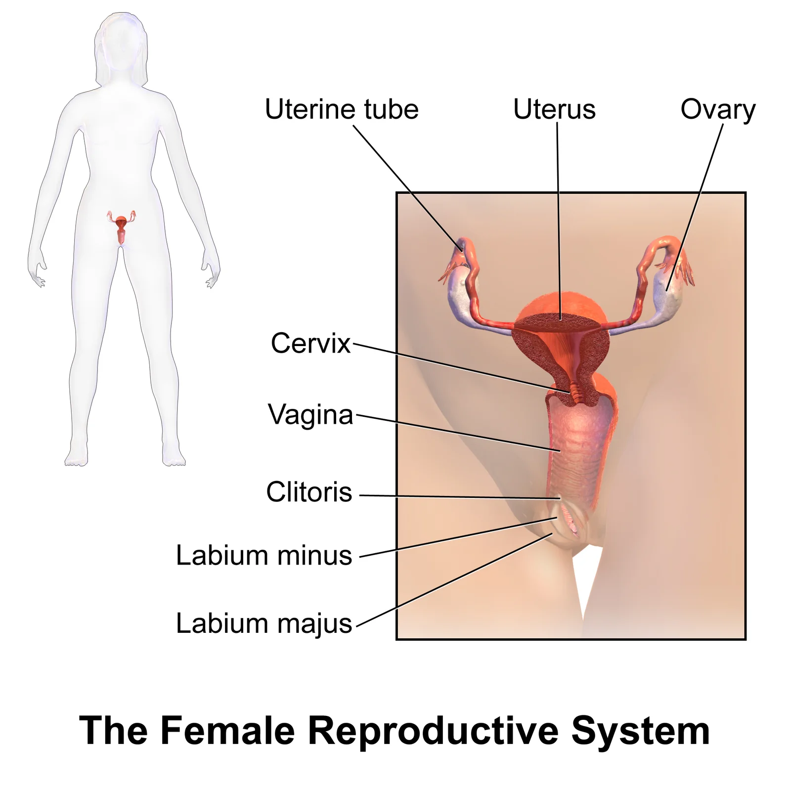
Female Reproductive System. See a full animation of this medical topic.

Figure 6: Female Reproductive System.
The female reproductive organs reside within the true pelvis, suspended by ligaments that provide structural support and vascular pathways. Understanding their spatial relationships is critical for surgical safety and diagnostic imaging. The uterus is a pear-shaped muscular organ, typically anteverted and anteflexed. It consists of the fundus, body, isthmus, and cervix. The fundus is the rounded superior portion, while the cervix projects into the vagina. The uterus is supported by the broad ligament, which extends laterally to the pelvic walls. This peritoneal fold encloses the uterine tubes, ovaries, and associated vessels. The broad ligament has three parts: the mesometrium, mesosalpinx, and mesovarium.
DefinitionBroad Ligament
A double layer of peritoneum extending from the lateral margins of the uterus to the pelvic sidewalls, enclosing the uterine tubes and ovaries.
The uterine tubes, or fallopian tubes, extend from the uterine cornua to the ovaries. Each tube has four parts: the interstitial part, isthmus, ampulla, and infundibulum. The infundibulum is the funnel-shaped lateral end with fimbriae that sweep over the ovary to capture the released oocyte. The ovaries are almond-shaped glands located in the ovarian fossa on the lateral pelvic wall. They are suspended by the suspensory ligament of the ovary, which contains the ovarian vessels and nerves. The proper ligament of the ovary connects the ovary to the uterus. [1]
| Structure | Attachment | Function |
|---|---|---|
| Broad Ligament | Uterus to pelvic wall | Encloses tubes and ovaries |
| Suspensory Ligament | Ovary to pelvic wall | Contains ovarian vessels |
| Proper Ligament | Ovary to uterus | Anchers ovary to uterus |
| Round Ligament | Uterus to labia majora | Maintains anteversion |
The vagina is a muscular canal extending from the cervix to the vestibule. It is anterior to the rectum and posterior to the bladder and urethra. The vaginal fornices are recesses around the cervix. The anterior fornix is shorter than the posterior fornix, which lies close to the rectouterine pouch (pouch of Douglas). This anatomical relationship is clinically significant for drainage of pelvic fluid.
The uterus is centrally located, with the fundus pointing anteriorly. The uterine tubes extend laterally, with the fimbriae near the ovaries. The ovaries are positioned in the ovarian fossa. The bladder is anterior to the uterus, and the rectum is posterior. Understanding these relationships helps in identifying lesion sites and injury patterns during imaging or surgery.
The primary oocyte within the growing secondary follicle secretes a thin acellular membrane called the zona pellucida, which is critical for fertilization. Concurrently, follicular fluid collects into a large pool known as the antrum, defining the tertiary or antral follicle. Several follicles reach this stage simultaneously, but most undergo atresia. The surviving follicle grows until ovulation, expelling the secondary oocyte surrounded by granulosa cells[1].
| Factor | Role in Dominance |
|---|---|
| Follicle size | Larger follicles have a competitive advantage |
| Granulosa cell number | Higher counts support estrogen production |
| FSH receptor density | More receptors enhance response to FSH |
Larger, more developed follicles produce more estrogen in response to LH stimulation. This rise in systemic plasma estrogen triggers a negative feedback loop, reducing hypothalamic GnRH and pituitary LH and FSH production. Because large tertiary follicles require FSH to survive, this decline causes most to die via atresia. Typically, only one follicle, the dominant follicle, survives this reduction and releases the oocyte. [1]
The post-ovulatory phase of progesterone secretion is termed the luteal phase. If pregnancy does not occur within 10 to 12 days, the corpus luteum stops secreting progesterone and degrades into the corpus albicans. This nonfunctional structure disintegrates over several months. During this period of reduced progesterone, FSH and LH are stimulated again, restarting the follicular phase with new early tertiary follicles. [1]
Exam trapCorpus Albicans Timing
The corpus luteum degrades into the corpus albicans only if pregnancy does not occur within 10 to 12 days.
High estrogen levels prompt the LH surge and increase uterine tube contractions to facilitate oocyte pick-up. Estrogen also slightly decreases vaginal acidity, making it more hospitable to sperm. In the ovary, luteinization of granulosa cells forms the progesterone-producing corpus luteum. In the uterus, progesterone initiates the secretory phase, preparing the endometrial lining for implantation. Over the next 10 to 12 days, endometrial glands secrete a fluid rich in glycogen. If fertilization [1]
Luteal Phase Resolution
- Corpus luteum formsSecretes progesterone
- Question: Pregnancy occurs?
Yes
- Maintain corpus luteum
No
- Wait 10 to 12 days
- Degrade to corpus albicans
- FSH and LH rise
Students frequently confuse the protective mechanisms of the lower genital tract. A common error is believing that douching maintains hygiene. In fact, douching disrupts the normal balance of healthy microorganisms and increases the risk for infections and irritation. The vagina is a self-cleansing organ because Lactobacillus bacteria secrete lactic acid, maintaining an acidic pH below 4.5 which prevents pathogen survival[1].
Another frequent mistake involves the hormonal regulation of the menstrual cycle. Students often assume high estrogen always suppresses gonadotropins. However, extremely high concentrations of systemic plasma estrogen trigger a regulatory switch in the anterior pituitary that responds by secreting large amounts of LH and FSH. This positive feedback loop occurs only when the dominant follicle produces more estrogen than all developing follicles did together. [1]
Misunderstanding the endometrial response to progesterone is also common. Some think the entire endometrium sheds during menstruation. Only the stratum functionalis layer of the endometrium sheds during menstruation, while the stratum basalis remains intact. The decline in progesterone triggers this shedding. [1]
Confusion often arises regarding the corpus luteum. Students may forget its origin. The surge of LH stimulates a change in granulosa and theca cells that remain in the follicle after ovulation, transforming it into the corpus luteum. This structure produces progesterone, which is critical for pregnancy maintenance. [1]
Regarding contraception, a misconception is that menstruation is physiologically required. Menstruation every 28 days is not required for health reasons, and there are no reported adverse effects of not having a menstrual period in an otherwise healthy individual. Continuous low-dose estrogen and progesterone pills prevent menses entirely. [1]
In breast anatomy, students often equate breast size with lactation capacity. Breast size differs between individuals and does not affect the amount of milk produced. Milk production depends on alveoli and myoepithelial cells, not fat tissue volume. [1]
Finally, regarding male reproductive secretions, it is a trap to assume pre-ejaculate is sterile. Bulbourethral fluid may pick up sperm already present in the urethra, and therefore it may be able to cause pregnancy. [3]
At the bedside, understanding vaginal flora guides management of discharge. Recognising that Lactobacillus maintains pH below 4.5 helps explain why antibiotic overuse can lead to yeast infections. Clinicians advise against douching to preserve this acidic barrier.
In the operating theatre, knowledge of the corpus luteum is vital during ovarian surgery. Surgeons must identify the luteinized granulosa and theca cells of the corpus luteum to distinguish it from other ovarian structures. This structure produces progesterone, which is critical for the establishment and maintenance of pregnancy. If a corpus luteum is removed prematurely, progesterone production ceases, risking early pregnancy loss.
On imaging, ultrasound identification of the endometrial layers is crucial. The stratum functionalis is the inner functional layer that provides the proper site of implantation for the fertilized egg. During the luteal phase, progesterone maintains a thick stratum functionalis. If no embryo implants, the corpus luteum degrades, and progesterone levels fall, leading to shedding of this layer. The menses phase averages approximately five days but can last from 2 to 7 days. [1]
In examinations, students must distinguish between feedback loops. The positive feedback loop by which more estrogen triggers release of more LH and FSH only occurs at this point in the cycle, specifically when the dominant follicle is mature. Progesterone triggers negative feedback at the hypothalamus and pituitary, which keeps GnRH, LH, and FSH secretions low. This ensures no new dominant follicles develop during the luteal phase. [1]
For contraception counselling, explaining the mechanism of combined oral contraceptives is key. Some birth control pills contain 21 active pills containing hormones, and 7 inactive pills. The decline in hormones during the week that the person takes the placebo pills triggers menses. This flow is typically lighter than a normal menstrual flow because of the reduced endometrial thickening. [1]
Breast examination relies on understanding lactiferous anatomy. Milk exits the breast through the nipple via 15 to 20 lactiferous ducts. These ducts connect to lactiferous sinuses and then to glandular lobes containing alveoli. Myoepithelial cells surrounding the alveoli contract to push milk to the sinuses. [1]
In male reproductive health, understanding accessory glands is important. The bulbourethral glands release a thick, salty fluid that lubricates the end of the urethra and the vagina. This fluid helps to clean urine residues from the penile urethra. Clinicians must warn patients that this pre-ejaculate can contain sperm.
Menstrual Cycle Hormonal Control
- Start: Follicular PhaseTertiary follicles secrete estrogen
- Question: Estrogen levels rise?
Yes, moderate
- Negative FeedbackInhibits GnRH, LH, FSH
Yes, very high
- Positive FeedbackTriggers LH/FSH surge
- OvulationLH surge causes oocyte release
- Question: Ovulation occurred?
Yes
- LuteinizationFollicle becomes corpus luteum
- Luteal PhaseCorpus luteum secretes progesterone
- Question: Pregnancy?
Yes
- Maintain Corpus LuteumProgesterone continues
No
- Corpus Luteum DegradesProgesterone drops
- MensesStratum functionalis sheds
Vaginal pH is maintained below 4.5 by Lactobacillus-derived lactic acid. Douching disrupts normal flora and increases infection risk. High estrogen triggers positive feedback, causing LH and FSH surge. Only the stratum functionalis sheds during menstruation. Corpus luteum forms from luteinized granulosa and theca cells. Progesterone maintains the endometrium for potential implantation. Breast size does not determine milk production capacity. Pre-ejaculate may contain sperm and can cause pregnancy.
Answer each one in your head first, then open the answer.
What pH level does Lactobacillus maintain in the vagina?
Show answerHide answer
Below 4.5Which layer of the endometrium sheds during menstruation?
Show answerHide answer
Stratum functionalisWhat hormone does the corpus luteum primarily produce?
Show answerHide answer
ProgesteroneHow many lactiferous ducts open on the nipple surface?
Show answerHide answer
15 to 20What effect does progesterone have on GnRH, LH, and FSH?
Show answerHide answer
Keeps secretions low
- Betts JG, Young KA, Wise JA, et al. Anatomy and Physiology of the Ovarian Reproductive System. OpenStax, Anatomy and Physiology 2e (CC BY-NC-SA 4.0). Accessed .
- Betts JG, Young KA, Wise JA, et al. Development of the Male and Female Reproductive Systems. OpenStax, Anatomy and Physiology 2e (CC BY-NC-SA 4.0). Accessed .
- Betts JG, Young KA, Wise JA, et al. Anatomy and Physiology of the Testicular Reproductive System. OpenStax, Anatomy and Physiology 2e (CC BY-NC-SA 4.0). Accessed .
Figure credits
- Figure 1: Henry Vandyke Carter, via Wikimedia Commons, Public domain, Public domain.
- Figure 2: Henry Vandyke Carter, via Wikimedia Commons, Public domain, Public domain.
- Figure 3: Henry Vandyke Carter, via Wikimedia Commons, Public domain, Public domain.
- Figure 4: OpenStax College, via Wikimedia Commons, CC BY 3.0, CC BY 3.0.
- Figure 5: Henry Vandyke Carter, via Wikimedia Commons, Public domain, Public domain.
- Figure 6: BruceBlaus. When using this image in external sources it can be cited as: Blausen.com staff (2014). "Medical gallery of Blausen Medical 2014". WikiJournal of Medicine 1 (2). DOI:10.15347/wjm/2014.010. ISSN 2002-4436., via Wikimedia Commons, CC BY 3.0, CC BY 3.0.
Educational content for medical students and doctors. It is not a substitute for clinical judgement or local guidelines.