Uterus and supporting ligaments
Anatomy and supporting structures of the human uterus, cervix, vaginal fornices, and congenital anomalies.
DefinitionFemale reproductive system
The female reproductive system is located primarily inside the pelvic cavity to produce gametes, support the developing fetus, and deliver it to the outside world. [1]
The vagina is a muscular canal approximately 10 cm long that serves as the entrance to the reproductive tract and the exit from the uterus during menses and childbirth. The superior portion of the vagina is called the fornix, which meets the protruding uterine cervix. The cervix is the narrow inferior portion of the uterus that projects into the vagina and produces mucus secretions that facilitate sperm movement. [1]
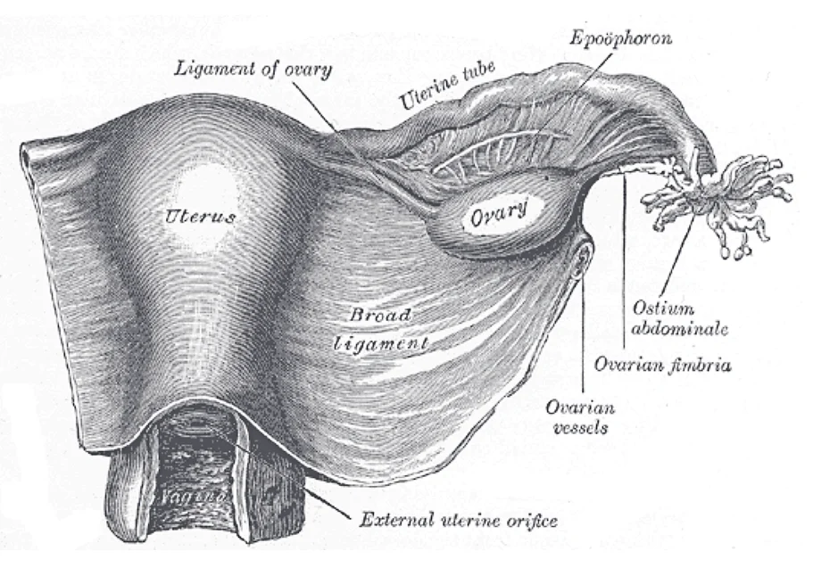
The uterus is the muscular organ that nourishes and supports the growing embryo. When a female is not pregnant, its average size is approximately 5 cm wide by 7 cm long. Several ligaments maintain the position of the uterus within the abdominopelvic cavity. The broad ligament is a fold of peritoneum extending laterally from both sides of the uterus and attaching it to the pelvic wall. The round ligament attaches to the uterus near the uterine tubes and extends to the labia majora. The uterosacral ligament stabilizes the uterus posteriorly by its connection from the cervix to the pelvic wall. The cardinal ligament also provides critical lateral support from the cervix to the pelvic wall. Pelvic organ prolapse occurs when these supportive ligaments and pelvic floor structures fail, leading to descent of the uterus or vaginal walls. [1]
Figure 1Gray1161
Long description
Uterus and right broad ligament, seen from behind. The broad ligament has been spread out and the ovary drawn downward.

Figure 1: Uterus and right broad ligament, seen from behind.
Congenital uterine anomalies arise from abnormal development or fusion of the Müllerian ducts. A septate uterus features a fibrous or muscular partition dividing the uterine cavity. A bicornuate uterus presents with two distinct horns sharing a common lower cavity. Uterus didelphys involves complete duplication of the uterus and cervix, sometimes associated with renal anomalies.
| Structure or Anomaly | Description | Key Feature |
|---|---|---|
| Broad ligament | Peritoneal fold | Attaches uterus laterally to pelvic wall |
| Round ligament | Fibromuscular cord | Extends from uterus near tubes to labia majora |
| Utersacral ligament | Connective tissue band | Stabilizes uterus posteriorly from cervix to pelvic wall |
| Septate uterus | Duplication defect | Presence of a dividing internal septum |
| Bicornuate uterus | Fusion defect | Two well-defined uterine horns |
| Uterus didelphys | Complete duplication | Double uterus and cervix |
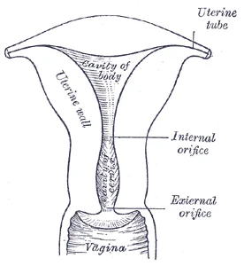
The uterus is a hollow muscular organ situated in the pelvic cavity, and its parts include the fundus, body, and cervix.
The wall of the uterus is made up of three layers. The most superficial layer is the serous membrane, or perimetrium, which consists of epithelial tissue covering the exterior portion of the uterus. The middle layer, or myometrium, is a thick layer of smooth muscle responsible for uterine contractions. Most of the uterus is myometrial tissue, and the muscle fibers run horizontally, vertically, and diagonally, allowing powerful contractions during labor and less powerful contractions or cramps that help expel menstrual blood during a period. Anteriorly directed myometrial contractions also occur near the time of ovulation and may facilitate sperm transport through the female reproductive tract. [1]
Figure 2Gray1169
Long description
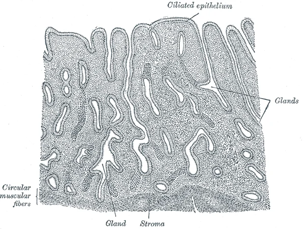
Vertical section of mucous membrane of human uterus. (Sobotta.)

Figure 2: Vertical section of mucous membrane of human uterus (Sobotta.).
Supporting structures maintain the position of the uterus in the pelvis. The broad ligament is a fold of peritoneum that connects the sides of the uterus to the pelvic wall. The cardinal and uterosacral ligaments provide key mechanical support, anchoring the cervix and upper vagina to the pelvic walls and sacrum. The round ligament extends from the uterine horns through the inguinal canal to the labia majora, maintaining the anteverted position of the uterus.
Congenital anomalies and structural variations can affect the uterus. Obstructions at the level of the uterus in congenital anomalies may increase intrauterine pressure and promote the penetration of endometrial cells within the myometrial layer and the growth of adenomyotic foci. Mayer-Rokitansky-Küster-Hauser syndrome is a rare congenital disorder characterized by a defect in the development of the paramesonephric or Müllerian ducts, leading to congenital aplasia of the upper two thirds of the vagina and the presence of two atrophic hemiuteri. Uterus didelphys involves two totally separated uteri and two cervices. Uterine prolapse occurs when supporting ligaments and pelvic floor muscles weaken, allowing the uterus to descend into or through the vaginal canal. [2]
Figure 3Gray1167
Long description
Posterior half of uterus and upper part of vagina.

Figure 3: Posterior half of uterus and upper part of vagina.
DefinitionMyometrium
The thick middle layer of smooth muscle in the uterine wall that generates labor and menstrual contractions.
The uterus lies centrally within the lesser pelvis between the urinary bladder anteriorly and the rectum posteriorly. Peritoneum drapes over the fundus and body of the uterus, reflecting anteriorly onto the bladder to form the vesicouterine pouch and posteriorly onto the rectum to form the rectouterine pouch. Viscerally, the uterus is divided into the fundus above the uterine tubes, the central body, and the inferior cervix.
The broad ligament (ligamentum latum uteri) is a double fold of peritoneum that extends from the lateral margins of the uterus to the pelvic sidewalls, enclosing the uterine tubes, blood vessels, and ureters between its layers. The round ligament of the uterus (ligamentum teres uteri) originates from the uterine horns just anterior and inferior to the uterotubal junction. It courses anterolaterally through the inguinal canal to terminate in the labia majora, maintaining the anteverted position of the uterus.
The cardinal ligament (transverse cervical ligament of Mackenrodt) is a dense condensation of endopelvic fascia located at the base of the broad ligament. It extends from the lateral cervix and vaginal fornix to the lateral pelvic wall, transmitting the uterine artery and serving as the primary mechanical support for the uterus against downward displacement. [1]
The uterosacral ligament runs posteriorly from the posterolateral cervix to the periosteum of the sacrum, passing on either side of the rectum to maintain the retroverted position of the cervix and anchoring the superior vagina.
| Ligament | Origin | Course | Major Relations |
|---|---|---|---|
| Broad ligament | Uterine lateral margin | Lateral fold to pelvic wall | Uterine tube, ureter, uterine vessels |
| Round ligament | Uterine horn (anteroinferior) | Anterolaterally through inguinal canal | Terminates in labia majora |
| Cardinal ligament | Lateral cervix and vagina | Transversely to lateral pelvic wall | Uterine artery, ureter superiorly |
| Uterosacral ligament | Posterolateral cervix | Posterorly to sacrum | Encircles rectum, anchors vaginal vault |
The outermost layer is the perimetrium, a serous covering continuous with the broad ligament. Beneath this lies the thick, muscular myometrium, which constitutes the bulk of the uterine wall. The innermost layer is the endometrium, a mucosal lining that undergoes cyclical changes. The uterus is divided into three regions: the fundus, the dome-shaped superior portion; the body, the main central region; and the cervix, the narrow inferior projection into the vagina. [1]
DefinitionUterine Layers
The uterus comprises three distinct layers: the outer perimetrium, the muscular myometrium, and the inner endometrium. [1]
The endometrium is functionally divided into two zones. The superficial functionalis layer responds to hormonal fluctuations and is shed during menstruation. The deep basalis layer remains intact and serves as the source for regeneration of the functionalis. The myometrium contains smooth muscle fibres arranged in longitudinal, circular, and oblique bundles. These layers facilitate powerful contractions during labour and menstruation. A specialized structure known as the junctional zone exists in the inner third of the myometrium. This zone is highly hormone-responsive and acts as an interface between the endometrium and the deeper myometrium.
NoteJunctional Zone
The junctional zone (JZ) is a highly specialized hormone-responsive structure found in the inner third of the myometrium. [2]
Tissue damage to the endometrial–myometrial interface increases the risk of adenomyosis and supports the concurrent diagnosis of the latter with previous cesarean section, multiparity, or uterine surgery. [2]
| Region | Description |
|---|---|
| Fundus | Dome-shaped superior portion of the uterus |
| Body | Main central region between fundus and cervix |
| Cervix | Narrow inferior projection into the vagina |
| Perimetrium | Outer serous covering |
| Myometrium | Thick muscular middle layer |
| Endometrium | Inner mucosal lining |
In the uterus, progesterone from the corpus luteum begins the secretory phase of the menstrual cycle, in which the endometrial lining prepares for implantation. [1]
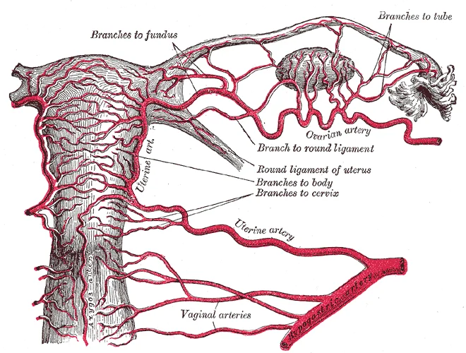
The arterial supply to the uterus is principally derived from the uterine artery, which typically arises from the anterior division of the internal iliac artery. From its origin, the uterine artery courses medially and anteriorly within the retroperitoneum across the lateral pelvic wall to reach the base of the broad ligament. Within the base of the broad ligament, the artery travels horizontally toward the lateral border of the cervix, lying superior and anterior to the ureter in the parametrium. Upon reaching the lateral margin of the supra-vaginal cervix, the uterine artery ascends tortuously alongside the uterus between the leaves of the broad ligament. Along this ascending course, it gives off multiple lateral cervical and vaginal branches to supply the lower uterine segment and upper vagina. Near the uterotubal junction, the uterine artery divides into its terminal branches: a prominent ovarian branch that anastomoses with the ovarian artery, and a tubal branch that supplies the uterine tube.
Figure 4Gray1170
Long description
The arteries of the internal organs of generation of the female, seen from behind. (After Hyrtl.)

Figure 4: The arteries of the internal organs of generation of the female, seen from behind (After Hyrtl.).
DefinitionSpiral arteries
Specialized branches off of the uterine artery called spiral arteries supply the thickened stratum functionalis. [1]
The paired internal iliac arteries are positioned laterally on the posterior pelvic wall, giving rise to the anterior divisions from which the tortuous uterine arteries originate. As the main trunk ascends along the lateral uterine wall, small straight radial branches penetrate the myometrium, ultimately terminating as the spiral arteries within the inner endometrial layer.
| Structure | Origin | Distribution |
|---|---|---|
| Uterine artery | Anterior division of internal iliac artery | Cervix, myometrium, and endometrium via ascending and descending branches |
| Ovarian branch | Terminal division of uterine artery | Ovary, anastomosing with the direct ovarian artery |
| Spiral arteries | Branches off of the uterine artery | Thickened stratum functionalis of the endometrium |
Uterine blood supply pathway
- Internal iliac arteryAnterior division gives rise to the main trunk.
- Uterine arteryCrosses superior to the ureter in the parametrium and ascends the lateral border.
- Question: Which terminal branch?
Ovarian branch
- Anastomosis with ovarian artery
Endometrial branches
- Spiral arteries supply the stratum functionalis
The vascular architecture of the uterus is critical for understanding both physiological function and pathological haemorrhage. The blood supply originates primarily from the uterine arteries, which are branches of the anterior division of the internal iliac arteries. These vessels enter the broad ligament and run along the lateral aspects of the uterus. Concurrently, the ovarian arteries, arising directly from the abdominal aorta, contribute to the upper uterine supply. The spiral arteries develop to provide blood to the thickened stratum functionalis. This dual supply ensures adequate perfusion for the endometrial changes required for potential implantation. [1]
The uterine veins are located within the broad ligament, running parallel to the uterine arteries. They drain the myometrium and endometrium. The uterine veins join to form the uterine venous plexus, which lies adjacent to the cervix and lower uterus. This plexus is extensive and communicates with the vaginal venous plexus. The ovarian veins ascend retroperitoneally. The right ovarian vein drains directly into the inferior vena cava. The left ovarian vein drains into the left renal vein. The uterine veins drain into the internal iliac veins. This arrangement allows for collateral circulation. The uterine veins are accompanied by the uterine arteries. The ovarian veins accompany the ovarian arteries. The round ligament contains small vessels that anastomose with the uterine veins. The broad ligament contains the uterine vessels and the ovarian vessels. Understanding this anatomy is essential for surgical procedures. The uterine veins are at risk during hysterectomy. The ovarian veins are at risk during oophorectomy. The uterine veins drain into the internal iliac veins. The ovarian veins drain into the inferior vena cava or left renal vein. This distinction is important for understanding metastatic spread. The lymph nodes are located along the vessels.
DefinitionUterine Veins
Paired veins that drain the uterus and run within the broad ligament alongside the uterine arteries, terminating in the internal iliac veins.
The venous drainage mirrors the arterial supply. The uterine veins form a plexus around the cervix and lower uterus. This plexus communicates with the vaginal veins. The vaginal veins drain into the internal iliac veins. The ovarian veins drain the fundus and upper body. The right ovarian vein enters the inferior vena cava. The left ovarian vein enters the left renal vein. This asymmetry is a key anatomical feature. It has clinical implications for thrombosis and metastasis. The uterine veins are valveless. This allows for retrograde flow. This is significant in endometriosis. Endometrial tissue can spread via venous channels. The supporting ligaments also have venous drainage. The broad ligament contains the uterine and ovarian veins. The cardinal ligament contains the uterine veins. The uterosacral ligament contains small venous channels. These channels drain into the internal iliac veins. The venous system is highly interconnected. Collateral circulation is robust. This protects against ischaemia. However, it also facilitates the spread of infection. The venous drainage is complex. It requires careful study.
DefinitionLymphatic Pathways
The lymphatic drainage of the uterus follows three main anatomical routes corresponding to the vascular supply and ligamentous attachments of the organ.
Fundus and upper body lymphatic vessels drain mainly via the ovarian vessels, ascending along the suspensory ligament of the ovary to reach the aortic or lumbar lymph nodes.
Uterine Lymphatic Drainage
- Uterine regionsFundus, upper body, lower body, and cervix drain to distinct nodal groups.
- Question: Which region?
Fundus/Upper Body
- Aortic and lumbar nodesAscend via suspensory ligament of ovary.
Lower Body/Cervix
- Internal and external iliac nodesPass through broad ligament and lateral pelvic walls.
Cervix/Lower Uterus
- Superficial inguinal nodesTravel via round ligament through inguinal canal.
Lower body and cervix lymphatic vessels pass laterally through the broad ligament, reaching the external iliac, internal iliac, and obturator lymph nodes along the lateral pelvic walls.
Cervical lymphatics also track posteriorly along the uterosacral ligaments to drain into the sacral lymph nodes situated on the anterior surface of the sacrum.
A specific anterior pathway originates from the fundus near the entrance of the uterine tube, where vessels travel along the round ligament of the uterus, pass through the inguinal canal, and terminate in the superficial inguinal lymph nodes.
Anteriorly, a distinct vessel is traced from the uterotubal junction entering the deep inguinal ring to reach the superficial inguinal nodes. Clinical understanding of these pathways is essential for staging gynaecological malignancies, as cervical and uterine carcinomas spread predictably along these established lymphatic channels to regional nodes.
The innervation of the uterus and its supporting ligaments involves a complex interplay of autonomic and somatic pathways, which dictates both physiological function and the nature of pain perception. Understanding this supply requires examining the sympathetic and parasympathetic contributions alongside the somatic nerves that traverse the pelvic floor.
The autonomic nervous system regulates uterine tone and vascular control. Sympathetic fibres originate from the lower thoracic and upper lumbar spinal segments. These fibres travel via the hypogastric plexus to reach the uterus. They are responsible for vasoconstriction and modulating myometrial contraction. Parasympathetic innervation arises from the pelvic splanchnic nerves. These fibres join the uterovaginal plexus. They generally promote vasodilation and may influence glandular secretion. The integration of these autonomic inputs allows for the coordinated changes in blood flow and muscle activity required during the menstrual cycle and pregnancy.
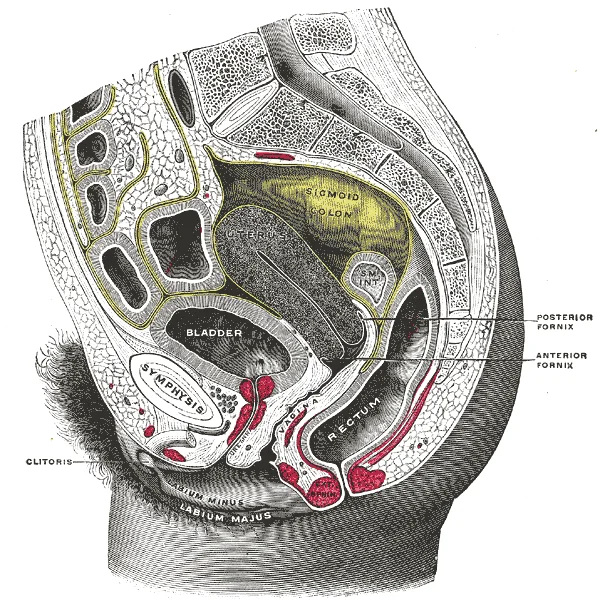
Figure 5Gray1166
Long description
Sagittal section of the lower part of a female trunk, right segment. SM. INT. Small intestine. (Testut.)

Figure 5: Sagittal section of the lower part of a female trunk, right segment (sm).
Somatic innervation is critical for understanding clinical presentations, particularly pain. The uterus itself lacks significant somatic sensory innervation in its fundus and body. However, the cervix and the upper vagina receive somatic fibres from the pudendal nerve and the pelvic splanchnic nerves. This distinction explains referred pain patterns. Pain from the uterine fundus is often perceived in the lower abdomen or back, following dermatomal distributions associated with the T10 to L1 spinal segments. In contrast, pain originating from the cervix or lower uterine segment is often felt in the perineum, sacrum, or thighs, reflecting the somatic pathways of the pudendal and sacral nerves. This differential innervation is vital for localising pathology during examination.
The lymphatic drainage of the uterus follows specific pathways that correlate with its vascular supply. Lymph from the fundus and upper body drains primarily to the para-aortic and external iliac nodes. This route is clinically significant in the staging of endometrial and ovarian cancers. Lymph from the lower uterine segment and cervix drains to the internal iliac and sacral nodes. The neurovascular bundle supplying the uterus runs within the broad ligament. The uterine artery crosses over the ureter "water under the bridge," a relationship that must be preserved during hysterectomy. The uterine veins accompany the arteries, draining into the internal iliac veins. The nerve supply accompanies these vessels, forming the uterovaginal plexus. This plexus lies in the parametrium, lateral to the cervix and upper vagina. It contains both sympathetic and parasympathetic fibres, as well as sensory fibres. Damage to this plexus during pelvic surgery can lead to bladder dysfunction or sexual dysfunction, highlighting the importance of anatomical precision.
Link to the histology page.
The uterus is a hollow, muscular organ situated in the true pelvis, between the bladder and the rectum. Its wall consists of three distinct layers, each with specific histological characteristics relevant to its function during the menstrual cycle and pregnancy.
The innermost layer is the endometrium, a mucous membrane that undergoes cyclic changes in response to ovarian hormones. It is composed of two main parts: the stratum functionalis and the stratum basalis. The stratum functionalis is the superficial layer that thickens during the proliferative phase and is shed during menstruation if fertilisation does not occur. It contains uterine glands and spiral arteries. The stratum basalis is the deep layer adjacent to the myometrium; it remains intact after menstruation and serves as the source for regeneration of the functionalis. Histologically, the endometrium is lined by simple columnar epithelium, which includes ciliated cells and secretory cells. The glands are simple tubular glands. The stroma is cellular and oedematous during the secretory phase.
The middle and thickest layer is the myometrium, composed primarily of smooth muscle fibres arranged in three poorly defined layers: an inner longitudinal layer, a middle circular or vascular layer, and an outer longitudinal layer. The myometrium is responsible for uterine contractions during labour. Histologically, the smooth muscle cells are spindle-shaped with central nuclei. The tissue is highly vascularised, particularly in the middle layer, which contains the largest blood vessels. The muscle fibres are interconnected by gap junctions, allowing coordinated contraction.
The outermost layer is the perimetrium, which is a serous membrane consisting of visceral peritoneum. It covers the fundus and the anterior and posterior surfaces of the uterus. In some areas, particularly near the cervix, it is thin or absent. Histologically, it consists of a single layer of mesothelial cells supported by a thin layer of connective tissue. [1]
DefinitionEndometrial Cycle
The endometrium undergoes cyclic changes driven by ovarian hormones, preparing for potential implantation.
The initiation of ovulation—the release of an oocyte from the ovary—marks the transition from puberty into reproductive maturity. From then on, throughout a the reproductive years, ovulation occurs approximately once every 28 days. Just prior to ovulation, a surge of luteinizing hormone triggers the resumption of meiosis in a primary oocyte. This initiates the transition from primary to secondary oocyte. However, as you can see in , this cell division does not result in two identical cells. Instead, the cytoplasm is divided unequally, and one daughter cell is much larger than the other. This larger cell, the secondary oocyte, eventually leaves the ovary during ovulation. The smaller cell, called the first polar body, may or may not complete meiosis and produce second polar bodies; in either case, it eventually disintegrates. [1]
How does the haploid secondary oocyte become an ovum—the haploid female gamete? Meiosis of a secondary oocyte is completed only if a sperm succeeds in penetrating its barriers. Meiosis II then resumes, producing one haploid ovum that, at the instant of fertilization by a (haploid) sperm, becomes the first diploid cell of the new offspring (a zygote). Thus, the ovum can be thought of as a brief, transitional, haploid stage between the haploid oocyte and diploid zygote. [1]
The number of FSH receptors on granulosa cells contributes to a follicle becoming the one surviving dominant follicle. [1]
The female reproductive system originates from the paramesonephric, or Müllerian, ducts during embryogenesis. These paired structures fuse to form the uterus, cervix, and upper vagina, while the unfused cranial portions develop into the fallopian tubes. The ovaries arise separately from the gonadal ridge. Congenital uterine anomalies result from altered development of these ducts, leading to diverse morphologies that may compromise reproductive potential. Such anomalies are linked to decreased fertility and higher risks of unfavorable maternal–fetal outcomes. Classification systems, such as those by the European Society of Human Reproduction and Embryology, categorize these malformations to guide clinical management. [2]
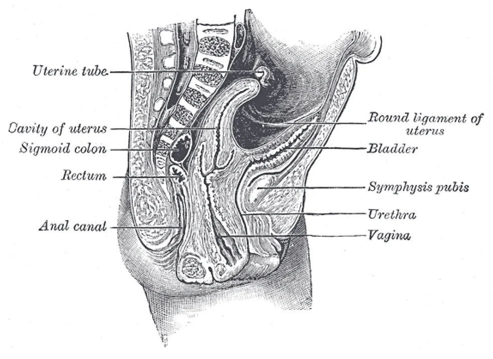
Figure 6Gray1168
Long description
Sagittal section through the pelvis of a newly born female. child.

Figure 6: Sagittal section through the pelvis of a newly born female (child).
NoteEmbryological Link
The uterus and supporting ligaments derive primarily from the fused paramesonephric ducts, with the ovaries originating from the genital ridge.
Oogenesis begins with oogonia formed during fetal development, which divide via mitosis. Unlike male gametogenesis, oogonia form primary oocytes prior to birth, arresting in meiosis I. This arrest persists until puberty, with the number of primary oocytes declining from one to two million in an infant to approximately 400,000 at puberty. The ovarian cycle involves predictable changes in oocytes and follicles, correlating with but distinct from the menstrual cycle. Folliculogenesis, the growth of ovarian follicles, takes approximately two months from primordial to early tertiary stage, with final maturation occurring over 28 days. [1]
DefinitionParamesonephric Ducts
Also known as Müllerian ducts, these embryonic structures fuse to form the uterus, fallopian tubes, and upper third of the vagina.
Supporting ligaments develop concurrently with the uterus, anchoring it within the pelvic cavity. The broad ligament, a fold of peritoneum, encloses the uterus, tubes, and ovaries. The round ligament maintains anteversion, while the uterosacral and cardinal ligaments provide major structural support. Disorders such as Mayer-Rokitansky-Küster-Hauser (MRKH) syndrome involve rudimentary uteri and vaginal agenesis, often linked to HOX gene alterations affecting Müllerian duct development. In MRKH, rudimentary horns may contain functional endometrium, leading to cyclic pain if adenomyosis develops.
| Structure | Embryological Origin |
|---|---|
| Uterus | Fused paramesonephric (Müllerian) ducts |
| Fallopian Tubes | Unfused paramesonephric ducts |
| Ovaries | Genital ridge (gonadal ridge) |
| Upper Vagina | Paramesonephric ducts |
Clinical evaluation of congenital anomalies often requires both hysteroscopy and laparoscopy for accurate classification. Understanding these developmental pathways is essential for diagnosing anomalies that impact fertility and reproductive health. [2]
The external female reproductive structures are referred to collectively as the vulva[1]. The mons pubis is a pad of fat that is located at the anterior, over the pubic bone[1]. After puberty, it becomes covered in pubic hair[1].
The labia majora are folds of hair-covered skin that begin just posterior to the mons pubis[1]. The thinner and more pigmented labia minora extend medial to the labia majora[1]. Although they naturally vary in shape and size from person to person, the labia minora serve to protect the urethra and the entrance to the reproductive tract[1].
DefinitionExternal Genitalia
The vulva encompasses all external female structures, with the labia minora specifically safeguarding the urethral opening and vaginal introitus.
Internal structures interact closely with congenital and acquired conditions. For instance, endometriosis and congenital uterine abnormalities are frequently found during infertility work-ups, and endometriosis is sometimes concurrent with obstructive and nonobstructive uterine abnormalities, such as septate uteri.
| Structure | Location and Features | Function |
|---|---|---|
| Mons pubis | Anterior over pubic bone, pad of fat | Becomes covered in pubic hair after puberty |
| Labia majora | Folds of hair-covered skin posterior to mons pubis | Protective outer folds |
| Labia minora | Medial to labia majora, thinner and pigmented | Protect urethra and reproductive tract entrance |
Pathological processes alter normal pelvic architecture. Adenomyosis is often diagnosed in women of reproductive age, mainly in those with menorrhagia and dysmenorrhea, abnormal uterine bleeding, dyspareunia, or infertility, although one third of them are asymptomatic. [2]
Clinical pearlAdenomyosis presentation
While many patients present with abnormal bleeding or pain, exactly one third of affected individuals remain entirely asymptomatic.
The external structures such as the mons pubis lie most anteriorly, while the internal uterine body and supporting ligamentous complexes occupy the true pelvis. The upper uterine segment relates superiorly to the peritoneal cavity and bowel, laterally to the broad ligaments and fallopian tubes, and inferiorly to the cervix and vagina. Understanding these spatial relationships is essential when evaluating pelvic organ prolapse and planning surgical approaches such as hysterectomy.
Clinical evaluation of uterine conditions
- Initial presentationAssess for menorrhagia, dysmenorrhea, or infertility.
- Question: Symptomatic?
Yes
- Investigate for adenomyosis or congenital abnormalities.
No
- Monitor incidentally discovered anatomical variants.
The uterus is a hollow muscular organ situated in the lesser pelvis between the urinary bladder anteriorly and the rectum posteriorly. It is divided into the fundus, body or corpus, and cervix. The walls are composed of an outer serosa or perimetrium, a thick middle smooth muscle layer or myometrium, and an inner mucosal lining or endometrium. [1]
Supporting the uterus and maintaining its position in the pelvic cavity are several condensation layers of pelvic fascia and peritoneal folds known as supporting ligaments. These include the paired broad ligaments, round ligaments, cardinal ligaments, and uterosacral ligaments.
The clinical anatomy of uterus and supporting ligaments: lesion sites, injury patterns or the imaging appearance. The central pear-shaped structure is the uterus, flanked on either side by the broad ligaments extending to the pelvic side walls. Within the upper free margin of each broad ligament lies the fallopian tube. The round ligament originates from the anterolateral uterine cornua just inferior to the uterotubal junction, courses anterolaterally across the pelvic brim, traverses the inguinal canal, and terminates in the labium majus. The cardinal or transverse cervical ligament extends from the lateral aspect of the cervix and superior vagina to the lateral pelvic wall, underlying the base of the broad ligament and transmitting the uterine artery. The uterosacral ligament passes posteriorly from the cervix and upper vagina to the anterior aspect of the sacrum, forming the lateral boundary of the rectouterine pouch or pouch of Douglas.
DefinitionCardinal Ligament
The cardinal ligament is the primary passive support of the uterus, preventing uterine prolapse by anchoring the cervix to the lateral pelvic wall.
| Ligament | Origin | Course | Termination |
|---|---|---|---|
| Broad ligament | Pelvic side wall | Extends from lateral uterine border | Double layer of peritoneum |
| Round ligament | Uterine cornua | Anterolaterally through inguinal canal | Labium majus |
| Cardinal ligament | Lateral cervix and vagina | Laterally across pelvic floor | Lateral pelvic wall |
| Uterosacral ligament | Posterior cervix and vagina | Posteriorly around rectum | Anterior sacrum |
Pelvic Support and Injury Assessment
- Identify uterine positionAssess anteverted and anteflexed orientation.
- Question: Is prolapse present?
Yes
- Evaluate cardinal and uterosacral ligament integrity
No
- Normal pelvic support demonstrated
The uterus is a hollow, muscular organ situated in the true pelvis, bounded by the bladder anteriorly and the rectum posteriorly. It is divided into the fundus, body, isthmus, and cervix. The supporting ligaments provide structural integrity and maintain anteversion.
The broad ligament is a double fold of peritoneum that extends from the lateral margins of the uterus to the pelvic walls. It does not provide significant mechanical support but encloses the uterine tubes, ovaries, and round ligaments. It contains the uterine vessels and nerves within its layers.
The round ligament originates from the uterine horns, inferior to the uterine tube attachments. It courses through the inguinal canal and terminates in the labia majora. Its primary function is to maintain the anteverted position of the uterus.
The uterosacral ligaments extend from the posterior aspect of the cervix to the sacrum. They are composed of smooth muscle and connective tissue. These ligaments pull the cervix posteriorly, contributing to the anteversion of the uterine body.
Also known as the transverse cervical ligament, the cardinal ligament extends from the cervix and upper vagina to the lateral pelvic wall. It contains the uterine artery and vein. This ligament is the primary support preventing uterine prolapse.
The ovarian ligament connects the inferior pole of the ovary to the lateral aspect of the uterus, just below the uterine tube. It is a fibrous cord that anchors the ovary to the uterus.
The uterine artery arises from the anterior division of the internal iliac artery. It travels medially within the base of the broad ligament. It anastomoses with the ovarian artery, providing collateral circulation to the uterus.
The ovarian artery arises directly from the abdominal aorta, inferior to the renal arteries. It descends into the pelvis within the suspensory ligament of the ovary. It supplies the ovary and anastomoses with the uterine artery.
Lymph from the fundus and body drains primarily to the external iliac and para-aortic nodes. Lymph from the cervix drains to the internal iliac, external iliac, and sacral nodes. This pattern is critical in staging cervical and endometrial cancers.
Uterine prolapse results from weakness in the pelvic floor muscles and ligaments, particularly the cardinal and uterosacral ligaments. It is more common in multiparous women. Symptoms include a sensation of heaviness or a visible bulge.
An ectopic pregnancy most commonly implants in the ampulla of the uterine tube. Rupture can cause severe haemoperitoneum. The round ligament pain is distinct from tubal pain, often referred to the groin.
While varicoceles are more common on the left due to venous anatomy, uterine varicosities can occur during pregnancy due to increased pelvic blood flow and compression by the gravid uterus.
Students frequently confuse the layers and attachments of the uterus, assuming that the entire endometrium sheds during menstruation rather than solely the inner functional layer, or stratum functionalis. Another common mistake is missing the hormonal control mechanisms, specifically believing that estrogen alone drives the entire cycle without acknowledging that progesterone maintains the thick stratum functionalis during the post-ovulatory luteal phase. [1]
Learners often misjudge follicular dynamics, mistakenly thinking that all ovarian follicles mature successfully, whereas roughly 99 percent of the follicles in the ovary will undergo atresia. It is also a frequent error to confuse the triggers of ovulation, incorrectly attributing the release of the secondary oocyte to estrogen peaks rather than the large burst of luteinizing hormone known as the LH surge. [1]
Students sometimes confuse the source and destination of gonadotropins, erroneously stating that the ovaries secrete follicle-stimulating hormone, whereas the hypothalamus produces gonadotropin-releasing hormone which signals the anterior pituitary gland to produce follicle-stimulating hormone and luteinizing hormone. Another frequent pitfall is misunderstanding the role of vaginal flora, mistakenly believing that douching promotes cleanliness, whereas the American College of Obstetricians and Gynecologists recommend that people do not douche. Finally, students often misunderstand hormonal contraceptives, incorrectly assuming that monthly bleeding during a 28-day cycle is an absolute physiological requirement for health, whereas menstruation every 28 days is not required for health reasons. [1]
Clinical pearlMenstrual shedding
Only the stratum functionalis sheds during menstruation, leaving the deeper basal layer intact to regenerate the tissue for the next cycle.
At the bedside, clinicians apply knowledge of uterine anatomy and supporting ligaments when assessing pelvic organ prolapse, evaluating abnormal uterine bleeding, and fitting pessaries that rely on the integrity of the cardinal and uterosacral ligaments for structural support. In the operating theatre, surgeons performing hysterectomies must navigate the complex fascial attachments of the parametrium and identify the ureters as they course close to the uterine artery, preventing accidental ureteric ligation.
On pelvic ultrasound and magnetic resonance imaging, radiologists examine the zonal anatomy of the uterus, distinguishing the inner endometrium, the junctional zone of the myometrium, and the outer myometrium to diagnose conditions such as adenomyosis and leiomyomas. In clinical examinations, students are routinely tested on the peritoneal reflections, pouch of Douglas, and the broad, round, and cardinal ligaments that maintain uterine anteversion and pelvic stability.
Clinical Evaluation of Uterine Support
- Pelvic ExaminationAssess degree of descent and ligamentous tension.
- Question: Prolapse present?
Yes
- Evaluate levator ani and cardinal ligament integrity
No
- Routine anatomical screening
- Roughly 99 percent of ovarian follicles undergo atresia.
- The hypothalamus produces gonadotropin-releasing hormone to stimulate anterior pituitary gonadotropins.
- The LH surge triggers the resumption of meiosis and ovulation.
- Only the stratum functionalis sheds during menstruation.
- Post-ovulatory progesterone maintains the thick stratum functionalis.
- The American College of Obstetricians and Gynecologists recommend that people do not douche.
- Menstruation every 28 days is not required for health reasons.
Answer each one in your head first, then open the answer.
What percentage of ovarian follicles undergo atresia?
Show answerHide answer
roughly 99 percentWhich structure produces gonadotropin-releasing hormone?
Show answerHide answer
hypothalamusWhat hormone surge induces ovulation?
Show answerHide answer
LH surgeWhich layer of the endometrium sheds during menstruation?
Show answerHide answer
stratum functionalisWhat professional body recommends that people do not douche?
Show answerHide answer
American College of Obstetricians and Gynecologists
- Betts JG, Young KA, Wise JA, et al. Anatomy and Physiology of the Ovarian Reproductive System. OpenStax, Anatomy and Physiology 2e (CC BY-NC-SA 4.0). Accessed .
- Feghali E, Etrusco A, Haydamous J, et al. Concurrent Diagnosis of Adenomyosis and Congenital Uterine Anomalies: A Review. Journal of personalized medicine 2023;13(5):716. doi:10.3390/jpm13050716. Accessed .
Figure credits
- Figure 1: Henry Vandyke Carter, via Wikimedia Commons, Public domain, Public domain.
- Figure 2: Henry Vandyke Carter, via Wikimedia Commons, Public domain, Public domain.
- Figure 3: Henry Vandyke Carter, via Wikimedia Commons, Public domain, Public domain.
- Figure 4: Henry Vandyke Carter, via Wikimedia Commons, Public domain, Public domain.
- Figure 5: Henry Vandyke Carter, via Wikimedia Commons, Public domain, Public domain.
- Figure 6: Henry Vandyke Carter, via Wikimedia Commons, Public domain, Public domain.
Educational content for medical students and doctors. It is not a substitute for clinical judgement or local guidelines.