Vagina
The vagina is a muscular canal serving as the entrance to the reproductive tract and exit for menses and childbirth.
The vagina is structured as a collapsed fibromuscular tube measuring approximately 10 cm long. It acts as an entrance to the reproductive tract and an exit from the uterus during menses and childbirth. [1] [2]
The organ is part of the female lower reproductive tract, which also includes the ectocervix and the external genitalia comprising the mons pubis, labia majora, labia minora, clitoris, and the vestibule of the vagina. The superior portion of the vagina, known as the fornix, meets the protruding uterine cervix. [2]
DefinitionFibromuscular tube
A collapsed tubular structure composed of fibrous and muscular tissue capable of considerable expansion.
The walls of the vagina consist of three distinct histological layers. The outer layer is formed by a fibrous adventitia, the middle layer consists of smooth muscle, and the inner layer is composed of stratified squamous epithelium resting on a lamina propria. The outer walls of both the anterior and posterior vagina feature longitudinal columns or ridges, while the vaginal epithelium forms transverse folds called rugae. These rugae increase the surface area and permit stretching, which along with the middle and inner layers allows the organ to accommodate intercourse and childbirth. [1]
| Layer or Feature | Description | Function |
|---|---|---|
| Adventitia | Outer fibrous layer | Structural support |
| Smooth muscle | Middle muscular layer | Expansion and contraction |
| Stratified squamous epithelium | Inner mucosal layer resting on lamina propria | Protection and lining |
| Rugae | Transverse folds of vaginal epithelium | Increase surface area and allow stretching |
| Longitudinal columns | Ridges on outer walls of anterior and posterior vagina | Structural architecture |
| Fornix | Superior portion of the vagina | Meets the protruding uterine cervix |
The healthy vagina maintains a normal population of microorganisms dominated by bacteria from the genus Lactobacillus. These beneficial bacteria secrete lactic acid, often alongside hydrogen peroxide and bacteriocin-liked substances, which keeps the vaginal pH below 4.5. Generally, the physiological pH value of the vagina ranges between 3.5 and 5.5. During pregnancy, the pH alters to around 3.8 to 4.4, and following menopause, it increases to 7.0 to 7.4. The thickness and cell layers of the epithelium also vary across the menstrual cycle, regulated by sex hormones such as oestrogen. [1][2]
Clinical pearlSelf-cleansing mechanism
Lactic acid combined with natural vaginal secretions creates a self-cleansing organ that suppresses harmful microorganisms, though douching disrupts this flora and increases infection risk.
Vaginal pH and Microbiota Regulation
- Healthy VaginaDominant Lactobacillus species produce lactic acid and hydrogen peroxide.
- Question: What is the life stage or cycle phase?
First 12 days of cycle
- EpitheliumConsists of 28 cell layers with pH generally between 3.5 and 5.5.
Following week of cycle
- EpitheliumReduces to 26 cell layers with a thickness of 261 plus or minus 16 micrometres.
Pregnancy
- GestationVaginal pH alters to around 3.8 to 4.4.
Post-menopause
- Post-menopauseVaginal pH increases to 7.0 to 7.4.
The vagina is an elastic, muscular canal extending from the external vaginal opening, or introitus, up to the uterine cervix. Superiorly, the vaginal vault surrounds the cervix, creating recesses known as the anterior, posterior, and lateral fornices. The posterior fornix is deeper than the anterior fornix and lies in close proximity to the rectouterine pouch. Inferiorly, the vagina pierces the urogenital diaphragm and opens into the vestibule between the labia minora.
An optimal host-microbiota interaction governs the reproductive health status of a woman. The American College of Obstetricians and Gynecologists recommend that people do not douche, and that they allow the vagina to maintain its normal healthy population of protective microbial flora. The marked depletion in the beneficial Lactobacillus species increases the risk of infection with sexually transmitted pathogens, resulting in gynaecological issues. [1] [2]
Clinical pearlMicrobial Health
Maintaining normal Lactobacillus flora without douching protects against sexually transmitted pathogens.
Vaginal infections that are becoming increasingly prevalent, especially among women of reproductive age, require an effective concentration of antimicrobial drugs at the infectious sites for complete disease eradication. Topical treatment is recommended as it allows direct therapeutic action, reduced drug doses and side effects, and self-insertion. However, the alterations in the physiological conditions of the vagina affect the effectiveness of vaginal drug delivery considerably. [2]
Mucin is the major component present in the mucus along with water, proteins, cells, and lipids to form a gel-like lining at the mucosal tissues. Successful delivery of the non-mucoadhesive formulations via the vagina remains a challenge, primarily due to poor drug retention and absorption across the vaginal epithelium as a result of the vaginal self-defence mechanism. To prolong the residence time and avoid leakage of the drug administered, mucoadhesion plays an important role in the formulation. Mucoadhesive polymers interact with the vaginal mucosa to improve the adhesion of the formulation. The stronger the mucoadhesive property of the polymer, the longer the period of the formulation remains in the vagina. [2]
The superior portion of the vagina surrounds the cervix of the uterus, creating recesses known as the vaginal fornices. These include the anterior, posterior, and lateral fornices. The posterior fornix is particularly significant as it is separated from the rectouterine pouch (pouch of Douglas) only by the vaginal wall and peritoneum. This close relationship is clinically relevant, as fluid in the pouch of Douglas may bulge into the posterior fornix.
Figure 1Figure 28 02 06
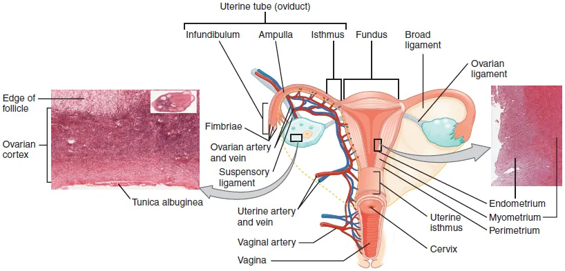
Long description
This anterior view shows the relationship of the ovaries, uterine tubes (oviducts), and uterus. Sperm enter through the vagina, and fertilization of an ovulated oocyte usually occurs in the distal uterine tube. From left to right, LM × 400, LM × 20. (Micrographs provided by the Regents of University

Figure 1: This anterior view shows the relationship of the ovaries, uterine tubes (oviducts), and uterus.
DefinitionVaginal Fornices
The vaginal fornices are the recesses surrounding the cervical os, with the posterior fornix being the deepest and lying adjacent to the rectouterine pouch.
The anterior wall of the vagina is related to the base of the urinary bladder and the entire length of the urethra. The urethra is embedded in the lower anterior vaginal wall, which explains why urinary incontinence can sometimes be associated with vaginal trauma or surgery. The vaginal opening is located between the opening of the urethra and the anus. It is flanked by outlets to the Bartholin’s glands, also known as the greater vestibular glands, which are situated posterolaterally. [1]
| Region | Anterior Relation | Posterior Relation | Lateral Relation |
|---|---|---|---|
| Superior | Base of bladder | Rectouterine pouch | Ureters and uterine vessels |
| Middle | Urethra | Rectum (ampulla) | Levator ani muscles |
| Inferior | Urethra (lower part) | Perineal body | Perineal membrane |
The posterior wall is related superiorly to the rectouterine pouch. In the middle third, it is related to the ampulla of the rectum. Inferiorly, the vagina is separated from the anal canal by the perineal body, a fibromuscular mass that provides support to the pelvic floor. Laterally, the upper third of the vagina is related to the ureters and uterine vessels, which pass close to the lateral fornices. The middle third is supported by the levator ani muscles, which form the pelvic diaphragm. The inferior third traverses the deep perineal pouch and is bounded by the perineal membrane.
Vaginal Relations Outline
- Superior RelationsCervix and rectouterine pouch posteriorly.
- Question: Which third?
Middle
- Anteriorly related to urethra; posteriorly to rectum.
Inferior
- Traverses perineal membrane; bounded by perineal body.
The hymen is a thin membrane that sometimes partially covers the entrance to the vagina. An intact hymen cannot be used as an indication of “virginity”; even at birth, this is only a partial membrane, as menstrual fluid and other secretions must be able to exit the body, regardless of penile–vaginal intercourse. The superior, anterior portions of the labia minora come together to encircle the clitoris, an organ that originates from the same cells as the glans penis and has abundant nerves that make it important in sexual sensation and orgasm. [1]
The vagina is an elastic, muscular canal serving as the internal passageway of the female reproductive tract. High systemic plasma estrogen concentrations can alter the chemical environment, slightly decreasing the acidity of the vagina and making it more hospitable to sperm. Conversely, the healthy vaginal environment is normally kept inhospitable to other pathogens because Lactobacillus sp. is determined to be the predominant microorganism in vaginal flora, producing lactic acid and competing for nutrients. [1] [2]
The cervix, which is the narrow inferior portion of the uterus, projects directly into the superior part of the vagina. Surrounding this projection are the anterior, posterior, and lateral fornices, which represent the recesses formed where the vaginal walls meet the protruding cervical os. Inferior to the fornices, the anterior and posterior walls of the vaginal canal lie in close apposition, marked by transverse mucosal folds known as vaginal rugae that allow for significant distension during childbirth and coitus. The lower extent of the internal canal terminates at the vaginal opening, or introitus, which is partially demarcated by the hymen before communicating with the vestibule of the external genitalia. [1]
Special populations, such as women experiencing bacterial vaginosis, suffer from an imbalance in normal vaginal microbiota that promotes pathogen colonization and bacterial biofilm formation. To address this, drug delivery systems like metronidazole-loaded nanofibers have been developed with polyvinylpyrrolidone to achieve controlled release and prolonged residence time via intermolecular hydrogen bonding between polyvinylpyrrolidone and mucin. Furthermore, vaginal films combining chitosan citrate and methyl polymethacrylate create a polyelectrolyte complex that improves stability, provides pH-dependent behaviour, and increases mucoadhesion capacity. Similarly, liquid crystal precursor systems exhibit low viscosity for easy administration before undergoing phase transition into liquid crystalline matrices upon contact with vaginal mucus, promoting sustained drug delivery. [2]
DefinitionVaginal Microbiota
Lactobacillus species dominate the healthy flora, producing lactic acid to protect against invading pathogens.
| Factor | Description | Effect |
|---|---|---|
| Lactobacillus sp. | Predominant microorganism | Renders environment inhospitable via lactic acid production |
| Estrogen | High systemic plasma levels | Slightly decreases acidity, making the canal more hospitable to sperm |
| Polyvinylpyrrolidone | Polymer used in nanofibers | Improves mucoadhesiveness via intermolecular hydrogen bonding with mucin |
| Methyl polymethacrylate | Component in vaginal films | Enhances stability, low swelling capacity, and mucoadhesion |
Figure 2Gray408
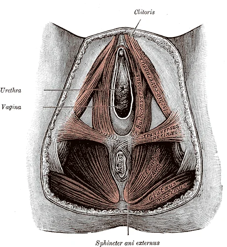
Long description
Structure of vagina. Labelled illustration from Wikimedia Commons, used under its open licence.

Figure 2: Structure of vagina.
The upper third of the vagina originates from the uterine artery, a branch of the internal iliac artery, which descends along the lateral fornix to give off the vaginal branches. The middle third receives blood from the vaginal artery, a direct branch of the internal iliac artery, running horizontally across the pelvic floor to supply the muscular wall. The lower third is supplied by the inferior vesical artery and the middle rectal artery, both arising from the anterior trunk of the internal iliac artery, coursing medially and anteriorly toward the rectovaginal septum. These vessels anastomost together to form a midline longitudinal channel known as the azygos artery of the vagina, positioned along both the anterior and posterior columns. Branches from these major trunks pierce the muscularis coat to form a dense submucosal capillary plexus. The vessels terminate by draining into a corresponding venous plexus that accompanies the arterial pathways.
DefinitionAzygos Artery
The longitudinal arterial anastomosis formed along the midline of the anterior and posterior vaginal walls by descending branches of the uterine and vaginal arteries.
| Segment | Primary Artery | Origin |
|---|---|---|
| Upper third | Uterine artery | Internal iliac artery |
| Middle third | Vaginal artery | Internal iliac artery |
| Lower third | Inferior vesical and middle rectal arteries | Internal iliac artery |
Vaginal Blood Supply Hierarchy
- Internal Iliac ArteryMain pelvic trunk supplying the vagina.
- Question: Which segment?
Upper third
- Uterine artery vaginal branches
Middle third
- Direct vaginal artery
Lower third
- Inferior vesical and middle rectal arteries
The venous drainage of the vagina is achieved through a rich plexus of thin-walled veins lying along the lateral walls of the organ, known as the vaginal venous plexus. This network communicates freely above with the uterine venous plexus and below with the vesical and rectal plexuses, creating an extensive interconnected pelvic pool of blood. Conventional vaginal products are associated with a few limitations such as poor adhesive property and short retention time due to the self-cleansing mechanism of the vagina. The cervicovaginal mucus mixture acts as a barrier to protect the vagina against infection by trapping pathogenic microbes. However, the properties of the vaginal fluid vary by age, vaginal flora, vaginal practices, sexual intercourse, pathological conditions and throughout the menstrual cycle. Variation in drug response arises due to the solubility of the drug affected by the water content in the vaginal fluid. Eventually, poorly water-soluble drugs will solubilize in vaginal fluid when there is higher water content, and more will be absorbed. [2]
Superiorly, the vaginal veins drain into the uterine veins alongside the uterine artery. The vagina has been the site for local delivery of various therapeutic agents including antimicrobial agents, spermicides, contraceptives, and labour inducers. Most vaginal dosage forms can be self-administered easily with minimal interference to daily life. However, these dosage forms have been proven to be associated with multiple limitations including the inadequate spreading of drugs over vaginal surfaces and low drug penetration to the submucosal layers. The possibility of leakage and expulsion of semi-solid dosage forms, due to vaginal fluid dynamics and self-cleaning action of the vaginal tract, may lead to incomplete delivery of sufficient doses. As a result, multiple doses are often needed daily to achieve the desired therapeutic effects, resulting in poor patient compliance and increased overall treatment costs. [2]
Clinical pearlClinical significance
The extensive interconnection of the vaginal plexus with the pelvic venous network explains why pelvic infections and malignant diseases can spread so readily to adjacent pelvic viscera.
From these regional channels, blood converges into the internal iliac veins, which subsequently ascend to join the common iliac veins and finally the inferior vena cava. Metronidazole vaginal gel is a standard treatment of bacterial vaginosis. Cure rates of 74.5% and 66.7% illustrated by the once-daily in situ MTZ gel after 1 week and 4 week. Vaginal gels contain [2]
The lymphatic drainage of the vagina is divided into three distinct anatomical tiers reflecting the embryological and segmental origins of the canal. The upper third of the vagina drains primarily to the obturator and internal iliac lymph nodes, as well as the external iliac nodes. The middle third of the vagina directs its lymph flow predominantly toward the internal iliac lymph nodes. The lower third of the vagina follows the route of the vulva, draining directly into the superficial inguinal lymph nodes.
DefinitionZonal lymphatic division
The vaginal canal is divided into upper, middle, and lower thirds, each directing lymph to specific regional nodal basins matching their pelvic and perineal relations.
The obturator lymph nodes lie within the obturator fossa, closely related to the obturator nerve and vessels lateral to the pelvic wall. The internal iliac lymph nodes sit along the internal iliac artery and its branches deep within the pelvis, receiving vessels from the upper and middle thirds of the vagina. The external iliac lymph nodes are positioned along the external iliac vessels superior and lateral to the pelvic brim. The superficial inguinal lymph nodes lie in the subcutaneous tissue of the femoral triangle inferior to the inguinal ligament, receiving direct channels from the lower third of the vagina.
| Vaginal Region | Primary Lymphatic Destination Nodes |
|---|---|
| Upper third | Obturator, internal iliac, and external iliac nodes |
| Middle third | Internal iliac nodes |
| Lower third | Superficial inguinal nodes |
Vaginal Lymphatic Drainage Pathways
- Vaginal CanalExamine by anatomical thirds.
- Question: Which third?
Upper third
- Obturator, internal iliac, and external iliac nodes
Middle third
- Internal iliac nodes
Lower third
- Superficial inguinal nodes
The nerve supply of the vagina is both autonomic and somatic, providing a dual pathway for visceral and sensory innervation. The autonomic supply reaches the upper and middle portions of the vagina via the uterovaginal plexus, an extension of the inferior hypogastric plexus. This autonomic network contains both sympathetic fibers derived from the superior hypogastric plexus and parasympathetic fibers arising from the pelvic splanchnic nerves. These autonomic fibers regulate vasomotor tone and glandular secretions within the vaginal wall.
The lower quarter of the vagina receives somatic innervation from the deep perineal branch of the pudendal nerve, conveying conscious somatic sensations including touch and temperature. This distinct somatic contribution accounts for the sharp, localized pain experienced in the inferior vaginal canal during injury or childbirth.
Inferiorly, the pudendal nerve enters the perineum and gives rise to the deep perineal branch, which courses medially to supply the lower quarter of the vaginal mucosa and surrounding vestibule.
Referred pain from the vagina follows predictable visceral pathways depending on the pelvic level. Afferent fibers from the upper and middle vagina ascend via sympathetic pathways through the uterovaginal and inferior hypogastric plexuses, entering the spinal cord at the lower thoracic and upper lumbar segments (T10 to L2), which explains referred pain to the lower abdomen and flanks. Conversely, visceral pain from the lower vagina travels along parasympathetic pathways through the pelvic splanchnic nerves to spinal segments S2 to S4, referring pain to the perineum and sacral regions.
DefinitionVaginal innervation
Autonomic fibers supply the upper and middle vagina via the uterovaginal plexus, while somatic fibers supply the lower quarter via the pudendal nerve.
The vaginal wall is a dynamic mucosal structure designed for distension and protection. It consists of four distinct layers, each serving specific physiological functions. Understanding these layers is fundamental to appreciating how the vagina accommodates childbirth and resists infection.
The innermost layer is the stratified squamous non-keratinised epithelium. This epithelium is thick and resilient, allowing the vagina to stretch significantly during intercourse and parturition. The cells are rich in glycogen, which is released upon desquamation. This glycogen serves as a substrate for lactobacilli, the predominant flora. Lactobacilli metabolise glycogen into lactic acid, maintaining an acidic pH. This acidity inhibits the growth of pathogenic organisms. The epithelium lacks blood vessels, relying on diffusion from the underlying lamina propria for nutrients.
DefinitionGlycogen-rich epithelium
The vaginal epithelium stores glycogen to support lactobacilli, which produce lactic acid to maintain a protective acidic environment.
Beneath the epithelium lies the lamina propria, a layer of loose connective tissue. It contains numerous elastic fibres, providing the tissue with recoil properties after stretching. This layer is highly vascularised, containing a rich network of capillaries and venules. The vascularity allows for rapid healing of minor abrasions. It also houses lymphatic vessels and immune cells, contributing to local immunity. The lamina propria merges with the muscularis layer, ensuring structural continuity.
The muscularis layer consists of smooth muscle fibres arranged in inner circular and outer longitudinal bundles. These muscles are not as distinct as in the gastrointestinal tract but allow for significant tonicity and peristaltic-like movements. The muscle layer provides the structural support for the vaginal canal. It also plays a role in expelling menstrual blood and semen. The tone of these muscles is influenced by hormonal changes throughout the menstrual cycle.
The outermost layer is the adventitia, composed of dense connective tissue. It blends with the surrounding pelvic fascia and connective tissue. This layer anchors the vagina to adjacent structures, such as the bladder anteriorly and the rectum posteriorly. It contains blood vessels and nerves that supply the vaginal wall. The adventitia ensures the vagina remains fixed in its anatomical position while allowing for necessary mobility.
NoteClinical Relevance
The rich vascularity of the lamina propria explains why vaginal tears during childbirth can bleed significantly, yet heal rapidly due to good blood supply.
The vaginal environment is also influenced by microbial communities. Biofilm formation can occur secondary to adaptation under diverse nutritional and environmental conditions. Such biofilms are composed of a group of pathogenic microbes adhered to one another on a surface enclosed by the self-produced extracellular polymeric substances. The extracellular matrix acts as a physical barrier that limits the accessibility of antimicrobial molecules and evades the immune response. Consequently, drug release will be altered and the pathogenic microbes have an increased persistence in a hostile environment and enhanced tolerance to the adverse conditions. Moreover, some antimicrobials are unable to exert their action due to the reduced metabolism within the biofilm. Hence, formulations indicated for vaginal delivery must not harm the normal vaginal flora and its environment. [2]
The female reproductive system originates from the intermediate mesoderm and the coelomic epithelium. The ovaries develop from the genital ridge, a thickened area of the coelomic epithelium and underlying mesenchyme. The primordial germ cells, which give rise to the oocytes, migrate from the yolk sac endoderm to the genital ridge during the fifth week of gestation. These cells are called oogonia. They undergo rapid mitotic divisions to increase their numbers. By the third month of fetal development, the oogonia begin to enter meiosis I and become primary oocytes. These primary oocytes become arrested in the prophase stage of meiosis I. This arrest is maintained by inhibitory factors from the surrounding somatic cells. The number of primary oocytes peaks at approximately one to two million in the infant ovary. This number declines significantly over time. By puberty, only approximately 400,000 primary oocytes remain. The remaining oocytes will either degenerate or be ovulated during the reproductive years. At menopause, the supply of primary oocytes is effectively zero.
The uterus develops from the paramesonephric ducts, also known as the Müllerian ducts. These ducts form from the invagination of the coelomic epithelium. The cranial parts of the ducts remain separate and form the uterine tubes. The caudal parts fuse in the midline to form the uterovaginal canal. This canal gives rise to the uterus and the upper part of the vagina. The lower part of the vagina develops from the urogenital sinus. The fusion of the paramesonephric ducts is a critical process. Failure of fusion can lead to uterine anomalies such as a bicornuate uterus. The vaginal plate forms from the sinovaginal bulbs, which are outgrowths of the urogenital sinus. This plate canalizes to form the vaginal lumen.
The external genitalia, or vulva, develop from the genital tubercle, urogenital folds, and labioscrotal swellings. The genital tubercle elongates to form the clitoris. The urogenital folds remain unfused in females and form the labia minora. The labioscrotal swellings form the labia majora. The vestibular bulbs develop from the same tissue as the corpus spongiosum in males. The hymen is a remnant of the vaginal plate. It may persist as a thin membrane or be absent.
The ovarian cycle involves predictable changes in oocytes and follicles. It is a roughly 28-day cycle during the reproductive years. This cycle includes oogenesis and folliculogenesis. Folliculogenesis is the growth and development of ovarian follicles. The process from primordial follicle to early tertiary follicle takes approximately two months. The final stages, ending with ovulation, occur over approximately 28 days. These changes are regulated by GnRH, LH, and FSH. The breast tissue also responds to hormonal fluctuations. Estrogen and progesterone levels cause swelling and tenderness, especially during the secretory phase. If pregnancy occurs, mammary tissue develops further.
DefinitionOogenesis
The production of female gametes, beginning with oogonia in fetal life and arresting in meiosis I until puberty.
| Stage | Timeframe | Key Feature |
|---|---|---|
| Oogonia Formation | Fetal Development | Mitotic division of primordial germ cells |
| Primary Oocyte Arrest | Fetal Life to Puberty | Arrested in prophase of meiosis I |
| Puberty | ~10-14 years | ~400,000 primary oocytes remain |
| Menopause | ~50 years | Primary oocytes depleted |
The radiological anatomy of the female pelvis requires a systematic approach to identify the vagina and its surrounding spaces on cross-sectional imaging. Magnetic resonance imaging provides superior soft-tissue contrast resolution compared with computed tomography, making it the primary imaging modality for delineating pelvic floor anatomy. Modern high-field magnets allow physicians to trace the fibromuscular canal from its origin at the uterine cervix down to its termination at the vaginal introitus in the perineum.
DefinitionImaging modality
Magnetic resonance imaging is the gold standard for assessing pelvic organ prolapse and anatomical relationships due to multiplanar capability.
The vagina appears as a flattened fibromuscular tube extending from the perineal membrane to the uterine cervix. Anteriorly, the upper third of the vagina is related to the base of the bladder and the urethra, while the lower two-thirds are separated from the urethra by the urethrovesical septum. Posteriorly, the upper fourth of the vagina relates to the rectouterine pouch, the middle half relates to the anterior wall of the rectum via the rectovaginal septum, and the lower fourth is separated from the anal canal by the perineal body.
Special considerations apply during pregnancy, where increased pelvic vascularity and hormonal engorgement cause marked thickening of the vaginal walls and alterations in signal intensity on T2-weighted sequences. In older adults, postmenopausal atrophy results in a thinner vaginal wall and reduced high-signal intensity in the mucosal layer due to diminished estrogen stimulation. In pediatric patients, high-resolution pelvic ultrasound is often preferred over magnetic resonance imaging to avoid sedation while still clearly demonstrating the fluid-filled or collapsed mucosal interface of the prepubertal vagina.
| Layer | T2-weighted MRI signal | Histological correlate |
|---|---|---|
| Inner mucosa | High signal intensity | Epithelium and subepithelial vascular plexus |
| Muscularis | Intermediate signal intensity | Smooth muscle coat |
| Outer adventitia | Low signal intensity | Dense connective tissue |
Vaginal Compartments on Cross-Section
- Anterior compartmentUrethra and urinary bladder base
- Question: Location along axis?
Upper fourth
- Posterior compartmentRectouterine pouch of Douglas
Middle half
- Posterior compartmentRectum via rectovaginal septum
The vagina is a muscular and membranous canal extending from the vulva to the uterus. It serves as an excretory channel for uterine secretions, the conduit for menstrual flow, and the lower part of the birth canal.
The upper vagina is related anteriorly to the base of the bladder and the ureters, and superiorly to the cervix uteri. Laterally, the vagina is supported by the pelvic fascia and the levator ani muscle. Inferiorly, the distal canal pierces the urogenital diaphragm and opens into the vestibule between the labia minora.
The muscular walls of the vagina rely on supportive pelvic floor muscles for structural integrity. Pelvic support maintains proper alignment against intra-abdominal pressures.
| Muscle | Origin | Insertion | Action | Nerve Supply |
|---|---|---|---|---|
| Levator ani (Pubococcygeus) | Posterior body of pubis | Urethra, vagina, and anal canal | Supports pelvic viscera | S2 to S4 nerve roots |
| Bulbospongiosus | Perineal body | Pubic arch and clitoris | Constricts vaginal orifice | Pudendal nerve |
DefinitionVaginal fornices
The recesses formed where the vaginal walls attach to the circumference of the cervix are divided into anterior, posterior, and lateral fornices.
The arterial supply derives mainly from the vaginal branch of the internal iliac artery, supplemented by branches from the uterine, middle rectal, and internal pudendal arteries. Venous drainage occurs via a dense vaginal plexus that surrounds the canal, ultimately draining into the internal iliac veins. Lymphatic drainage follows regional pathways: the upper third drains to the external and internal iliac nodes, the middle third to the internal iliac nodes, and the lower third to the superficial inguinal nodes. Sensory and autonomic innervations arise from the uterovaginal nerve plexus, conveying sympathetic, parasympathetic, and visceral afferent fibers.
Vaginal Anatomy and Lesion Evaluation
- Examine anatomical relationsAssess anterior bladder and posterior rectal relations.
- Question: Identify lesion site?
Upper third
- Check internal iliac lymph drainage
Lower third
- Check superficial inguinal lymph drainage
Ovulation marks the transition from puberty into reproductive maturity, initiating the release of an oocyte from the ovary. This event occurs approximately once every 28 days throughout the reproductive years. Just prior to ovulation, a surge of luteinizing hormone triggers the resumption of meiosis in a primary oocyte. This hormonal surge initiates the transition from a primary to a secondary oocyte. The resulting cell division is unequal, producing one large secondary oocyte and one small first polar body. The secondary oocyte is the larger daughter cell that eventually leaves the ovary during ovulation. The first polar body may complete meiosis to produce second polar bodies or disintegrate. Although oogenesis produces up to four cells, only one survives to become the functional gamete. Ovarian follicles consist of oocytes and their supporting cells, growing via folliculogenesis. Folliculogenesis typically leads to the ovulation of one follicle every 28 days. The death of ovarian follicles is termed atresia and can occur at any developmental stage. A female infant is born with one to two million oocytes within ovarian follicles. This number declines throughout life until menopause, when no follicles remain. Follicles progress from primordial to primary, secondary, and tertiary stages prior to ovulation. The oocyte remains as a primary oocyte until right before ovulation occurs. [1]
Exam trapPolar Body Fate
The first polar body may or may not complete meiosis; it eventually disintegrates regardless of completion. [1]
DefinitionAtresia
The death of ovarian follicles, which can occur at any point during follicular development.
Clinical pearlOocyte Count
A female infant at birth will have one to two million oocytes within the ovarian follicles.
NoteHormonal Trigger
A surge of luteinizing hormone triggers the resumption of meiosis in a primary oocyte just prior to ovulation.
Exam trapContraceptive Adherence
Skipping even just one or two pills at certain points of the cycle can lead to an increase in FSH and LH and result in ovulation. Being several hours late taking the pill can also disrupt negative feedback and cause ovulation. Birth control pills function by providing constant estrogen and progesterone levels. It is important, therefore, to follow the directions on the birth control pill package to successfully prevent pregnancy. [1]
DefinitionEndometrium Layers
The endometrium consists of two layers: the stratum basalis and the stratum functionalis. The stratum basalis layer is part of the lamina propria and is adjacent to the myometrium. This layer does not shed during menses. The thicker stratum functionalis layer contains the glandular portion of the lamina propria. It is the stratum functionalis that grows and thickens in response to increased levels of estrogen and progesterone. In the luteal phase of the menstrual cycle, spiral arteries supply the thickened stratum functionalis. These are special branches off of the uterine artery. [1]
Clinical pearlSpiral Arteries
In the luteal phase of the menstrual cycle, special branches off of the uterine artery called spiral arteries supply the thickened stratum functionalis. [1]
Exam trapBasalis vs Functionalis
The stratum basalis layer is part of the lamina propria and is adjacent to the myometrium; this layer does not shed during menses.
NoteEndometrial Structure
The innermost layer of the uterus is called the endometrium, containing a connective tissue lining, the lamina propria. [1]
Students often mistake the vagina for the vulva, failing to separate the internal fibromuscular canal from the external female genitalia. Another frequent error is assuming the vaginal axis is vertical; in anatomical position, the vagina curves superiorly and posteriorly, forming an angle of roughly forty-five degrees with the horizontal plane.
Learners also confuse vaginal fornices with the pelvic peritoneal pouches. The posterior fornix is deeper and sits in direct relation to the rectouterine pouch, whereas the anterior and lateral fornices are shallower.
A common histological mistake is describing the vaginal epithelium as keratinized. The lining is a non-keratinized stratified squamous epithelium that lacks glands entirely, relying instead on cervical secretions and transudation for lubrication.
Students occasionally misattribute the arterial supply of the vagina to a single vessel. The upper vagina receives blood from the uterine and vaginal branches of the internal iliac artery, while the middle and lower sections are supplied by branches of the middle rectal and internal pudendal arteries.
Another error involves venous drainage; learners often forget that vaginal veins form a dense plexus around the vagina that drains directly into the internal iliac veins. Finally, students commonly misidentify the nerve supply, forgetting that the upper vagina is innervated by the autonomic inferior hypogastric plexus, whereas the lower quarter receives somatic innervation via the deep perineal branch of the pudendal nerve.
Exam trapEpithelial confusion
Do not confuse the non-keratinized stratified squamous epithelium of the vagina with the simple columnar epithelium of the endocervix.
At the bedside, knowledge of vaginal anatomy is essential for speculum insertion, bimanual pelvic examinations, and high vaginal swab collection to diagnose infections. Clinicians rely on the correct identification of the vaginal fornices when performing procedures such as culdocentesis or assessing pelvic organ prolapse.
In the operating theatre, surgeons performing hysterectomies or pelvic floor repairs must navigate the close anatomical relations of the vagina, specifically the ureters running anterior and lateral to the lateral fornices, and the rectum lying directly against the posterior wall.
On cross-sectional imaging such as magnetic resonance imaging, the normal vagina appears as a high-signal central fluid or mucus line surrounded by a low-signal muscular wall, allowing radiologists to stage cervical and vaginal malignancies accurately.
In examinations, questions frequently test the lymphatic drainage of the vagina: the upper two-thirds drain to the internal and external iliac nodes, the middle third drains to the internal iliac nodes, and the lower third drains directly to the superficial inguinal lymph nodes.
Vaginal Lymphatic Drainage
- Upper two-thirdsDrains to internal and external iliac lymph nodes
- Middle thirdDrains to internal iliac lymph nodes
- Lower thirdDrains to superficial inguinal lymph nodes
| Region | Anterior Relation | Posterior Relation |
|---|---|---|
| Upper third | Base of bladder and ureters | Rectouterine pouch (pouch of Douglas) |
| Middle third | Trigone of bladder and urethra | Anterior wall of rectum |
| Lower third | Perineal body and urethra | Anal canal |
- The vagina is a muscular canal extending from the cervix to the vulva.
- The epithelium is non-keratinized stratified squamous and contains no glands.
- The posterior fornix is deeper than the anterior and lateral fornices.
- Arterial supply is derived from the uterine, vaginal, middle rectal, and internal pudendal arteries.
- Venous drainage flows via vaginal plexuses into the internal iliac veins.
- Upper vaginal innervation is autonomic, while the lower quarter is somatic via the pudendal nerve.
- Lymphatic drainage is divided between iliac nodes and superficial inguinal nodes.
Answer each one in your head first, then open the answer.
What type of epithelium lines the normal adult vagina?
Show answerHide answer
non-keratinized stratified squamous epitheliumWhich fornix is in direct relation to the rectouterine pouch?
Show answerHide answer
posterior fornixWhere do the lymphatics of the lower third of the vagina drain?
Show answerHide answer
superficial inguinal lymph nodesWhich nerve supplies somatic sensation to the lower quarter of the vagina?
Show answerHide answer
deep perineal branch of the pudendal nerveWhat is the primary arterial supply to the superior vagina?
Show answerHide answer
uterine and vaginal arteries
- Betts JG, Young KA, Wise JA, et al. Anatomy and Physiology of the Ovarian Reproductive System. OpenStax, Anatomy and Physiology 2e (CC BY-NC-SA 4.0). Accessed .
- Pandey M, Choudhury H, Abdul-Aziz A, Bhattamisra SK, Gorain B, Carine T, Wee Toong T, Yi NJ, Win Yi L. Promising Drug Delivery Approaches to Treat Microbial Infections in the Vagina: A Recent Update. Polymers 2020. Accessed .
Figure credits
Educational content for medical students and doctors. It is not a substitute for clinical judgement or local guidelines.