Prostate
Anatomical overview of the male reproductive accessory glands and prostate.
The male reproductive system relies on accessory glands to produce the bulk of semen, a thick, milky fluid ejaculated during sexual activity. Sperm themselves constitute only a small fraction of this volume, specifically five percent of the final ejaculate. The remaining ninety-five percent is generated by three critical glands: the seminal vesicles, the prostate, and the bulbourethral glands. [1]
The paired seminal vesicles are located posterior to the bladder and superior to the prostate. They contribute approximately sixty percent of the total semen volume. The fluid they secrete is rich in fructose, a sugar substrate that sperm mitochondria utilise to generate adenosine triphosphate (ATP). This energy production is essential for sperm motility as they traverse the female reproductive tract. [1]
As sperm travel through the ampulla of the ductus deferens, they mix with seminal vesicle fluid. This mixture enters the ejaculatory duct, a short structure formed by the union of the ampulla and the duct of the seminal vesicle. The paired ejaculatory ducts transport this combined seminal fluid directly into the prostate gland. [1]
The prostate is a walnut-sized gland situated inferior to the bladder and surrounding the urethra. It receives the ejaculatory ducts and adds its own secretions to the seminal fluid. While benign conditions like benign prostatic hyperplasia (BPH) are common, the prostate is also the site of prostate cancer. Prostate cancer is the second most common cancer in males according to the CDC. Some forms grow slowly and may never require treatment, whereas aggressive forms can metastasise to organs such as the lungs and brain. Diagnosis typically involves a digital rectal examination to palpate for masses, blood tests for prostate-specific antigen (PSA), and biopsy for confirmation. [1] [2]
The bulbourethral glands, also known as Cowper’s glands, are small pea-sized structures located inferior to the prostate. They secrete a clear, mucus-like fluid that lubricates the urethra and neutralises residual acidity from urine before ejaculation. This preparation facilitates the smooth passage of sperm through the urethra.
PSA levels provide diagnostic clues. Patients with PSA levels between four ng/mL and ten ng/mL have an approximately one in four chance of having prostate cancer. If the PSA exceeds ten ng/mL, the possibility of cancer rises to over fifty percent. However, PSA is not cancer-specific; it can also indicate benign pathologies such as BPH or prostatitis. A tissue biopsy is usually required to confirm the presence of cancer. [2]
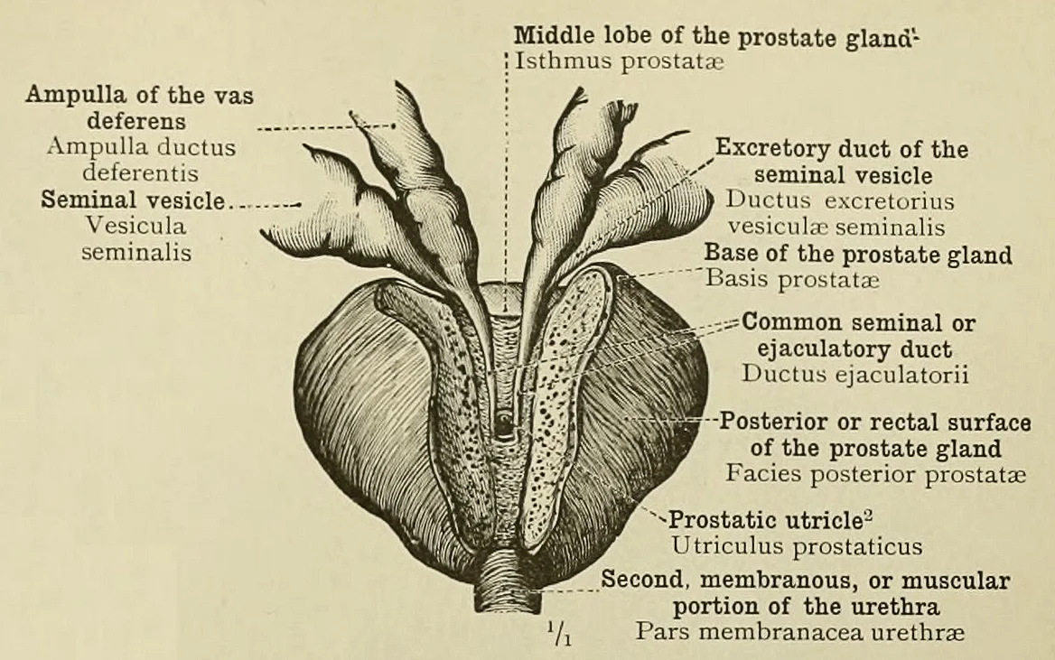
Figure 1Prostate. Common seminal or ejaculatory ducts and the blind extremity of the prostatic utricle
Long description
Fig. 860.—THE COMMON SEMINAL OR EJACULATORY DUCTS AND THE BLIND EXTREMITY OF THE PROSTATIC UTRICLE, LAID BARE FROM BEHIND BY THE REMOVAL OF A PORTION OF THE PROSTATE GLAND.

Figure 1: Fig.
The prostate is a walnut-sized gland situated in the male pelvis, inferior to the bladder and anterior to the rectum. It surrounds the prostatic urethra, which passes through its central axis. The gland is divided into distinct zones that are critical for understanding clinical presentations and imaging findings.
Spermatogenesis begins with mitosis of spermatogonia, producing two identical diploid cells; one remains a spermatogonium, while the other becomes a primary spermatocyte. Primary spermatocytes undergo meiosis I, where 23 pairs of chromosomes separate, resulting in two secondary spermatocytes with half the chromosome number. Meiosis II involves the division of 23 replicated chromosomes, resulting in a total of four haploid spermatids. Tightly packed mitochondria in the mid-piece produce ATP to power the flagellum, which extends from the neck and the mid-piece through the tail. [1]
The zonal anatomy directly informs diagnostic strategies. Since the peripheral zone is the most frequent origin of malignant tumors, imaging techniques like multiparametric magnetic resonance imaging are used to detect lesions in this area before biopsy. The transition zone is the site of benign enlargement, which can lead to severe symptoms requiring surgical removal of a portion of the prostate tissue. The central zone is less commonly involved in both benign and malignant processes. The gland’s position anterior to the rectum means that abnormalities in the peripheral zone can often be detected by assessing the size and texture of the gland through the rectal wall. The prostatic urethra’s course through the gland means that any mass effect in the transition or central zones will initially present with lower urinary tract symptoms due to urethral compression. The capsule of the prostate is a fibromuscular layer that encloses the peripheral zone, acting as a barrier that, if breached, indicates extracapsular extension of disease. The seminal vesules lie posterior and superior to the prostate, draining into the ejaculatory ducts which then pierce the central zone to enter the urethra. This anatomical arrangement explains why seminal vesicle involvement is a marker of advanced local disease. The neurovascular bundles run laterally to the prostate, within the surgical plane between the prostate and the pelvic sidewall, making them critical structures to preserve during radical prostatectomy to maintain erectile function. The relationship between the peripheral zone and the rectum is also why digital rectal examination remains a conventional method for assessing gland size and detecting nodular abnormalities. [1] [2]
The prostate is composed of glandular and stromal tissue. The glandular component consists of acini and ducts lined by epithelial cells, which secrete prostatic fluid. The stromal component provides structural support and contains smooth muscle fibers. The peripheral zone is predominantly glandular, whereas the central and transition zones have a higher proportion of stroma. This histological difference influences how the gland responds to hormonal changes and how it appears on imaging. The epithelial cells of the prostate secrete prostate-specific antigen, a glycoprotein that is normally found in semen but can also be detected in the bloodstream. The level of this antigen in the blood is used as a screening marker, with a cut-off point of 4 ng/mL suggesting the need for further investigation. The gland’s innervation is primarily autonomic, with parasympathetic fibers from the pelvic splanchnic nerves and sympathetic fibers from the hypogastric plexus. The blood supply comes from the prostatic arteries, branches of the internal iliac artery, which enter the gland laterally. Venous drainage is via the prostatic venous plexus, which communicates with the internal iliac veins and the vertebral venous plexus. This rich vascular supply and venous plexus are important considerations during surgical procedures to minimize bleeding. The lymphatic drainage is to the internal iliac and sacral lymph nodes. Understanding these vascular and lymphatic relationships is crucial for staging prostate cancer, as metastasis often occurs via these routes. The gland’s proximity to the bladder neck and the urethra means that surgical interventions must carefully preserve the sphincter mechanism to maintain continence. The external features of the prostate, including its smooth, symmetrical shape in a healthy individual, can be altered by nodular changes in benign or malignant conditions. The consistency of the gland, whether soft, firm, or nodular, is a key finding on digital examination that guides further management. [2] [1]
The prostate is a fibromuscular gland situated in the male pelvis, inferior to the bladder and anterior to the rectum. It surrounds the prostatic urethra, which is the initial segment of the urethra passing through the gland. The organ is roughly the size of a chestnut and weighs approximately 20 grams in the adult male. Its position is critical for clinical examination and surgical access.
DefinitionProstate Anatomy
The prostate is a walnut-sized gland located below the bladder, surrounding the urethra, and lying anterior to the rectum.
Anteriorly, the prostate is separated from the pubic symphysis by the retropubic space of Retzius, which contains fat and the dorsal venous complex. This space allows for surgical access during procedures such as a retropubic prostatectomy. Posteriorly, the prostate is closely related to the rectum, separated only by the rectoprostatic fascia (Denonvilliers’ fascia). This intimate posterior relationship permits the assessment of the prostate via digital rectal examination (DRE). Through the anterior wall of the rectum, a clinician can palpate the posterior surface of the prostate, including the median sulcus which divides the right and left lobes.
Digital Rectal Examination
- Perform DREInsert gloved, lubricated finger into rectum
- Question: Palpate posterior surface
Yes
- Assess size, consistency, and nodules
No
- Reposition finger
Superiorly, the base of the prostate is continuous with the neck of the bladder. The seminal vesicles lie posterior and superior to the base of the prostate, separated by the rectovesical pouch. The ejaculatory ducts pass through the posterior aspect of the prostate to empty into the prostatic urethra at the seminal colliculus. This arrangement is crucial for understanding the pathways of semen and potential obstructions.
| Structure | Relation to Prostate |
|---|---|
| Bladder | Superior (Base) |
| Rectum | Posterior |
| Pubic Symphysis | Anterior (via Retropubic Space) |
| Seminal Vesicles | Posterior-Superior |
| Urogenital Diaphragm | Inferior (Apex) |
The prostate is divided into zones rather than distinct anatomical lobes in modern terminology, though the terms anterior, posterior, and lateral lobes are still used clinically. The peripheral zone constitutes the majority of the gland and is located posteriorly, making it accessible to DRE. The central zone surrounds the ejaculatory ducts. The transition zone surrounds the urethra and is the site of benign prostatic hyperplasia. The anterior fibromuscular stroma contains no glandular tissue.
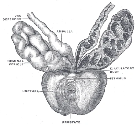
Figure 2Gray1160
Long description
Prostate with seminal vesicles and seminal ducts, viewed from in front and above. (Spalteholz.)

Figure 2: Prostate with seminal vesicles and seminal ducts, viewed from in front and above (Spalteholz.).
The zones of the prostate are shaded differently to distinguish the peripheral, central, and transition zones, clarifying their spatial relationships to the urethra and ejaculatory ducts.
The prostate is a fibromuscular glandular organ situated inferior to the bladder and anterior to the rectum. It surrounds the proximal urethra, specifically the prostatic urethra, which passes through its centre. The organ is divided into distinct zones that dictate its anatomical and clinical behaviour. The peripheral zone constitutes the posterior and lateral aspects of the gland. This region is the most common site for the development of adenocarcinoma. The central zone surrounds the ejaculatory ducts. It is less frequently involved in malignancy compared to the peripheral zone. The transition zone encircles the proximal urethra. Benign prostatic hyperplasia typically originates here, potentially compressing the urethra and causing obstructive symptoms. The anterior fibromuscular stroma forms the front of the gland. It contains no glandular tissue, consisting mainly of smooth muscle and connective tissue.
The central zone is located centrally, surrounding the ejaculatory ducts. The anterior fibromuscular stroma is the non-glandular tissue at the front. These structures are arranged concentrically around the urethral lumen.
The prostatic ducts drain into the prostatic urethra. There are numerous small ducts opening along the prostatic crest. The ejaculatory ducts enter the posterior aspect of the gland. They pass through the central zone to join the urethra. This arrangement ensures the mixing of semen components during ejaculation. The glandular tissue is supported by a fibromuscular stroma. This stroma provides structural integrity and allows for contraction during ejaculation. The vascular supply is derived from the inferior vesical artery. Venous drainage occurs via the prostatic venous plexus. This plexus communicates with the vertebral venous system. This connection is clinically significant for the spread of metastatic disease. [1]
DefinitionProstatic Zones
The prostate is anatomically divided into the peripheral, central, transition, and anterior fibromuscular zones, each with distinct clinical implications for pathology and surgery.
The internal structure supports the dual functions of urinary passage and reproductive secretion. The smooth muscle in the stroma contracts under sympathetic nervous control. This action expels prostatic fluid and sperm into the urethra. The glandular epithelium secretes prostatic fluid, which forms part of the semen. This fluid contains enzymes and ions that support sperm viability. The anatomical arrangement ensures efficient mixing and expulsion. Understanding these internal structures is essential for interpreting imaging and biopsy results. The peripheral zone's posterior location makes it accessible for digital rectal examination. This clinical feature highlights the importance of anatomical knowledge in diagnosis.
The prostate gland is centrally located, sitting anterior to the rectum at the base of the bladder, where it surrounds the prostatic urethra. This anatomical position places the gland in close proximity to the rectal wall, a relationship critical for digital examination and surgical planning. The organ itself is approximately the size of a walnut and comprises both muscular and glandular tissues. [1]
The primary arterial supply to the prostate arises from branches of the internal iliac artery. In some individuals, contributions may also come from the middle rectal artery. These vessels enter the prostate at its base and posterior aspect, distributing blood to the glandular and muscular components. The venous drainage follows a similar pattern, forming the prostatic venous plexus, which communicates with the vertebral venous system. This c
The prostate excretes an alkaline, milky fluid into the seminal fluid, now known as semen. This secretion is critical for the initial coagulation of semen following ejaculation, which helps retain it within the female reproductive tract. Subsequently, the semen regains its fluid state, allowing sperm to pass further into the female reproductive tract. [1]
DefinitionProstatic Urethra
The portion of the urethra that runs within the prostate, surrounded by the glandular and muscular tissue.
The scrotum contains the dartos muscle, a subcutaneous layer that forms the scrotal septum, dividing the cavity into two compartments, each housing one testis. The cremaster muscles, descending from the internal oblique, cover each testis like a muscular net and contract to elevate the testes in cold weather. Simultaneous contraction of the dartos and cremaster muscles moves the testes closer to the body, decreasing scrotal surface area to retain heat. Conversely, increased environmental tempera. Externally, the scrotum has a raised medial thickening on the surface called the raphae. [1]
| Structure | Relation | Clinical Significance |
|---|---|---|
| Rectum | Posterior | Allows digital examination of posterior surface |
| Bladder | Superior | Base of bladder surrounds prostatic urethra |
| Pubic Symphysis | Anterior | Separated by retropubic space |
| Levator Ani | Lateral | Supports pelvic floor and prostate |
Understanding the arterial supply is essential for procedures such as radical prostatectomy, which involves the medical removal of the prostate gland by open and/or laparoscopic surgery. The procedure requires making small incisions on the abdomen or via the perineum. Precise knowledge of vascular anatomy minimizes blood loss and preserves surrounding structures. [2]
The venous drainage of the prostate is integral to understanding the local vascular anatomy and its clinical implications, particularly regarding the spread of pathology and the mechanics of adjacent structures. The prostate gland is drained by a complex network of veins that ultimately connect to the systemic circulation.
The prostatic venous plexus drains into the internal iliac veins. [1]
Understanding the blood supply is essential to contextualising the venous drainage. The prostate receives its arterial supply from branches of the internal iliac artery. Additional contributions may come from the middle rectal artery and the internal pudendal artery. The arterial blood flows through the capillary beds within the prostate gland, where exchange of nutrients and waste occurs. The deoxygenated blood then enters the venules, which coalesce to fo [1]
The prostatic venous plexus communicates with the vertebral venous plexus (also known as the internal vertebral venous plexus or Batson’s plexus). This anatomical feature is clinically significant because it provides a route for the metastatic spread of prostate cancer cells to the vertebral column and brain, bypassing the pulmonary circulation.
Clinical pearlClinical Pearl
The valveless nature of the connection between the prostatic venous plexus and the vertebral venous plexus allows for metastatic spread of prostate cancer to the spine without passing through the lungs.
| Structure | Origin | Termination | Clinical Significance |
|---|
The venous drainage of the prostate is integral to understanding the local vascular anatomy and its clinical implications, particularly regarding the spread of pathology and the mechanics of adjacent structures. The prostate gland is drained by a complex network of veins that ultimately connect to the systemic circulation. | Prostatic Plexus | Prostate Gland | Internal Iliac Veins | Valveless connection to vertebral plexus | | Internal Iliac Veins | Prostatic Plexus | Common Iliac Veins | Systemic return to heart |
The lymphatic drainage of the prostate is a critical anatomical pathway that determines the spread of disease and guides surgical planning. Understanding these routes requires examining the specific nodal groups involved and their spatial relationships.
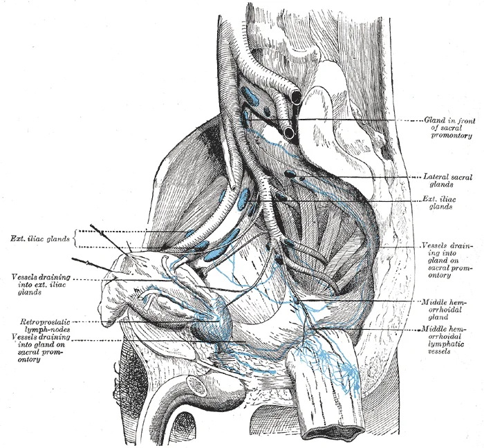
Figure 3Lymphatics of the prostate-Gray619
Long description
Lymphatics of the prostate (lateral view)

Figure 3: Lymphatics of the prostate (lateral view).
The primary efferent lymphatic vessels emerge from the posterior and lateral aspects of the gland. These vessels travel alongside the inferior vesical arteries. They drain primarily into the internal iliac lymph nodes. Secondary drainage occurs into the external iliac lymph nodes. Tertiary drainage may reach the common iliac nodes. The obturator nodes are also frequently involved due to their proximity to the lateral prostatic surface. Recognising this pattern is essential for staging prostate cancer, as metastasis typically follows this predictable anatomical route. [1]
DefinitionLymphatic Drainage
The prostate drains primarily into the internal iliac, external iliac, and obturator lymph nodes, following the vascular supply.
The nerves associated with the prostate are part of the pelvic autonomic plexus. The prostatic plexus lies on the lateral aspect of the prostate. It receives sympathetic fibres from the superior hypogastric plexus and parasympathetic fibres from the pelvic splanchnic nerves. These fibres travel within the neurovascular bundles. The neurovascular bundles are located posterolateral to the prostate. They contain the cavernous nerves responsible for erectile function. Preservation of these nerves during surgery is vital to prevent erectile dysfunction. The urethral sphincter is also innervated by somatic fibres from the pudendal nerve. Damage to these structures during radical prostatectomy can lead to urinary incontinence.
| Structure | Origin | Course | Function |
|---|---|---|---|
| Prostatic Plexus | Superior Hypogastric & Pelvic Splanchnic | Lateral prostate | Autonomic regulation |
| Cavernous Nerves | Prostatic Plexus | Posterolateral prostate | Erectile function |
| Pudendal Nerve | Sacral Plexus | Perineum | Somatic sphincter control |
Salvage radical prostatectomy is usually recommended to patients with local recurrence in the absence of metastases after undergoing external beam radiation therapy, brachytherapy, or cryotherapy. This approach carries risks of morbidity. Patients younger than age 70 with organ-confined prostate cancer, with a life expectancy higher than 10 years who have little to no comorbidities, are best suited for radical prostatectomy. Complications include incontinence and erectile dysfunction arising from surgical damage to the urinary sphincter and erectile nerves. [2]
Hormonal therapy is also known as androgen deprivation therapy (ADT). This technique is applied in the treatment of advanced and/or metastasized prostate cancer. Its therapeutic mechanism is based on the blockage of testosterone production and other male hormones, preventing them from fueling prostate cancer cells. Therefore, significantly decreased male hormonal levels are responsible for inhibition of the action of androgen on the androgen receptor. This is often achieved using bilateral orchiectomy or medical castration via administration of luteinizing hormone-releasing hormone (LHRH) analogs or antagonists. [2]
Amongst all the available treatment strategies available for metastatic prostate cancer, androgen deprivation therapy (ADT) has more potential combination treatment compared to other therapeutic strategies for prostate cancer. Approved and currently ongoing clinical trials with ADT treatment include ADT with radiation therapy, which often treats high-risk patients to delay or prevent the disease from progressing to CRPC. ADT and chemotherapy, which in several clinical studies has shown to increase patient survival but results in adverse side effects and sometimes death. Immunotherapy and ADT, which has been reported to increase patient survival by 8.5 months. [2]
The innervation of the prostate is complex, involving both autonomic and somatic pathways that regulate function and mediate sensation. Understanding this supply requires examining the pelvic plexus and the sacral roots.
The autonomic nervous system provides the primary motor and secretory control. The pelvic splanchnic nerves, arising from the second, third, and fourth sacral spinal nerves (S2–S4), carry parasympathetic fibres. These fibres join the inferior hypogastric plexus, also known as the pelvic plexus. From this plexus, prostatic plexuses branch off to supply the gland. Parasympathetic stimulation is responsible for vasodilation of the penile arteries and erection, but it also influences prostatic secretion. The sympathetic nervous system arises from the lower thoracic and upper lumbar segments (T10–L2). These fibres travel through the lumbar splanchnic nerves to the superior hypogastric plexus, then descend to the inferior hypogastric plexus. Sympathetic fibres mediate ejaculation by causing contraction of the smooth muscle in the prostate and the bladder neck, preventing retrograde ejaculation. They also provide vasoconstrictor tone. [1]
Somatic innervation is provided by the pudendal nerve, which arises from S2–S4. This nerve supplies the external urethral sphincter and the perineal muscles. It also carries sensory information from the skin of the perineum and the distal urethra.
Referred pain from the prostate is a critical clinical concept. Because the prostate shares sensory innervation with other pelvic structures, pain can be perceived in distant areas. The prostate is innervated by the pelvic splanchnic nerves and the pudendal nerve. Pain from the prostate often refers to the lower back, the sacral region, the perineum, and the tip of the penis. This referral pattern occurs because the sensory fibres from the prostate and these skin areas converge on the same spinal cord segments (S2–S4). For example, inflammation of the prostate (prostatitis) may present as pain in the perineum or the testicles, rather than directly in the gland itself. This is because the visceral afferent fibres travel with the autonomic nerves to the spinal cord, where they interact with somatic afferent fibres from the skin.
The inferior hypogastric plexus is located on the lateral wall of the pelvis, adjacent to the rectum and the prostate. The pelvic splanchnic nerves enter this plexus from the sacral roots. The prostatic plexuses branch from the inferior hypogastric plexus and wrap around the prostate gland. The sympathetic fibres from the superior hypogastric plexus descend to join the inferior hypogastric plexus.
DefinitionReferred Pain
Pain perceived in a location other than the site of the painful stimulus, due to convergence of sensory pathways in the spinal cord.
| Nerve | Origin | Type | Function |
|---|---|---|---|
| Pelvic Splanchnic | S2–S4 | Parasympathetic | Vasodilation, secretion |
| Lumbar Splanchnic | T10–L2 | Sympathetic | Ejaculation, vasoconstriction |
| Pudendal | S2–S4 | Somatic | External sphincter, sensation |
Link to the histology page.
DefinitionProstate tissue architecture
The prostate gland is composed of tubuloalveolar glands embedded in a fibromuscular stroma, featuring a distinct luminal epithelial layer surrounded by basal cells and a basement membrane.
Modern biological markers, such as the prostate health index, the TMPRSS2-ERG fusion gene, 4K tests, and PCA3, have proven to increase prostate-specific antigen specificity and sensitivity, resulting in patients avoiding biopsies and reducing over diagnosis[1]. Chronic stresses such as direct infection, urine reflux, a high-fat diet, and estrogens affect the prostate ability to become inflamed on a long-term basis[1]. High-grade prostate cancer is linked to enhanced stromal immune cell infiltrates with a variety of cellular types[1]. The stromal compartment experiences an inflow of several immune cells, including CD3+ T-cells, macrophages, and mast cells, amid ongoing inflammation[1]. [2]
Flavonoids are polyphenolic compounds characterized by a benzene ring condensed with a six-member phenyl ring attached to the carbon 2 and carbon 3 carbon positions[1]. Flavonols, myricetin, fisetin, and kaempferol are commonly found in several fruits and vegetables and display anti-inflammatory, antiviral, antineoplastic, antibacterial, and antioxidant activity[1]. [2]
Cabazitaxel is a novel antineoplastic semi-synthetic derived from the needles of various species of yew trees of the genus Taxus[1]. It is metabolized in the hepatic tissues by CYP3A4/5 and CYP2C8 at a rate of 10 to 20 percent[1]. Multidrug resistance protein transporters including MRP2, MRP3, MRP4, and multidrug resistance protein 1 have been reported in prostate cancer[1]. [2]
The prostate gland originates from the urogenital sinus during embryonic development. This process is driven by hormonal signals, specifically androgens. The primary androgen responsible for this differentiation is testosterone. Testosterone is a steroid hormone produced by Leydig cells. These cells are also known as interstitial cells because they are located between the seminiferous tubules in the testes. [1]
In male embryos, Leydig cells begin secreting testosterone by the seventh week of development. This early release of testosterone results in the anatomical differentiation of the male sexual organs. Peak concentrations of testosterone are reached in the second trimester. This surge is critical for the formation of the prostate and other male reproductive structures. [1]
After birth, testosterone concentrations drop to low levels during childhood. They remain low until puberty begins. During puberty, testosterone concentrations increase significantly. This increase activates characteristic physical changes in the male body. It also initiates spermatogenesis within the testes. [1]
The continued presence of testosterone is necessary to keep the male reproductive system working properly. Leydig cells produce approximately 6 to 7 mg of testosterone per day. Testicular steroidogenesis results in testosterone concentrations that are 100 times higher in the testes than in the circulation. Maintaining these normal concentrations of testosterone promotes spermatogenesis. Low levels of testosterone can lead to infertility. [1] Sperm are smaller than most cells in the body; in fact, the volume of a sperm cell is 85,000 times less than that of the female gamete.
In addition to intratesticular secretion, testosterone is released into the systemic circulation. It plays an important role in muscle development and bone growth. It is also involved in the development of secondary sex characteristics. Furthermore, it maintains libido in both males and females. In females, the ovaries secrete small amounts of testosterone. Most of this female testosterone is converted to estradiol. [2] [1]
For a detailed overview of the embryological origins and hormonal regulation of the reproductive system, please refer to the embryology page. This resource provides further context on the urogenital sinus and the role of androgens in organogenesis. Understanding these developmental principles is essential for grasping adult prostate anatomy and physiology. The prostate develops as an outgrowth of the urogenital sinus under the influence of dihydrotestosterone, a potent metabolite of testosterone. This developmental pathway explains the gland's location and its susceptibility to hormonal influences throughout life.
The prostate is a fibromuscular gland situated inferior to the bladder and anterior to the rectum. Its posterior surface is smooth and convex, allowing direct palpation via the anterior wall of the rectum. This anatomical relationship makes the digital rectal examination a primary method for assessing prostate size, consistency, and the presence of nodules. The gland surrounds the prostatic urethra, which exits the bladder and passes through the centre of the prostate before continuing as the membranous urethra.
The peripheral zone, located posteriorly and laterally, appears as a high-signal intensity region and is the most common site for adenocarcinoma. The seminal vesicles are identified posterior and superior to the prostate base. The rectum lies immediately posterior to the prostate, separated by the rectoprostatic fascia. The bladder neck is superior to the prostate, marking the transition from the prostatic urethra.
They are surrounded by the tunica vaginalis and the tough tunica albuginea[1]. Spermatogenesis occurs in the seminiferous tubules within the testicular lobules[1]. Sperm mature in the epididymis and are stored in its tail[1]. During ejaculation, sperm travel through the ductus deferens, joining with secretions from the seminal vesicles and prostate to form semen[1].
It contains three columns of erectile tissue. The urethra runs through the penis, conveying both urine and semen. The prostate contributes alkaline secretions to semen, which help sperm survive the acidic environment of the female reproductive tract[1]. Understanding these relationships is essential for interpreting imaging and performing clinical examinations.
The prostate gland is a walnut-sized organ located in the male pelvis, situated directly below the bladder and in front of the rectum. It surrounds the urethra, which passes through its center, effectively acting as a sphincter for the urinary stream. Understanding the anatomical zones of the prostate is critical for interpreting imaging and planning surgical interventions. The gland is divided into distinct zones: the peripheral zone, the central zone, and the transition zone. The peripheral zone forms the posterior and lateral aspects of the gland and is the most common site for prostate cancer development. The transition zone, located anteriorly and laterally, is the primary site for benign prostatic hyperplasia, which causes urinary obstruction by compressing the urethra. [2]
Conversely, the transition zone is better assessed via transperineal approaches due to its anterior location.
The anatomical distribution of disease dictates clinical presentation and management strategies. Benign prostatic hyperplasia primarily affects the transition zone, leading to bladder outlet obstruction. As the gland enlarges, it can compress the bladder neck, resulting in incomplete emptying and potential bladder decompensation. In contrast, prostate cancer most frequently arises in the peripheral zone. Tumors in this region may remain asymptomatic until they invade the seminal vesicles or the neurovascular bundles, which run laterally along the prostate. These bundles contain the cavernous nerves responsible for erectile function, explaining why radical prostatectomy carries a risk of erectile dysfunction. The central zone is less commonly involved in early-stage disease but is significant in advanced pathology. [2]
DefinitionZonal Anatomy
The prostate is anatomically divided into the peripheral, central, and transition zones, each with distinct histological features and disease predilections.
The surgical anatomy of the prostate is further defined by its fascial planes. The endopelvic fascia surrounds the gland, and the surgical capsule is a fibromuscular layer that is not a true anatomical capsule but is used as a landmark during surgery. Preserving the neurovascular bundles requires precise dissection of the lateral fascial planes. The anterior aspect of the prostate is related to the pubic symphysis and the space of Retzius. Injury to the dorsal venous complex during surgery can lead to significant hemorrhage. The posterior aspect is related to the rectum, and the superior aspect connects to the bladder neck. Understanding these relations is essential for minimizing complications such as rectal injury or urinary incontinence.
| Zone | Location | Primary Pathology | Clinical Relevance |
|---|---|---|---|
| Peripheral | Posterior/Lateral | Prostate Cancer | Most common cancer site; accessible via transrectal biopsy |
| Transition | Anterior/Lateral | Benign Hyperplasia | Causes urinary obstruction; target for TURP |
| Central | Superior/Midline | Less common | Surrounds ejaculatory ducts; involved in advanced disease |
The urethra is divided into the preprostatic, prostatic, and postprostatic segments. The prostatic urethra runs through the center of the gland and is the site of the verumontanum, a small elevation where the ejaculatory ducts open. This structure is a key landmark for identifying the midline during surgical procedures. The bladder neck, located superiorly, is the junction between the bladder and the prostatic urethra. Stenosis at this level can occur post-operatively, leading to obstructive symptoms. The anatomical integrity of the external urethral sphincter, located just distal to the prostate, is crucial for maintaining continence. Damage to this structure during radical prostatectomy is a major cause of post-operative incontinence.
Prostate Disease Localization
- Identify SymptomObstruction vs. Cancer signs
- Question: Is it Obstructive?
Yes
- Transition ZoneLikely BPH; assess bladder outlet
No
- Peripheral ZoneLikely Cancer; assess for metastasis
The vascular supply of the prostate is derived from the internal iliac artery, specifically the prostatic branches. These vessels run along the lateral surfaces of the gland. Lymphatic drainage follows the vessels, primarily to the internal iliac and presacral lymph nodes. Metastatic spread of prostate cancer often follows this lymphatic pathway. The anatomical proximity of the prostate to the rectum and bladder also facilitates local invasion in advanced stages. The seminal vesicles, located superior and posterior to the prostate, drain into the prostatic urethra via the ejaculatory ducts. Involvement of these structures is a sign of locally advanced disease.
Exam trapSurgical Landmarks
The surgical capsule is not a true anatomical capsule; it is a fibromuscular layer used as a landmark, and its misinterpretation can lead to tumor spillage.
In summary, the clinical anatomy of the prostate is defined by its zonal structure and its relations to the bladder, rectum, and neurovascular bundles. The peripheral zone is the primary site for malignancy, while the transition zone is the site for benign enlargement. Mastery of these anatomical relationships is essential for accurate diagnosis, effective surgical planning, and the management of complications such as incontinence and erectile dysfunction.
The scrotum contains the dartos muscle, a subcutaneous layer that forms the scrotal septum, dividing the cavity into two compartments, each housing one testis. The cremaster muscles, descending from the internal oblique, cover each testis like a muscular net and contract to elevate the testes in cold weather. Simultaneous contraction of the dartos and cremaster muscles moves the testes closer to the body, decreasing scrotal surface area to retain heat. Conversely, as the environmental temperature increases, the scrotum relaxes, moving the testes farther from the body core and increasing scrotal surface area, which promotes heat loss. [1]
Spermatogenesis begins with mitosis of spermatogonia, producing two identical diploid cells; one remains a spermatogonium, while the other becomes a primary spermatocyte. Primary spermatocytes undergo meiosis I, where 23 pairs of chromosomes separate, resulting in two secondary spermatocytes with half the chromosome number. Meiosis II involves the division of 23 replicated chromosomes, resulting in a total of four haploid spermatids. The sperm head is only 5 μm long and contains a compact haploid nucleus covered by an acrosome filled with lysosomal enzymes. [1]
Immature sperm move from the seminiferous tubules to the epididymis, a coiled tube approximately 6 m long if straightened. Sperm acquire motility as they move along the epididymis, driven by smooth muscle contractions. The bulbourethral glands release a thick, salty pre-ejaculate fluid that lubricates the urethra and vagina. This fluid may pick up residual sperm in the urethra, potentially causing pregnancy. [1]
Testosterone regulation begins with pulsatile release of gonadotropin-releasing hormone (GnRH) from the hypothalamus. GnRH stimulates the anterior pituitary to release luteinizing hormone (LH) and follicle-stimulating hormone (FSH). FSH binds to Sertoli cells in the seminiferous tubules to promote spermatogenesis. [1]
Students frequently mistake the zonal anatomy of the prostate, assuming the transition zone is the primary site for malignancy; in reality, most adenocarcinomas arise within the peripheral zone, whereas benign prostatic hyperplasia typically develops in the transition zone.
Another common error is misjudging the venous drainage of the gland, believing it empties solely into the systemic venous system; the prostatic venous plexus actually communicates directly with the vertebral venous plexus of Batson, providing a well-known anatomical route for pelvic and spinal metastasis.
Learners often confuse the levator ani muscle with the pelvic diaphragm floor, overlooking the specific puboprostatic ligaments that anchor the prostate anteriorly to the pubic bones and maintain crucial support for the urinary sphincter mechanism.
A further pitfall involves misinterpreting the course of the prostatic urethra, picturing it as a straight conduit; it actually forms an anteriorly convex curve as it traverses the gland from the base to the apex.
Students also misplace the ejaculatory ducts, incorrectly thinking they open into the spongy urethra; they traverse the central zone to open specifically into the prostatic urethra upon the seminal colliculus or verumontanum.
Finally, learners mistake the capsule of the prostate for a true anatomical barrier, whereas the true capsule is simply the condensed outer fibromuscular stroma of the gland itself, distinct from the surrounding pelvic fascia.
At the bedside, knowledge of prostate anatomy guides digital rectal examinations, where clinicians palpate the posterior surface of the gland through the anterior rectal wall, assessing symmetry, consistency, and the presence of nodules.
In the operating theatre during radical prostatectomy or transurethral resection of the prostate, surgeons rely on precise anatomical boundaries to preserve the neurovascular bundles of the pelvis running posterolaterally, thereby minimizing the risks of postoperative erectile dysfunction and urinary incontinence.
On cross-sectional imaging such as multiparametric magnetic resonance imaging, radiologists use standardized sector maps to localize lesions across the peripheral, transition, and central zones, correlating imaging findings directly with histological landmarks.
In examinations, students must accurately describe the relations of the prostate, remembering that it sits inferior to the neck of the bladder, superior to the urogenital diaphragm, anterior to the rectum, and posterior to the pubic symphysis.
| Zone | Proportion of Gland | Clinical Significance |
|---|---|---|
| Peripheral zone | 70 percent | Primary site of prostatic adenocarcinoma |
| Transition zone | 5 percent | Primary site of benign prostatic hyperplasia |
| Central zone | 25 percent | Site wrapping around ejaculatory ducts |
Prostatic evaluation and anatomical orientation
- Digital Rectal ExaminationPalpate posterior surface through the anterior rectal wall.
- Question: Palpable abnormality?
Yes
- Multiparametric MRIMap lesion to peripheral or transition zone.
No
- Routine SurveillanceMonitor baseline prostate-specific antigen.
- The prostate gland surrounds the prostatic urethra immediately inferior to the urinary bladder.
- The peripheral zone constitutes seventy percent of the glandular tissue and is the commonest site for carcinoma.
- The transition zone accounts for five percent of tissue and gives rise to benign prostatic hyperplasia.
- The central zone surrounds the ejaculatory ducts as they approach the prostatic urethra.
- Venous drainage flows via the prostatic plexus into Batson and internal iliac pathways.
- The true capsule is a condensed layer of fibromuscular stroma rather than a distinct surgical barrier.
- Surgical preservation of neurovascular bundles prevents postoperative erectile dysfunction.
Answer each one in your head first, then open the answer.
Which zone of the prostate is the primary site for benign prostatic hyperplasia
Show answerHide answer
transition zoneWhat percentage of glandular tissue does the peripheral zone represent
Show answerHide answer
seventy percentWhich venous plexus provides a direct metastatic route to the vertebral column
Show answerHide answer
Batson vertebral venous plexusWhere do the ejaculatory ducts open within the urethra
Show answerHide answer
seminal colliculusWhich structure lies immediately anterior to the prostate gland
Show answerHide answer
pubic symphysis
- Betts JG, Young KA, Wise JA, et al. Anatomy and Physiology of the Testicular Reproductive System. OpenStax, Anatomy and Physiology 2e (CC BY-NC-SA 4.0). Accessed .
- Sekhoacha M, Riet K, Motloung P, et al. Prostate Cancer Review: Genetics, Diagnosis, Treatment Options, and Alternative Approaches. Molecules (Basel, Switzerland) 2022;27(17):5730. doi:10.3390/molecules27175730. Accessed .
Figure credits
Educational content for medical students and doctors. It is not a substitute for clinical judgement or local guidelines.