Pelvic blood supply
Pelvic blood supply encompasses the venous drainage and lymphatic pathways of reproductive and pelvic organs.
Figure 1Gray544
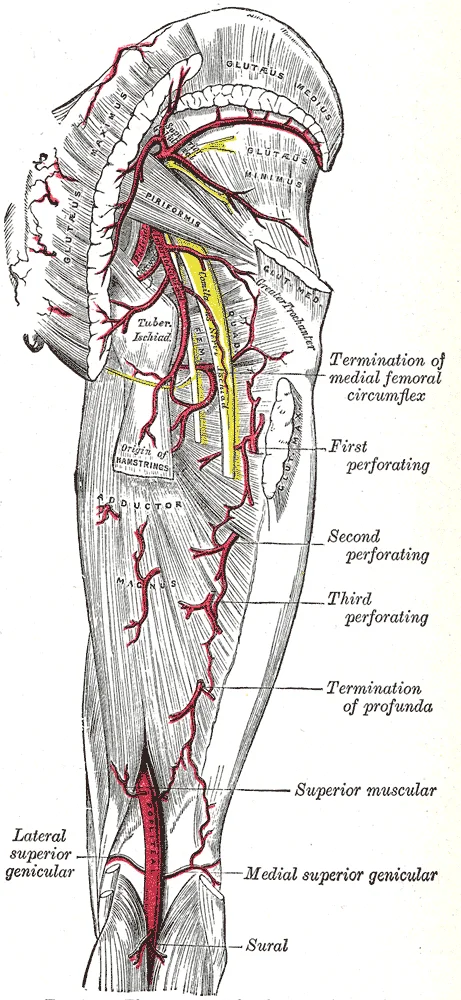
Long description
The arteries of the gluteal and posterior femoral regions.

Figure 1: The arteries of the gluteal and posterior femoral regions.
Figure 2Gray589
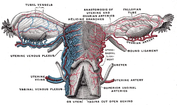
Long description
Vessels of the uterus and its appendages, rear view. (Testut.)

Figure 2: Vessels of the uterus and its appendages, rear view (Testut.).
Systemic veins contain approximately 64 percent of the blood volume at any given time[1]. Venous networks within the liver, bone marrow, and integument hold approximately 21 percent of the venous blood, which is referred to as venous reserve[1].
Venules and veins withstand a much lower pressure from the blood that flows through them compared to arteries[1]. Their walls are considerably thinner and their lumens are correspondingly larger in diameter, allowing more blood to flow with less vessel resistance[1]. Many veins contain valves that assist the unidirectional flow of blood toward the heart and prevent backflow[1]. The vasomotor center located in the medulla oblongata sends sympathetic stimulation to the smooth muscles in the walls of the veins, causing venoconstriction[1].
Figure 3Gray588
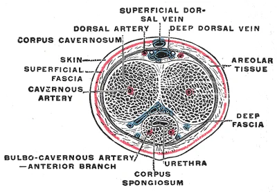
Long description
The penis in transverse section, showing the bloodvessels. (Testut.)

Figure 3: The penis in transverse section, showing the bloodvessels (Testut.).
The space enclosed by the bony pelvis is divided into the greater pelvis, which is occupied by portions of the small and large intestines, and the lesser pelvis, which contains the bladder and other pelvic organs[1]. The pelvic brim separates the lesser pelvis from the greater pelvis[1]. The female pelvic inlet often has a more rounded or oval shape compared to males, and the lesser pelvic cavity of females is wider and more shallow than the narrower, deeper lesser pelvis of males. [2]
Arteries and arterioles have thicker walls than veins and venules because they receive blood surging at a far greater pressure from proximity to the heart[1]. Each vessel type has a lumen, and arteries possess smaller lumens than veins to help maintain the pressure of blood moving through the system[1]. These thicker walls and smaller diameters give arterial lumens a more rounded appearance in cross section than the lumens of veins[1]. By the time blood passes through capillaries and enters venules, the pressure exerted by heart contractions has diminished considerably, requiring veins to rely on larger lumens and valves to manage lower pressures and counteract the pull of gravity in the extremities[1].
When blood flow redistribution is necessary, sympathetic stimulation from the vasomotor center in the medulla oblongata triggers venoconstriction, effectively stiffening the vessel wall to increase pressure on the blood and speed its return to the heart[1]. This mechanism allows the venous reserve stored in the liver, bone marrow, and integument to quickly reenter active circulation[1]. After circulating through this complex network, blood completes its journey through arteries, arterioles, capillaries, venules, and veins before returning to the heart[1].
DefinitionCapacitance vessels
Systemic veins act as capacitance vessels because their large lumens and thin walls allow them to distend readily and store high blood volume at low pressure[1].
The pelvic blood supply originates from the main systemic arterial tree within the abdominal cavity. The lower abdominal aorta descends and bifurcates into the left and right common iliac arteries. Each common iliac artery travels downward and laterally along the medial border of the psoas major muscle to reach the pelvic brim. At this boundary, the common iliac artery divides into the external iliac artery, which continues along the brim to supply the lower limb, and the internal iliac artery, which enters the lesser pelvis to supply the pelvic viscera, walls, and perineum. [1]
The reader can trace the abdominal aorta inferiorly as it bifurcates into the left and right common iliac arteries lying against the lumbar spine. Moving laterally and inferiorly, follow each common iliac artery to its bifurcation at the pelvic brim.
DefinitionPelvic Brim
The pelvic brim is defined by a line formed by the upper margin of the pubic symphysis anteriorly, and the pectineal line of the pubis, the arcuate line of the ilium, and the sacral promontory posteriorly. [2]
The internal iliac artery represents the primary vascular inflow for the lesser pelvis. It descends posteriorly across the pelvic inlet and divides into anterior and posterior divisions, which give off numerous visceral and parietal branches. These branches course through the pelvic connective tissue and fascial planes to reach their respective target organs, including the urinary bladder, reproductive organs, and rectum, before terminating in capillary beds within the pelvic structures.
| Division | Branch | Target Region |
|---|---|---|
| Posterior | Iliolumbar | Posterior abdominal wall and iliacus |
| Posterior | Lateral sacral | Sacrum and meninges of sacral canal |
| Posterior | Superior gluteal | Gluteal muscles through greater sciatic foramen |
| Anterior | Obturator | Medial compartment of thigh |
| Anterior | Inferior vesical | Urinary bladder, prostate or vagina |
| Anterior | Internal pudendal | Perineum and external genitalia |
Pelvic Arterial Pathways
- Abdominal AortaDescends on posterior abdominal wall.
- Common Iliac ArteryBifurcates at pelvic brim.
- Question: Which branch?
Internal Iliac
- Lesser pelvisSupplies viscera, walls and perineum.
External Iliac
- Lower limbPasses deep to inguinal ligament.
The pelvic blood supply originates from the internal iliac artery, which arises from the common iliac artery. This vessel is the primary source of arterial blood to the pelvic viscera, walls, and gluteal region. Understanding its branching pattern is essential for surgical planning and interpreting imaging.
The anterior division gives rise to branches supplying the pelvic viscera, such as the umbilical artery and the inferior vesical artery. The posterior division supplies the gluteal region and pelvic wall, including the superior gluteal artery.
The anterior division supplies the pelvic viscera and perineum. The posterior division supplies the gluteal region, pelvic wall, and medial thigh.
| Structure | Origin | Course | Relations | Branches/Termination |
|---|---|---|---|---|
| Internal Iliac Artery | Common Iliac Artery | Descends into pelvis | Posterior to ureter, anterior to sacroiliac joint | Anterior and posterior divisions |
| Superior Gluteal Artery | Posterior division | Exits greater sciatic notch above piriformis | Between lumbosacral trunk and sacral plexus | Divides into superficial and deep branches [1] |
| Inferior Gluteal Artery | Posterior division | Exits greater sciatic notch below piriformis | Between sacral plexus and coccygeus muscle | Supplies gluteus maximus and surrounding muscles |
| Internal Pudendal Artery | Anterior division | Exits lesser sciatic notch | Accompanied by internal pudendal vein and nerve | Supplies perineum and external genitalia |
Pelvic veins form extensive plexuses that drain into the internal iliac veins. These veins lack valves, allowing for bidirectional flow, which is clinically significant in the spread of infection or malignancy. The pelvic venous plexuses include the vesical, prostatic, uterine, and rectal plexuses.
Gonadal veins (testicular or ovarian) drain directly into the systemic circulation, not the internal iliac system. The right gonadal vein drains into the inferior vena cava, while the left gonadal vein drains into the left renal vein. This anatomical difference explains why left-sided varicoceles are more common than right-sided ones.
The walls of arteries and veins are largely composed of living cells and their products (including collagenous and elastic fibers); the cells require nourishment and produce waste. Larger arteries and veins contain small blood vessels within their walls known as the vasa vasorum—literally “vessels of the vessel”—to provide them with this critical exchange. [1]
In larger arteries, there is also a thick, distinct layer of elastic fibers known as the internal elastic membrane (also called the internal elastic lamina) at the boundary with the tunica media. The internal elastic membrane is not apparent in veins. [1]
The arterial system is a relatively high-pressure system, so arteries have thick walls that appear round in cross section. The venous system is a lower-pressure system, containing veins that have larger lumens and thinner walls. [1]
The internal iliac artery arises from the common iliac artery and descends into the pelvis. It divides into anterior and posterior divisions. The anterior division supplies pelvic viscera and the perineum. The posterior division supplies the gluteal region and lateral pelvic wall. Key branches include the superior gluteal artery, which exits via the greater sciatic foramen above the piriformis muscle. The inferior gluteal artery exits below the piriformis. The internal pudendal artery also exits the greater sciatic foramen, then loops around the sacrospinous ligament to enter the lesser sciatic foramen.
The pelvic girdle is formed by the hip bones, sacrum, and coccyx. Each hip bone consists of the fused ilium, ischium, and pubis. These bones provide attachment for muscles and transmit body weight to the lower limbs. The pelvis has a pronounced anterior tilt. The primary function is to support the upper body. The right and left hip bones articulate posteriorly at the sacroiliac joint with the sacrum. Anteriorly, they meet at the pubic symphysis. This bony structure protects the pelvic viscera. [2]
Blood flow distribution in the pelvis is regulated by neural and chemical controls. The precise diameter of arterioles determines local perfusion. Vasoconstriction and vasodilation in arterioles are the primary mechanisms for distributing blood flow. Small vascular nerves, known as nervi vasorum, run within vessel walls. These are generally sympathetic fibers. Some trigger vasodilation, while others induce vasoconstriction. Parasympathetic stimulation triggers vasodilation and erection during sexual arousal in external genitalia. Nervous control is often generalized. Local controls account for specific targeting.
Capillaries supply blood to tissues in a process called perfusion. Exchange of gases and substances occurs between capillary blood and interstitial fluid. Capillary lumen diameter ranges from 5–10 micrometers. The smallest are just wide enough for an erythrocyte to squeeze through. Flow through capillaries is described as microcirculation. In the pelvic viscera, capillary types vary. Fenestrated capillaries have pores in the endothelial lining. They are permeable to larger molecules. These are common in endocrine structures. Sinusoid capillaries are flattened with extensive intercellular gaps. They allow passage of plasma proteins and cells. Blood flow through sinusoids is very slow. This allows more time for exchange. Sinusoids are found in bone marrow and many endocrine glands.
Venules are extremely small veins, generally 8–100 micrometers in diameter. Postcapillary venules join multiple capillaries exiting from a capillary bed. Multiple venules join to form veins. The walls of venules consist of endothelium, a thin middle layer with a few muscle cells and elastic fibers, plus an outer layer of connective tissue fibers. Venules and capillaries are the primary sites of emigration or diapedesis. White blood cells adhere to the endothelial lining and squeeze through adjacent cells to enter tissue fluid. This mechanism is crucial for immune response within the pelvic tissues.
DefinitionPerfusion
The process by which blood supplies tissues, allowing exchange of gases and substances between capillaries and interstitial fluid.
Clinical pearlCapillary Diameter
Capillary lumen diameter ranges from 5–10 micrometers, with the smallest being just wide enough for an erythrocyte to squeeze through.
| Structure | Description | Relation to Vessels |
|---|---|---|
| Greater Sciatic Foramen | Formed by greater sciatic notch, sacrum, and sacrospinous ligament | Exit for superior/inferior gluteal and internal pudendal arteries |
| Lesser Sciatic Foramen | Formed by lesser sciatic notch, sacrospinous and sacrotuberous ligaments | Entry for internal pudendal artery into perineum |
| Sacrospinous Ligament | Connects sacrum to ischial spine | Defines boundaries of sciatic foramina |
The pelvis possesses an extensive network of vascular anastomoses that protect pelvic organs from ischaemia if a major vessel is interrupted. The internal iliac artery, which supplies the pelvic viscera and walls, communicates freely with neighbouring vascular territories through several classic anatomical pathways.
The paired common iliac arteries descend from the abdominal aorta and bifurcate into external and internal iliac branches over the pelvic brim. Medially, the superior rectal artery, the direct continuation of the inferior mesenteric artery, descends into the sigmoid mesocolon to anastomose on the surface of the rectum with the middle rectal branches derived from the internal iliac artery. This provides a direct collateral bridge between the systemic internal iliac system and the portal-systemic mesenteric circulation.
Figure 4Gray587
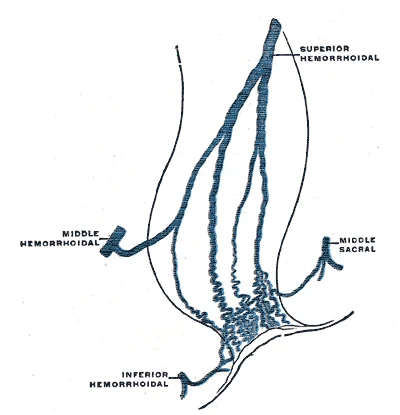
Long description
Scheme of the anastomosis of the veins of the rectum. (Poirier and Charpy.)

Figure 4: Scheme of the anastomosis of the veins of the rectum (Poirier and Charpy.).
Clinical pearlUterine and ovarian anastomoses
The uterine artery runs within the broad ligament and forms a prominent anastomosis near the lateral margin of the uterus with the ovarian artery, which descends directly from the abdominal aorta.
Further collateral pathways exist around the obturator foramen and the pelvic floor. The obturator artery, a branch of the internal iliac, forms an anastomosis with the inferior epigastric artery derived from the external iliac system. This connection, when large, is clinically referred to as the aberrant obturator artery or corona mortis. Posteriorly, the superior and inferior gluteal branches of the internal iliac system anastomose extensively with branches of the deep femoral artery, specifically the lateral circumflex femoral artery, establishing a major collateral route between the pelvis and the lower limb.
| Anastomotic site | Vessel 1 (Internal Iliac source) | Vessel 2 (Collateral source) |
|---|---|---|
| Rectum | Middle rectal artery | Superior rectal artery (Inferior mesenteric) |
| Uterus | Uterine artery | Ovarian artery (Abdominal aorta) |
| Pubic region | Obturator artery | Inferior epigastric artery (External iliac) |
| Gluteal region | Superior and inferior gluteal arteries | Lateral circumflex femoral artery (Deep femoral) |
Pelvic collateral pathways
- Internal iliac occlusionMain pelvic conduit obstructed.
- Question: Which region?
Superior / Rectal
- Inferior mesenteric via superior rectal artery
Lateral / Pubic
- External iliac via inferior epigastric to obturator
Inferior / Gluteal
- Deep femoral via lateral circumflex femoral
DefinitionAnatomical variation
Vascular branching patterns in the pelvis demonstrate significant deviation from textbook descriptions in clinical practice.
Pelvic arterial anatomy exhibits considerable divergence from standard classical descriptions, a fact that becomes critical during complex surgical interventions and interventional radiology procedures. The internal iliac artery, also known as the hypogastric artery, typically divides into anterior and posterior trunks in only seventy percent of individuals. In the remaining thirty percent, major branches arise independently from the main stem without forming distinct trunks. This variation is n [1]
The obturator artery originates most frequently as a branch of the internal iliac artery, yet it can arise from the external iliac or inferior epigastric arteries in up to twenty percent of cases. When the obturator artery stems from the inferior epigastric artery, it is termed the corona mortis. The term 'corona [1]
Figure 5Gray543
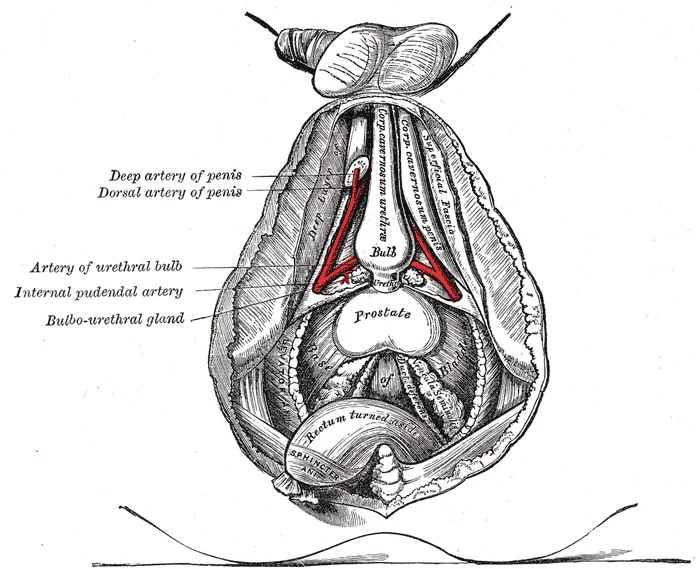
Long description
The deeper branches of the internal pudendal artery.

Figure 5: The deeper branches of the internal pudendal artery.
Figure 6Gray542
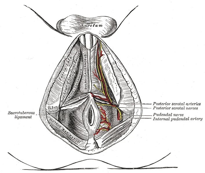
Long description
The superficial branches of the internal pudendal artery.

Figure 6: The superficial branches of the internal pudendal artery.
Figure 7Gray541
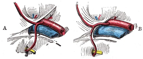
Long description
Variations in origin and course of obturator artery.

Figure 7: Variations in origin and course of obturator artery.
Figure 8Gray540
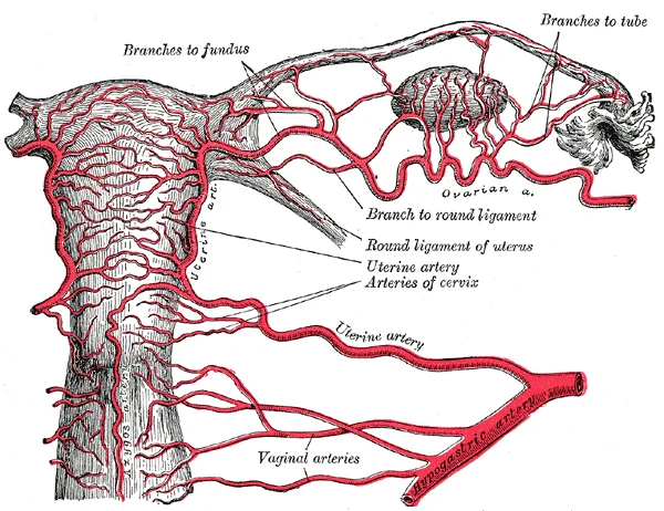
Long description
The arteries of the internal organs of generation of the female, seen from behind. (After Hyrtl.)

Figure 8: The arteries of the internal organs of generation of the female, seen from behind (After Hyrtl.).
The superior gluteal artery represents the largest branch of the internal iliac artery. Variations involve its origin directly from the posterior division or as a dual vessel piercing the sacral plexus differently. Its close proximity to the lumbosacral trunk renders it susceptible to compression or injury during posterior pelvic approaches. In cases of pelvic fracture, the superior gluteal artery is a common source of
| Structure | Normal Origin | Variant Origin | Clinical Impact |
|---|---|---|---|
| Obturator artery | Internal iliac artery | Inferior epigastric artery | Vulnerable during femoral hernia repair |
| Superior gluteal artery | Posterior trunk | Main internal iliac stem | Risk of hemorrhage during pelvic surgery |
| Internal iliac division | Bifurcation into trunks | Trifurcation or absent trunks | Alters angiographic embolization planning |
Pelvic Artery Assessment
- Identify internal iliac arteryLocate bifurcation at pelvic brim.
- Question: Are trunks distinct?
Yes
- Proceed with standard anterior and posterior division mapping.
No
- Map individual branches originating from main stem.
Access points for endovascular pelvic interventions rely on palpable arterial pulse points. The common femoral artery is the standard vascular access point in the groin, identified by its pulse point located halfway between the anterior superior iliac spine and the pubic symphysis. Ligation of the internal iliac artery, also known as hypogastric artery ligation, was historically performed to control intractable pelvic haemorrhage. When ligating this vessel, surgeons must place the ligature at least 5 centimetres distal to the common iliac bifurcation to preserve the superior gluteal and obturator branches when collateral flow is desired, though proximal control reduces pulse pressure within the true pelvis.
Clinical pearlVascular access landmark
The surface landmark for the femoral pulse lies directly over the femoral head, lateral to the femoral vein and medial to the femoral nerve.
Pelvic arterial aneurysms frequently involve the internal iliac artery and its major divisions, particularly the gluteal arteries within the posterior compartment of the pelvis. Aneurysm sites in this region present diagnostic challenges due to deep retroperitoneal positioning. Traumatic injury patterns commonly occur during high-energy pelvic ring fractures, where sharp bone spicules from the ilium or sacrum lacerate the posterior division branches or the presacral venous plexus.
The internal iliac artery gives rise to the superior gluteal artery exiting above the piriformis muscle, and the inferior gluteal artery exiting below it. Lesion sites are marked near the sacral ala and the greater sciatic notch, demonstrating where bony displacement during pelvic fractures tears the adjacent arterial walls.
| Structure | Access or Lesion Site | Clinical Significance |
|---|---|---|
| Common femoral artery | Inguinal crease | Standard endovascular access point |
| Internal iliac artery | Posterior pelvis | Primary site for pelvic aneurysm formation |
| Superior gluteal artery | Greater sciatic notch | Vulnerable to laceration in pelvic ring fractures |
The pelvic blood supply is derived primarily from the internal iliac artery, which arises from the common iliac bifurcation. This vessel is the principal source of oxygenated blood for the pelvic viscera, perineum, and gluteal region. Understanding its branching pattern is critical for surgical interventions and interpreting imaging studies.
The anterior division supplies the pelvic viscera and perineum, while the posterior division supplies the gluteal region and pelvic walls. This division is not always distinct, and variations in branching are common.
The anterior division gives rise to several key branches. It supplies the medial compartment of the thigh and the hip joint.
It exits the pelvis through the greater sciatic foramen below the piriformis muscle. This artery supplies the gluteus maximus muscle and surrounding structures.
Arteries supplying the pelvis share common histological features. The smooth muscle layers of the tunica media are supported by a framework of collagenous fibers that also binds the tunica media to the inner and outer tunics. Along with the collagenous fibers are large numbers of elastic fibers that appear as wavy lines in prepared slides. Separating the tunica media from the outer tunica externa in larger arteries is the external elastic membrane (also called the external elastic lamina), which also appears wavy in slides. This structure is not usually seen in smaller arteries, nor is it seen in veins. [1]
Arterioles, which are very small arteries leading to capillaries, have the same three tunics as larger vessels, but the thickness of each is greatly diminished. The critical endothelial lining of the tunica intima is intact. The tunica media is restricted to one or two smooth muscle cell layers in thickness. The tunica externa remains but is very thin. [1]
Knowledge of pelvic anatomy extends beyond vessels. Several ligaments unite the bones of the pelvis. The largely immobile sacroiliac joint is supported by a pair of strong ligaments that are attached between the sacrum and ilium portions of the hip bone. These are the anterior sacroiliac ligament on the anterior side of the joint and the posterior sacroiliac ligament on the posterior side. Also spanning the sacrum and hip bone are two additional ligaments. These ligaments help to support and imm [2]
The pubis forms the anterior portion of the hip bone. The body of the pubis articulates with the pubis of the opposite hip bone at the pubic symphysis. The superior margin of the pubic body has the pubic tubercle. The pubis is joined to the ilium by the superior pubic ramus, the superior surface of which forms the pectineal line. The inferior pubic ramus projects inferiorly and laterally. The pubic arch is formed by the pubic symphysis, the bodies of the adjacent pubic bones, and the two inferior pubic rami.
Clinical pearlObturator Artery Trap
The obturator artery may arise from the inferior epigastric artery in up to 20% of cases, a variation known as an accessory or aberrant obturator artery.
DefinitionInternal Iliac Artery
The main arterial supply to the pelvic organs, perineum, and gluteal region, branching into anterior and posterior divisions.
Exam trapExternal Elastic Lamina
Do not assume the external elastic lamina is present in all pelvic arteries; it is typically absent in smaller arteries and veins.
NoteSacrospinous Ligament
The sacrospinous ligament converts the greater sciatic foramen into two separate openings, influencing the passage of nerves and vessels.
Clinical pearlArteriole Structure
Arterioles have a tunica media restricted to one or two smooth muscle cell layers, allowing for significant regulation of blood flow.
DefinitionPubic Arch
Formed by the pubic symphysis, adjacent pubic bodies, and the two inferior pubic rami, defining the inferior boundary of the pelvic outlet.
Exam trapUmbilical Artery
The proximal part of the umbilical artery becomes the superior vesical artery; the distal part becomes the medial umbilical ligament.
Clinical pearlInferior Gluteal Artery
Often the largest branch of the anterior division, it exits the pelvis below the piriformis muscle to supply the gluteus maximus.
NoteSacroiliac Ligaments
The anterior and posterior sacroiliac ligaments provide strong support to the largely immobile sacroiliac joint.
DefinitionIschiopubic Ramus
Formed by the junction of the inferior pubic ramus and the ischial ramus, contributing to the structure of the pubic arch.
Clinical pearlTunica Media
The smooth muscle layers of the tunica media are supported by collagenous fibers that bind the tunica media to the inner and outer tunics.
Exam trapPelvic Venous Drainage
While not covered in detail here, note that pelvic veins often accompany arteries and form extensive plexuses, which can be a source of bleeding.
Students frequently confuse the structural components of blood vessels with the bony landmarks of the pelvis, leading to errors in both anatomy and physiology questions. A primary error is attributing the strength and flexibility of large arteries to the capillary wall structure. Capillaries are the site of exchange, not structural support. The wall of a capillary consists of the endothelial layer surrounded by a basement membrane with occasional smooth muscle fibers. In contrast, the strength of larger vessels comes from the tunica intima and media. Next to the endothelium is the basement membrane, or basal lamina, that effectively binds the endothelium to the connective tissue. The thin outer layer of the tunica intima contains a small amount of areolar connective tissue that consists primarily of elastic fibers to provide the vessel with additional flexibility; it also contains some collagenous fibers to provide additional strength. Confusing these layers leads to incorrect answers regarding permeability and vessel integrity. [1]
Another significant pitfall involves the identification of pelvic bones and their specific contributions to the hip joint and weight bearing. Students often misidentify the ilium as the primary weight-bearing structure during sitting. The ilium is the fan-like, superior region that forms the largest part of the hip bone. It is firmly united to the sacrum at the largely immobile sacroiliac joint. The actual weight-bearing structure when sitting is the ischium. The ischium forms the posteroinferio [2]
Misidentifying the boundaries of the sciatic notches and foramina is also common. The ischial spine is a critical landmark that separates the greater and lesser sciatic notches. The bony projection separating the lesser sciatic notch and greater sciatic notch is the ischial spine. The large, inverted U-shaped indentation located on the posterior margin of the lower ilium is called the greater sciatic notch. The slightly curved posterior margin of the ischium above the ischial tuberosity is the lesser sciatic notch. The sacrospinous and sacrotuberous ligaments contribute to the formation of the greater and lesser sciatic foramina. The sacrum is also joined to the hip bone by the sacrospinous ligament, which attaches to the ischial spine, and the sacrotuberous ligament, which attaches to the ischial tuberosity. [2]
Confusion regarding the pubic arch and subpubic angle often leads to errors in forensic or obstetric anatomy questions. The pubic arch is the bony structure formed by the pubic symphysis, and the bodies and inferior pubic rami of the adjacent pubic bones. The inverted V-shape formed as the ischiopubic rami from both sides come together at the pubic symphysis is called the subpubic angle. The inferior pubic ramus extends downward to join the ischial ramus. Together, these form the single ischiopubic ramus, which extends from the pubic body to the ischial tuberosity. [2]
Finally, students often overlook the role of the obturator foramen in muscle attachment. The large opening in the anteroinferior hip bone between the ischium and pubis is the obturator foramen. This space is largely filled in by a layer of connective tissue and serves for the attachment of muscles on both its internal and external surfaces. The three areas of each hip bone, the ilium, pubis, and ischium, converge centrally to form a deep, cup-shaped cavity called the acetabulum. This is located on the lateral side of the hip bone and is part of the hip joint. [2]
Exam trapCapillary Structure
Do not confuse capillary walls with arterial walls. Capillaries lack the elastic and collagenous fibers found in the tunica intima of larger vessels.
DefinitionIschial Tuberosity
The ischial tuberosity is the roughened area of the inferior ischium that supports body weight when sitting and attaches posterior thigh muscles. [2]
Knowledge of pelvic anatomy and blood vessel histology is essential for interpreting imaging, performing surgeries, and understanding clinical pathologies. In the operating theatre, surgeons must identify the ischial spine and sciatic notches to avoid nerve damage during pelvic procedures. The ischial spine separates the greater and lesser sciatic notches. The sacrospinous ligament attaches to the ischial spine. The sacrotuberous ligament attaches to the ischial tuberosity. These ligaments stabilize the sacroiliac joint and form the boundaries of the sciatic foramina. The sacroiliac joint is supported by the anterior and posterior sacroiliac ligaments.
On imaging, radiologists use the acetabulum and obturator foramen to assess hip joint integrity and pelvic fractures. The lateral side of the hip bone has the cup-like acetabulum, which is part of the hip joint. The large anterior opening is the obturator foramen. The obturator foramen is largely filled in by a layer of connective tissue and serves for the attachment of muscles on both its internal and external surfaces. Understanding the bony landmarks helps in localizing pathology. The ilium is the fan-like, superior region that forms the largest part of the hip bone. The ischium forms the posteroinferior region of each hip bone. The pubis forms the anterior portion of the hip bone. [2]
At the bedside, clinicians assess the pubic arch and subpubic angle in obstetric evaluations. The inverted V-shape formed as the ischiopubic rami from both sides come together at the pubic symphysis is called the subpubic angle. The pubic arch is the bony structure formed by the pubic symphysis, and the bodies and inferior pubic rami of the adjacent pubic bones. The pubic body is joined to the pubic body of the opposite hip bone by the pubic symphysis. This knowledge aids in predicting vaginal delivery outcomes.
In examinations, students must distinguish between the layers of blood vessels to answer questions on permeability and strength. The basement membrane provides strength while maintaining flexibility, and it is permeable, allowing materials to pass through it. The thin outer layer of the tunica intima contains a small amount of areolar connective tissue that consists primarily of elastic fibers to provide the vessel with additional flexibility; it also contains some collagenous fibers to provide additional strength. This distinction is crucial for understanding vascular diseases. [1]
Pelvic Landmark Identification
- Identify Hip Bone RegionSuperior, Posterior, or Anterior?
- Question: Superior Region?
Yes
- IliumForms sacroiliac joint
No
- Question: Posterior Region?
Yes
- IschiumContains ischial tuberosity and spine
No
- PubisForms pubic symphysis
Clinical pearlWeight Bearing
The ischial tuberosity carries the body weight when sitting, not the ilium or pubis.
NoteLigament Attachments
The sacrospinous ligament attaches to the ischial spine, and the sacrotuberous ligament attaches to the ischial tuberosity.
The capillary wall consists of an endothelial layer surrounded by a basement membrane with occasional smooth muscle fibers. The basement membrane binds the endothelium to connective tissue and provides strength while maintaining flexibility. The tunica intima contains elastic fibers for flexibility and collagenous fibers for strength. The ilium is the fan-like superior region of the hip bone united to the sacrum. The ischial tuberosity supports body weight when sitting and attaches posterior thigh muscles. The ischial spine separates the greater and lesser sciatic notches. The sacrospinous and sacrotuberous ligaments contribute to the formation of the sciatic foramina. The subpubic angle is formed by the inverted V-shape of the ischiopubic rami at the pubic symphysis.
Answer each one in your head first, then open the answer.
What structure serves as the attachment for the posterior thigh muscles and carries body weight when sitting?
Show answerHide answer
Ischial tuberosityWhich bony projection separates the lesser sciatic notch and greater sciatic notch?
Show answerHide answer
Ischial spineWhat provides strength and flexibility to the vessel wall while being permeable?
Show answerHide answer
Basement membraneWhich ligament attaches to the ischial spine?
Show answerHide answer
Sacrospinous ligamentWhat is the cup-shaped cavity formed by the convergence of the ilium, pubis, and ischium?
Show answerHide answer
Acetabulum
- Betts JG, Young KA, Wise JA, et al. Structure and Function of Blood Vessels. OpenStax, Anatomy and Physiology 2e (CC BY-NC-SA 4.0). Accessed .
- Betts JG, Young KA, Wise JA, et al. The Pelvic Girdle and Pelvis. OpenStax, Anatomy and Physiology 2e (CC BY-NC-SA 4.0). Accessed .
Figure credits
- Figure 1: Henry Vandyke Carter, via Wikimedia Commons, Public domain, Public domain.
- Figure 2: Henry Vandyke Carter, via Wikimedia Commons, Public domain, Public domain.
- Figure 3: Henry Vandyke Carter, via Wikimedia Commons, Public domain, Public domain.
- Figure 4: Henry Vandyke Carter, via Wikimedia Commons, Public domain, Public domain.
- Figure 5: Henry Vandyke Carter, via Wikimedia Commons, Public domain, Public domain.
- Figure 6: Henry Vandyke Carter, via Wikimedia Commons, Public domain, Public domain.
- Figure 7: Henry Vandyke Carter, via Wikimedia Commons, Public domain, Public domain.
- Figure 8: Henry Vandyke Carter, via Wikimedia Commons, Public domain, Public domain.
Educational content for medical students and doctors. It is not a substitute for clinical judgement or local guidelines.