Pelvic nerves
Essential topographical anatomy and surgical preservation of the pelvic autonomic and somatic nerves
A prerequisite for nerve-sparing pelvic surgery is a thorough understanding of the topographic anatomy of the fine and intricate pelvic nerve networks, and their connections to the central nervous system. Insights into the functions of pelvic nerves help to interpret disease symptoms correctly and improve treatment. Autonomic pelvic nerves include the superior hypogastric plexus, the hypogastric nerves, the inferior hypogastric plexus, and the pelvic splanchnic nerves. Somatic nerves encountered in the pelvis include the obturator and genitofemoral nerves, as well as the sciatic, the pudendal, the lumbosacral trunk, and the sacral plexus. [2] [3]
DefinitionNerve-sparing pelvic surgery
Surgical techniques designed to prevent autonomic and somatic pelvic nerves from getting significantly damaged during radical oncology and benign pelvic procedures.
The pelvic fascia consists of the parietal and visceral pelvic fascia. Both sympathetic and parasympathetic nerves are embedded within the dual-lamellar parietal pelvic fasciae, also known as the pre-hypogastric sheath or fascia. The inferior hypogastric plexus extends sagittally at the level of the fourth and fifth sacral segments and is located lateral to the pelvic organs but medial to the pelvic blood vessels. The rectum lies anterior to the parietal pelvic fascia and is surrounded by perirectal fatty lymphovascular tissue wrapped in the mesorectal fascia. The retrorectal space extends between the mesorectal fascia and the parietal pelvic fascia, corresponding to the nerve-sparing dissection plane during total mesorectal excision, which is a safe plane for dissection to avoid nerve damage. [2]
Figure 1Gray834
Long description
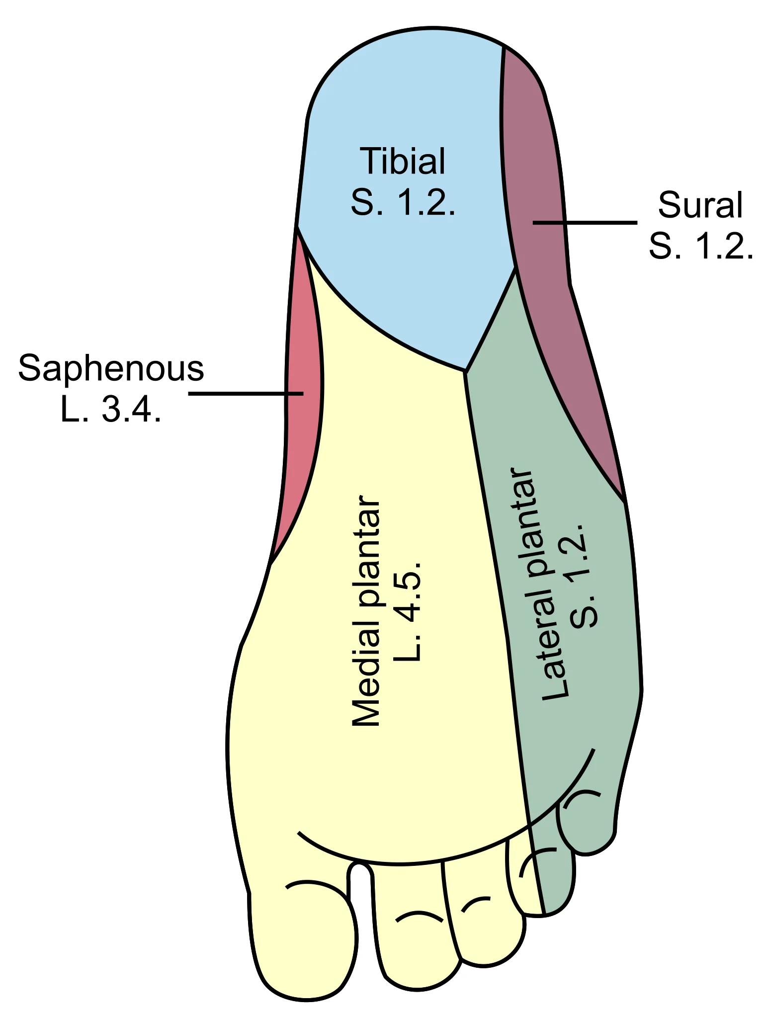
Diagram of the segmental distribution of the cutaneous nerves of the sole of the foot.

Figure 1: Diagram of the segmental distribution of the cutaneous nerves of the sole of the foot.
Figure 2Gray833
Long description
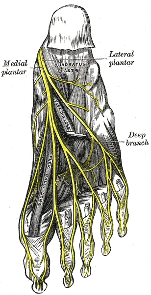
The plantar nerves. Labelled illustration from Wikimedia Commons, used under its open licence.

Figure 2: The plantar nerves.
| Nerve structure | System | Key anatomical relations | Primary function |
|---|---|---|---|
| Superior hypogastric plexus | Sympathetic | Bifurcation of the aorta | Mediates proprioception and visceral pain |
| Hypogastric nerves | Sympathetic | Retroperitoneal, medial to internal iliac vessels | Convey upper pelvic pain and sympathetic efferents |
| Inferior hypogastric plexus | Mixed | Lateral to pelvic organs, medial to vessels | Innervates pelvic viscera, sphincters, sexual function |
| Pelvic splanchnic nerves | Parasympathetic | S2 to S4 roots to inferior hypogastric plexus | Visceral smooth muscle contraction and nociception |
Pelvic nerves are closely related to the mechanisms of defecation and micturition, as well as chronic pelvic and visceral pain. They are also responsible for complex sexual activity involving genital arousal, orgasm, and ejaculation. Sympathetic pelvic nerves are involved in the mediation of proprioceptive sensation and pain from the upper pelvis via the hypogastric nerves and the superior hypogastric plexus to the sympathetic trunk. Efferent sympathetic nerve fibers evoke contraction of the internal urethral and anal sphincters and exert relaxation on the visceral nonsphincter smooth muscle. Parasympathetic pelvic nerves also carry nociceptive sensations to the sacral dorsal root ganglia from the pelvic viscera, such as the vagina, cervix, and bladder, and mediate contraction of visceral smooth muscles. [2]
Pelvic Nerve Dissection Guide
- Identify Fascial PlanesLocate parietal and visceral pelvic fasciae and the pre-hypogastric sheath.
- Question: Performing TME?
Yes
- Enter retrorectal holy plane between mesorectal and parietal fascia.
No
- Identify inferior hypogastric plexus lateral to organs and medial to vessels.
During gynaecological and pelvic procedures, unnecessary manipulation and grasping of the nerves should be avoided because the force generated between the jaws of grasping forceps is sufficient to cease the circulation in the intrafascicular vascular system. While a degree of elasticity and the capacity to resist compression maintains constant intrafascicular pressure, excessive traction leads to significant postoperative genitourinary and anorectal dysfunctions. [3]
Somatic nerves supplying the pelvic floor muscles, lower abdominal wall, and limbs arise from the lumbar and sacral spinal cord. Ventral branches of lumbar and sacral spinal nerves form the lumbosacral plexus. The lumbar plexus comprises the ilioinguinal (L1, L2) and iliohypogastric nerves (T12, L1), the genitofemoral nerve (L1, L2) with its genital and femoral branches, the lateral femoral cutaneous nerve (L2, L3), the femoral nerve (L2, L3, L4), and the obturator nerve (L2, L3, L4). The L4 and L5 roots of the lumbar plexus join the first sacral nerve root (S1) to form the lumbosacral trunk. [2]
Figure 3Gray836
Long description
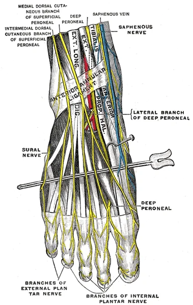
Nerves of the dorsum of the foot. (Testut.)

Figure 3: Nerves of the dorsum of the foot (Testut.).
Figure 4Gray835
Long description
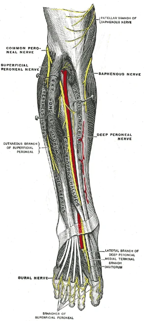
Deep nerves of the front of the leg. (Testut.)

Figure 4: Deep nerves of the front of the leg (Testut.).
The sacral nerve roots leave the sacrum via the ventral sacral foramina to form the sacral plexus and its peripheral nerves on both sides in front of the piriformis muscle. The lumbosacral trunk and sacral plexus give rise to the pudendal nerve (S2, S3, S4), the superior gluteal nerve (L4, L5, S1), the inferior gluteal nerve (L5, S1, S2), the sciatic nerve (L4, L5, S1, S2, S3), and the posterior femoral cutaneous nerve (S1, S2, S3). [2]
The sacral nerve roots S1 to S4 emerge from the ventral sacral foramina anterior to the piriformis muscle to construct the sacral plexus. Lying superficially on the posterior pelvic wall, the sympathetic trunk runs medially to the sacral foramina, while the pelvic splanchnic nerves project from the sacral roots to join the inferior hypogastric plexus.
DefinitionAutonomic innervation
The pelvic viscera receive a dual autonomic supply from the sympathetic and parasympathetic systems, coordinating visceral motor and sensory functions.
The sympathetic system comprises the sympathetic trunk and the preaortic plexus. The superior hypogastric plexus (SHP) extends anterior to the aorta and the sacral promontory, receiving branches from sympathetic ganglia L1 to L4, and occasionally L5. The SHP divides into the left and right hypogastric nerves (HN). The inferior hypogastric plexus (IHP) is an abundant nerve meshwork located adjacent to the vaginal fornix in females and close to the lateral rectal wall in males. Within each pelvic compartment, nerve bundles diverge from the IHP to form organ-specific plexuses, namely the rectal, uterovaginal, vesicourethral, prostatic, and cavernous plexuses. [2]
Medial to the lateral pelvic wall, the inferior hypogastric plexus is situated in the fibro-connective tissue between the rectum medially and the pelvic wall laterally, with its posterior part supplying the rectum, its anterosuperior part supplying the bladder, and its anteroinferior part supplying the internal anal sphincter. [2]
| Nerve structure | Spinal root origin | Main distribution |
|---|---|---|
| Superior hypogastric plexus | L1-L4 (occasionally L5) | Pelvic viscera via hypogastric nerves |
| Pelvic splanchnic nerves | S2-S4 | Left colon, anorectum, uterus/vagina, prostate/seminal vesicles, bladder |
| Pudendal nerve | S2, S3, S4 | Pelvic floor muscles, external urethral and anal sphincter |
| Obturator nerve | L2, L3, L4 | Medial compartment of the thigh and pelvic structures |
DefinitionPelvic nerve organisation
Pelvic organs and lower limb muscles receive their innervation from a dense accumulation of peripheral nerves and plexuses located within the lesser pelvis. [3]
The lesser pelvis, also known as the true pelvis, is the narrow, rounded space inferior to the pelvic brim that contains the bladder and other pelvic organs. The pelvic brim forms the superior margin of the lesser pelvis and is defined by a line formed by the upper margin of the pubic symphysis anteriorly, and the pectineal line of the pubis, the arcuate line of the ilium, and the sacral promontory posteriorly. The inferior limit of the lesser pelvis is the pelvic outlet, which is defined by the inferior margin of the pubic symphysis anteriorly, and the ischiopubic ramus, the ischial tuberosity, the sacrotuberous ligament, and the inferior tip of the coccyx posteriorly. Because of the anterior tilt of the pelvis, the lesser pelvis is angled from an anterosuperior pelvic inlet to a posteroinferior pelvic outlet. [1]
Superiorly, the abdominal aorta bifurcates into the common iliac arteries, over which the superior hypogastric plexus courses as a dense network of nerve fibers overlying the sacral promontory. Tracing the pelvic nerves inferiorly and laterally from this plexus, the right and left hypogastric nerves descend along the posterolateral wall of the lesser pelvis, medial to the internal iliac artery and ureter, toward the pelvic floor. The hypogastric nerves then merge with the pelvic splanchnic nerves, which arise directly from the anterior rami of spinal nerves S2, S3, and S4, to form the inferior hypogastric plexus (pelvic plexus). This flattened network of ganglia and nerve fibers lies directly upon the lateral surfaces of the rectum, cervix, and urinary bladder, embedded within the endopelvic fascia. From the inferior hypogastric plexus, subsidiary plexuses extend along the branches of the internal iliac artery to innervate the pelvic viscera. [2]
Figure 5Gray832
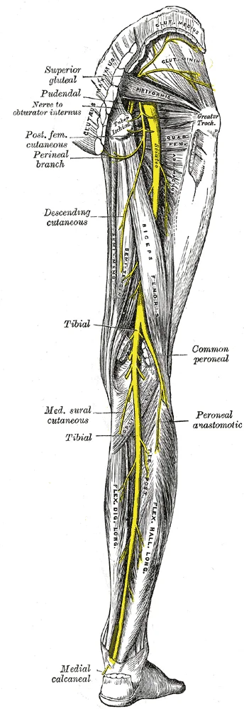
Long description
Nerves of the right lower extremity Posterior view.

Figure 5: Nerves of the right lower extremity Posterior view.
Peripheral nerves within the pelvis are organised to withstand mechanical stress during physiological movements. The peripheral nerve fibers are bundled into fascicles defined and organised by the perineurium sheath. The perineurium provides tensile strength and elasticity, and together with the epineurium, acts as a diffusion barrier that maintains constant intrafascicular pressure and resists external compression. Along their course, peripheral nerves are not straight; rather, they present an undulating pattern, and the individual fascicles and nerve fibers inside the fascicles share this same undulating organisation. This structural arrangement allows a degree of extension along the nerves when changing the position of innervated tissues and during movements of the respective organs. Traumatising these nerves during advanced gynaecological procedures must be avoided, as injury can lead to severe postoperative disabilities. [3]
DefinitionPelvic Nerve Complexity
Pelvic nerves are complex, requiring comprehension as a complete unit to understand the pathophysiology of pelvic diseases. [2]
The pelvic autonomic nervous system comprises mixed components that do not fit neatly into traditional sympathetic or parasympathetic categories. The usual understanding of hypogastric nerves as purely sympathetic and pelvic splanchnic nerves as purely parasympathetic is not entirely correct due to mixed components found in both. Similarly, the pudendal nerve, often considered purely somatic, is not entirely true in its physiological and pathological implications. [2]
| Structure | Type | Supply / Function |
|---|---|---|
| Pelvic Splanchnic Nerves | Parasympathetic (mixed) | Motor and sensory to pelvic viscera. |
| Inferior Hypogastric Plexus | Mixed Autonomic | Main source of sympathetic supply to rectum, uterus/vagina, bladder, prostate/seminal vesicles. [2] |
| Pudendal Nerve | Somatic (mixed) | Somatic muscle groups and pelvic floor,. |
| Vagus Nerve | Parasympathetic | Source of parasympathetic supply to remaining visceral organs. |
| Hypogastric Nerves | Sympathetic (mixed) | Afferent sensory pathway for sexual responses. |
The vascular supply to these nerves is critical for their function. A higher concentration of vessels is observed at the level of the perineurial sheath, which gives branches to the endoneurial compartment. The fascicular segmental vascular plexus is the main vascular unit of the peripheral nerves. Traction on these nerves can impair blood supply, so nerves should not be pulled, and traction should not be exerted, especially over a long period of time. [3]
Exam trapNerve Injury Risk
In neurotmesis, the continuity of the nerve is lost, and severe sensory, motor and autonomic disturbances are present distally from the injury site. [3]
Damage to these nerves often occurs during deep dissection for conditions like deep infiltrating endometriosis, which frequently calls for deep dissection of pararectal and paravaginal spaces in order to remove nodules lying adjacent to autonomic nerves. [2]
NoteClinical Consequence
Since the motor and sensory pathways from pelvic organs converge at the spinal level, neurotransmitters can be released onto the visceral targets through both pathways. [2]
This convergence means multiple sensory and motor symptoms may exist simultaneously in patients with pain and complaints in the bowel or bladder. Lower limb symptoms such as pain at a specific point or area while moving, and motor dysfunction, can also arise from pelvic nerve pathology. Any treatment of chronic pelvic pain must be based on a comprehensive understanding of anatomy and physiology. [2]
Neuropelveology deals with pelvic nerves, the generation of pain, and putative sites of lesions along the nerve pathway. Knowledge of these aspects enables the clinician to provide specific treatment. The diagnosis is confirmed by clinical investigation and imaging modalities. [2]
The hypogastric nerve lies approximately 5 to 20 mm dorsomedially to the ureter and extends posterolaterally to the rectum.
| Nerve or Plexus | Lesion Site | Resulting Deficit |
|---|---|---|
| Hypogastric nerve | Pelvic sidewall or ureteric crossing | Autonomic dysfunction and visceral pain |
| Pelvic splanchnic nerves | Caudal to the deep uterine vein | Bladder and sexual dysfunction |
| Obturator nerve | Paravesical fossa and lumbosacral space | Adductor weakness and medial thigh sensory loss |
| Genitofemoral nerve | Level of the psoas muscle | Inguinal sensory loss and cremasteric deficit |
Somatic and visceral nociception mediated by unmyelinated or lightly myelinated fibers often converges at the same spinal level. This viscerosomatic and viscerovisceral convergence could explain the diffuse nature of visceral pain, muscular spasms, and overlapping symptoms of chronic pelvic pain. Painful sensations carried from viscera stimulate somatic afferents and efferents, causing pain in the corresponding dermatome and muscle contraction in the corresponding myotome. Visceral organ pain evokes [2]
DefinitionNeuropelveology
A branch of neurological sciences focused on pelvic nerves, pain generation, and lesion sites along nerve pathways.
In gynaecological practice, severe deep endometriosis may require neurolysis because endometriotic lesions can envelope and infiltrate pelvic nerves. External neurolysis requires precise dissection between the epineurium and adjacent tissues using fine instrumentation. The loose unspecialised connective tissue surrounding the peripheral nerves is defined as paraneurium. Dissection at the paraneurium level, keeping the epineurium intact, is the preferred safe technique. [3]
Pelvic Nerve Dissection Guide
- Identify nerve pathwayLocate landmarks such as the deep uterine vein or ureter.
- Question: Is tissue infiltrating the nerve?
Yes
- Perform external neurolysisFind the plane between paraneurium and epineurium.
No
- Apply non-touch dissectionExert no mechanical force with instruments.
Pelvic autonomic nerves are fragile, and advanced gynaecological procedures may damage their fibers. Surgeons should avoid unnecessary manipulation and apply non-touch dissection where feasible, exerting no mechanical force with instruments. Laparoscopic neuronavigation reduces postoperative morbidity and preserves bladder function in high-risk pelvic surgeries. [3]
The clinical assessment of pelvic nerve integrity relies on identifying specific functional deficits resulting from anatomical disruption. A primary method for localising the site of pathology is dermatomal testing. This technique elucidates the area of sensory affect, which directly indicates which nerve root is affected. By mapping sensory loss or hyperaesthesia to specific dermatomes, clinicians can infer the level of neural involvement within the pelvic plexus or sacral roots. [2]
Nerve injuries are categorised using established classifications that correlate structural damage with clinical prognosis. Two classifications are widely recognized in medical practice. The first was defined by Seddon and includes three types of injuries: neuropraxia, axonotmesis and neurotmesis. Later, in 1951 Sunderland further developed the classification by dividing the neurotmesis into three different degrees of injuries, totaling 5 classes of nerve lesions. Each type of injury represents a [3]
DefinitionNerve Injury Classification
Seddon’s classification defines neuropraxia, axonotmesis, and neurotmesis, while Sunderland expanded this to five degrees based on the extent of connective tissue involvement.
The mechanical resilience of peripheral nerves is a critical factor in both injury mechanisms and surgical handling. When the nerve is pulled and put under traction, the fibers are first strengthened, then stretched and finally rupture if the traction does not cease. The nerve retains its elasticity as long as the perineurium stays intact. Although mechanically resistant to traction, the vascularisation of the nerve gets impaired, when the nerve is stretched. On average the elongation of the nerve of more than 15.7% causes complete arrest of blood flow. Another study by Lundborg shows similar results for cessation of the circulation when stretching to more than 15% of the nerve’s initial length. [3]
Exam trapTraction Ischaemia
Stretching a nerve by more than 15.7% arrests blood flow, causing ischaemic damage even before mechanical rupture occurs at 69.3% elongation.
Compression is another major mechanism of nerve injury. Nerve compression is one of the major etiologic factors for peripheral neuropathies and it has been widely examined in different studies. According to Rydevik et al. compression of 20 to 30 mmHg on the nerve leads to cessation of the venular blood flow. Despite its rich vascular network, the nerve’s microvasculature is susceptible to external damage with traction, elongation, extensive dissection in the adjacent tissues and particularly vulnerable to compression. The external injuries could have a detrimental effect on the nerve function, impairing the normal conduction of signals, and if applied with sufficient force and duration could have long-term effects. [3]
| Mechanism | Threshold/Effect | Consequence |
|---|---|---|
| Traction | >15.7% elongation | Complete arrest of blood flow |
| Traction | >30% elongation | Rupture of epineurial blood vessels |
| Traction | 69.3% elongation | Limit of elasticity |
| Compression | 20-30 mmHg | Cessation of venular blood flow |
In clinical practice, particularly during advanced gynaecological procedures, the peripheral nerves in the female pelvis are frequently encountered. Surgeries like promontofixation for pelvic organ prolapse, lymphadenectomy in the oncological cases or the treatment of deep endometriosis with nerve involvement could lead to severe postoperative morbidity and important deficits. In recent years, the concept of “nerve sparing” surgery has been widely incorporated in the different fields of gynaecology. The nerve sparing techniques have shown excellent results in terms of decreased morbidity and shorter functional recovery after surgery compared to the standard techniques. [3]
Clinical pearlNerve Sparing
Nerve sparing techniques reduce postoperative morbidity and accelerate functional recovery compared to standard surgical approaches in pelvic procedures.
When intraoperative injury occurs, repair may be necessary. When injured intraoperatively, the peripheral nerves may be repaired by the surgeon using a microsurgical technique. In cases of sharp transection with clear lines, an end-to-end anastomosis may be achieved. When the nerve is injured on a larger scale, the damaged part is excised; the proximal and distal nerve ends are connected either using an end-to-end anastomosis or using a nerve graft. Regardless of the method, it is of paramount importance to keep the nerve and the anastomosis tension free, otherwise the blood flow and respectively the healing process, will be impaired. The end-to-end anastomosis may be completed by performing an epineurial repair or even a perineurial repair, connecting the individual fascicles in a larger nerve. [3]
| Injury Type | Technique | Key Requirement |
|---|---|---|
| Sharp transection | End-to-end anastomosis | Tension-free connection |
| Large defect | Nerve graft or anastomosis | Tension-free connection |
| Microsurgery | Epineurial or perineurial repair | Preservation of fascicles |
For nerves involved by fibrosis, such as endometriotic nodules, neurolysis is indicated. The technique for nerve dissection and neurolysis is well known and is part of the training of plastic, orthopedic and neurosurgeons; however, it is not recognised and implemented in gynaecological practice. The concept of neurolysis was defined by Seddon et al. as an operation to liberate an injured nerve from scar tissue, fibrosis or other adjacent disease process that interferes with the normal nerve func [3]
NoteNeurolysis Tools
Simple instruments like cold scissors should be the preferred tools, as using other more sophisticated devices with electric or ultrasonic energy could cause thermal damage to the nerve. [3]
Internal neurolysis carries significant risks. Internal neurolysis may severely disrupt the normal function and organisation of the fascicles as well as the vascularisation, potentially causing more harm than the disease itself. As previously described, the fascicles inside the nerve are interconnected and dissecting even a single fascicle could impair the whole nerve structure. Internal neurolysis consists of a few separate steps. The first step is epifascicular epineurotomy, which consists of one of few longitudinal incisions across the disease thickened portion of the nerve. The compressed fascicles could start expanding from the incision site. If after this epineurotomy the nerve continues to look deformed, the surgeon must remove the epifascicular epineurium around the circumference of the nerve in the diseased area so that the fascicles can be released: this is an epifascicular epineurectomy. [3]
| Step | Description | Risk |
|---|---|---|
| Epifascicular epineurotomy | Longitudinal incisions across thickened nerve | Disruption of fascicle organisation |
| Epifascicular epineurectomy | Removal of epineurium around circumference | Impairment of whole nerve structure |
| General Principle | Preserve nerve integrity | Avoid additional trauma |
In cases when the somatic nerves are involved by fibrosis, as with endometriotic nodules, the surgeon must follow the main principles for neurolysis, defined by Millesi. As nerve dissection and repair techniques are usually not part of standard gynaecological training, it should be emphasised that our recommendations for neurolysis in pelvic surgeries are taken from other specialties and modified in such a way that gynecologists may implement them in their practice. The dissection should preserve the integrity of the nerve as much as possible and should not itself cause additional trauma. As recommended by Graf and Petruzelli, simple instruments like cold scissors should be the preferred tools, as using other more sophisticated devices with electric or ultrasonic energy could cause thermal damage to the nerve. [3]
Clinical pearlParasympathetic origin
Pelvic splanchnic nerves (PSN) arise from sacral spinal nerve segments S2 to S4. [2]
Clinical pearlAutonomic components
The autonomic nervous system supplying the pelvis comprises both sympathetic and parasympathetic divisions. [2]
Clinical pearlVisceral pain convergence
Visceral afferents exhibit extensive spinal arborization, causing diffuse pain and referred symptoms due to organ crosstalk. [2]
Clinical pearlErection pathway
Genital arousal pathways involve psychogenic centers at T11 to L2 and reflexogenic centers at S2 to S4. [2]
DefinitionGreater sciatic foramen
The superior posterolateral pelvic opening formed by the greater sciatic notch, sacrum, and sacrospinous ligament. [1]
DefinitionLesser sciatic foramen
The inferior posterolateral pelvic opening bounded by the lesser sciatic notch, sacrospinous ligament, and sacrotuberous ligament. [1]
NoteNerve-sparing surgery
Nerve-sparing and preservation is a key factor in reducing postoperative morbidity during advanced gynaecological procedures. [3]
Exam trapWater-jet pressure limits
Water-jet pressure must remain at or below 30 bar to prevent permanent structural and functional nerve damage. [3]
Clinical pearlBony pelvis formation
The bony pelvis is formed by the two hip bones, the sacrum, and the coccyx attached inferiorly. [1]
Clinical pearlPelvic splanchnic junction
Pelvic splanchnic nerves join the inferior hypogastric plexus 3 to 4 centimetres lateral and 2 to 4 centimetres caudal to the pouch of Douglas. [2]
The anatomy of the pelvic nerves is essential for understanding clinical presentations of chronic pelvic pain and the technical requirements of nerve-sparing surgery. Students must recognise that visceral afferent fibers are distinct from somatic fibers; specifically, visceral afferents exhibit more extensive arborization in the spinal cord. This anatomical arrangement explains why visceral pain is often diffuse and why patients may experience referred pain or organ crosstalk, where two afferent fibers from different organs converge on the same dorsal root ganglion.
When performing surgical dissections, surgeons must be aware that nerve identification is the prerequisite for preservation. Techniques such as laparoscopic neuro-navigation (LANN) rely on precise electrical stimulation parameters to map nerves intraoperatively.
Illustrative example: A patient presents with pelvic pain. The clinician considers the role of the autonomic nervous system, which modulates impulses at the central and peripheral levels. If the patient is undergoing a radical hysterectomy, the surgeon must identify the pelvic splanchnic nerves, which join the inferior hypogastric plexus at a specific location: 3 to 4 cm lateral and 2 to 4 cm caudal to the pouch of Douglas. Failure to identify these structures can lead to increased postoperative morbidity.
The bony pelvis provides the framework for these nerves, with the greater and lesser sciatic foramina serving as critical exit points for neurovascular structures. The sacrum and hip bones form the primary structure, with the coccyx attached inferiorly. Understanding these bony landmarks is vital for interpreting pelvic imaging and surgical access.
Finally, consider the physiology of genital arousal. This process involves a complex interplay between psychogenic input (T11–L2) and reflexogenic input (S2–S4). The final common pathway involves the pelvic nerves, which facilitate smooth muscle relaxation via neurotransmitters like nitric oxide and vasoactive intestinal peptide to override sympathetic detumescence. Chronic pelvic pain, in contrast, involves a complex array of mechanisms including peripheral and central sensitization, and the growth of new nerve fibers.
Students often mistake the structural organisation of peripheral nerves, assuming individual fascicles run in isolation from origin to termination. In reality, individual fascicles inside the nerve are not isolated from each other[1]. They are interconnected, constantly splitting and merging together along the nerve length, so that mobilisation of a single fascicule, without causing damage to the rest, with whom they are connected, is impossible[1]. [3]
Another frequent error is believing that nerve fascicular counts remain constant throughout their anatomical course. The number of fascicles is changing along the course of the nerve[1]. It is considered that the larger the number of fascicles at a particular level, the larger the resistance capacity of the nerve to external compression[1]. The high number of fascicles at certain areas designates a higher amount of interfascicular epineurium, which allows reorganisation of the fascicular pattern when external compression is applied, without increasing the pressure inside the fascicles[1]. [3]
A third misconception involves underestimating the mechanical thresholds required to cause physiological injury during compression. Minor repetitive compression could be one of the risk factors for nerve damage and the breakdown of the blood-brain barrier[1]. Venous flow impairment starts at compression between 0.15 to 0.30 N, further compression of more than 0.30 N impairs the arteriolar flow, and complete cessation of the circulation is present at 0.46 N, which corresponds to 48.7 mmHg[1]. Furthermore, disturbances in the conduction of electrical impulses start even with a lower degree of compression, approximately at 0.2 N, and the compression force of more than 0.3 N leads to complete conduction blockage and intraneural anoxia[1]. [3]
Students also confuse the grading and reversibility of nerve injuries. Neuropraxia, which is classified as type I injury according to Sunderland, represents the mildest form of nerve injury[1]. It is a temporary conduction block with preserved axonal continuity at a specific area of the nerve[1]. The connective tissues in the structure of the nerve, including the endoneurium, perineurium, and epineurium, are preserved[1]. Because the axons are damaged, but not destroyed, there is no wallerian degeneration, and clinically there is full regeneration and recovery of function a few days to weeks after the injury[1]. [3]
A fifth pitfall relates to surgical technique and tissue handling. During gynaecological operations, the force generated between the jaws of laparoscopic instruments when grasping tissues is sufficient to completely stop the blood flow in the neural vascular system[1]. Grip force differs between surgical experience levels, measuring 3 N for experienced surgeons and 7 N for novice surgeons[1]. [3]
Finally, learners often fail to differentiate between external and internal neurolysis. The dissection follows the course of the normal nerve towards the lesion from proximal and distal directions, so that the surgeon can recognise where the lesion starts and from where the neurolysis should begin[1]. External neurolysis involves dissection at the most superficial layer between the disease and the epifascicular epineurium to completely remove the disease[1]. If infiltration expands deep into the nerve beyond the epifascicular epineurium, internal neurolysis becomes necessary, requiring careful consideration of risks and benefits[1]. [3]
At the bedside, clinicians evaluate pelvic nerve integrity through sensory and motor examinations. Mild sensory or motor disturbances below the level of injury point toward a physiological conduction block without axonal destruction[1]. Understanding that orgasm involves complex neural circuits linking both sympathetic and parasympathetic systems at peripheral and central levels helps clinicians assess autonomic dysfunction following pelvic trauma or surgery. [2][3]
In the operating theatre, surgeons apply detailed anatomical knowledge of pelvic nerve pathways during complex pelvic resections and gynaecological procedures. Surgeons execute neurolysis by dissecting from proximal and distal directions towards the lesion[1]. When scar tissue or disease involves the paraneurium, external neurolysis is performed, whereas deeper infiltration necessitates internal neurolysis[1]. Advanced non-touch dissection methods such as water-jet streams are utilised to preserve vascular integrity and minimise thermal damage during fine neural dissection[1].
On imaging and in physiological evaluations, clinicians interpret the physical dimensions and bony boundaries of the pelvis that protect these neural networks. The greater sciatic notch of the male hip bone is narrower and deeper than the broader notch of females. Because the female pelvis is adapted for childbirth, it is wider, featuring a larger subpubic angle greater than 80 degrees, compared to less than 70 degrees in males. [1]
In examinations, students must recall the precise mechanical limits of nerve tissue. Questions frequently test that electrical impulse conduction disturbances begin at an approximate compression force of 0.2 N, while complete conduction blockage and intraneural anoxia occur above 0.3 N[1]. Candidates must also recognise that Sunderland type I injury corresponds to neuropraxia, featuring preserved connective tissue sheaths and spontaneous recovery within days to weeks[1].
- Individual nerve fascicles constantly split and merge, preventing the isolation of a single fascicle without damage.
- Higher fascicular counts at specific levels provide greater resistance to external compression through interfascicular epineurium.
- Venous flow impairment starts at 0.15 to 0.30 N, while complete circulatory cessation occurs at 0.46 N or 48.7 mmHg.
- Electrical conduction disturbances begin at 0.2 N of compression, with complete blockage occurring above 0.3 N.
- Laparoscopic instrument grip forces differ between experienced surgeons at 3 N and novices at 7 N.
- Neuropraxia or Sunderland type I injury is a temporary conduction block with preserved axonal and connective tissue continuity.
- External neurolysis addresses disease superficial to the epifascicular epineurium, whereas internal neurolysis breaches this layer.
- Female pelvic architecture features a wider subpubic angle exceeding 80 degrees compared to under 70 degrees in males.
Answer each one in your head first, then open the answer.
What is the Sunderland classification type for neuropraxia?
Show answerHide answer
Type IAt what compression force do electrical conduction disturbances begin in peripheral nerves?
Show answerHide answer
Approximately 0.2 NWhat pressure corresponds to complete cessation of circulation in compressed nerve tissue?
Show answerHide answer
48.7 mmHgWhat is the laparoscopic instrument grip force generated by novice surgeons?
Show answerHide answer
7 NWhat is the minimum subpubic angle measurement characteristic of the female pelvis?
Show answerHide answer
Greater than 80 degrees
- Betts JG, Young KA, Wise JA, et al. The Pelvic Girdle and Pelvis. OpenStax, Anatomy and Physiology 2e (CC BY-NC-SA 4.0). Accessed .
- Alkatout I, Wedel T, Pape J, et al. Review: Pelvic nerves - from anatomy and physiology to clinical applications. Translational neuroscience 2021;12(1):362-378. doi:10.1515/tnsci-2020-0184. Accessed .
- Aleksandrov A, Smith AV, Botchorishvili R, Rabischong B. How to dissect the pelvic nerves: from microanatomy to surgical rules. An evidence-based clinical review. Facts, views & vision in ObGyn 2022;14(1):17-29. doi:10.52054/fvvo.14.1.011. Accessed .
Figure credits
- Figure 1: Henry Vandyke Carter, via Wikimedia Commons, Public domain, Public domain.
- Figure 2: Henry Vandyke Carter, via Wikimedia Commons, Public domain, Public domain.
- Figure 3: Henry Vandyke Carter, via Wikimedia Commons, Public domain, Public domain.
- Figure 4: Henry Vandyke Carter, via Wikimedia Commons, Public domain, Public domain.
- Figure 5: Henry Vandyke Carter, via Wikimedia Commons, Public domain, Public domain.
Educational content for medical students and doctors. It is not a substitute for clinical judgement or local guidelines.