Perineum
Perineum anatomy: boundaries, triangles, pouches, erectile tissues, external genitalia, trauma, autonomic supply.
The perineum is the diamond-shaped region inferior to the pelvic diaphragm, lying between the thighs. Its bony framework is formed by the pubic symphysis anteriorly, the coccyx posteriorly, and the ischial tuberosities laterally. The deep limit is the pelvic floor (levator ani and coccygeus), while the superficial limit is skin. The region is divided into two triangles by a transverse line joining the ischial tuberosities. [1]
The urogenital triangle contains the external genitalia and urethra; the anal triangle contains the anal canal and ischiorectal fossae. The perineal membrane is a thick triangular fascial sheet spanning the anterior half of the pelvic outlet, deep to the superficial perineal pouch. The superficial perineal pouch lies below the membrane and contains the erectile tissues, superficial perineal muscles, and in males the bulb of t [1]
The perineal body is a fibromuscular node at the midpoint of the line between the ischial tuberosities, where several muscles and fasciae converge: the bulbospongiosus, superficial and deep transverse perinei, external anal sphincter, and levator ani. It is clinically important as a support for the pelvic organs; in females it may be torn during childbirth. [1][3]
The erectile tissues of the penis are three columns: two corpora cavernosa laterally and the corpus spongiosum ventrally, which surrounds the penile urethra and expands distally as the glans penis. The clitoris has two corpora cavernosa and a glans, but no corpus spongiosum. Erection results from nitric oxide (NO) release causing arterial dilation, increased blood inflow, and venous compression.
External genitalia in males: penis (root, shaft, glans, prepuce) and scrotum containing testes, which are kept 2 to 4°C below core temperature for efficient spermatogenesis. In females: labia majora, labia minora, clitoris, vestibule, and vaginal orifice. [3]
Figure 1Gray1230
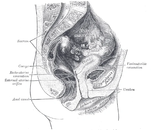
Long description
Median sagittal section of female pelvis.

Figure 1: Median sagittal section of female pelvis.
Figure 2Gray1229
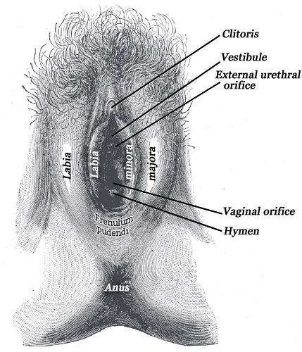
Long description
External genital organs of female. The labia minora have been drawn apart.

Figure 2: External genital organs of female.
Genitourinary trauma commonly involves straddle injuries (blunt trauma to the perineum) causing urethral injury, especially in males where the bulbar urethra is crushed against the pubic symphysis. Penetrating trauma may involve the anal canal.
| Muscle | Origin | Insertion | Action | Nerve |
|---|---|---|---|---|
| Bulbospongiosus | Perineal body | Corpus spongiosum (male) / clitoris (female) | Compresses bulb, aids erection and ejaculation | Pudendal (S2–S4) |
| Ischiocavernosus | Ischial tuberosity | Corpus cavernosum | Maintains erection | Pudendal (S2–S4) |
| Superficial transverse perinei | Ischial tuberosity | Perineal body | Fixes perineal body | Pudendal (S2–S4) |
| External anal sphincter | Coccyx | Perineal body | Closes anal canal | Pudendal (S2–S4) |
| Levator ani | Pubis, ischial spine | Coccyx, perineal body | Supports pelvic organs | Pudendal and S3–S4 |
Figure 3Gray542
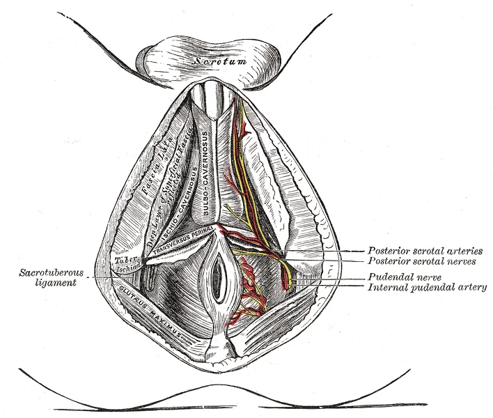
Long description
Gray's anatomy of the perineum. Labelled illustration from Wikimedia Commons, used under its open licence.

Figure 3: Gray's anatomy of the perineum.
The perineum is the diamond-shaped region located between the thighs, bounded anteriorly by the pubic symphysis, posteriorly by the coccyx, and laterally by the ischial tuberosities. This area can be divided into two triangles: the anterior urogenital triangle and the posterior anal triangle, separated by an imaginary line connecting the ischial tuberosities. The surface anatomy of the perineum can be identified by several palpable landmarks that are crucial for clinical examination and surgical approaches.
When standing in the anatomical position, the pelvis is tilted anteriorly, causing the anterior superior iliac spines and the pubic tubercles to lie in the same vertical plane. The pubic tubercles are small, palpable projections located on the superior margin of the pubic body and serve as important attachment points for ligaments. The ischial tuberosities are prominent bony structures that can be palpated on the lateral aspects of the perineum and serve as weight-bearing points when sitting. These structures are approximately 4-5 cm from the midline in adults and are particularly prominent in individuals with less subcutaneous fat. [1] [3]
Figure 4Gray1228
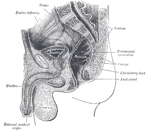
Long description
Median sagittal section of male pelvis. Labelled illustration from Wikimedia Commons, used under its open licence.

Figure 4: Median sagittal section of male pelvis.
The perineal body is particularly important in maintaining pelvic support and is a common site of injury during childbirth, which can lead to pelvic organ prolapse.
In males, the scrotum has a raised medial thickening on the surface called the raphae, which continues anteriorly as the raphe of the penis. In females, the raphe extends from the perineal body to the posterior commissure of the labia majora. This structure represents the line of fusion of the embryonic labioscrotal swellings and is an important anatomical landmark for identifying the midline of the perineum. [3]
The perineal body is located at the center of this diamond, and the perineal raphe extends along the midline from the perineal body to the anus. The ischial tuberosities are palpable as bony prominences on the lateral aspects of the perineum, approximately 4-5 cm from the midline.
The perineum is the diamond-shaped region inferior to the pelvic diaphragm. Its bony framework is provided by the pelvis, which is formed by the two hip bones, the sacrum)Skip? No, the perineum itself is bounded by specific bony landmarks. Each hip bone is formed by the fusion of three bones: the ilium, ischium, and pubis, which fuse during late teenage years[1]. These three components define the three regions of the adult hip bone.
The ilium forms the superior, flared portion of the hip bone. Its curved superior margin, the iliac crest, can be felt along your waistline[1]. The crest ends anteriorly at the anterior superior iliac spine (ASIS), a palpable landmark at the anterolateral hip; inferior to this lies the anterior inferior iliac spine[1]. Posteriorly, the crest terminates at the posterior superior iliac spine, which may appear as a “dimple” on the lower back, and below it is the posterior inferior iliac spine[1]. These spines serve as attachment points for thigh muscles[1].
The ischium forms the posterolateral portion of the hip bone[1]. Its large, roughened inferior area, the ischial tuberosity, carries body weight when sitting and is palpable when you wiggle your pelvis against a chair seat[1]. Projecting superiorly and anteriorly from the tuberosity is the ischial ramus[1]. Above the tuberosity, the slightly curved posterior margin is the lesser sciatic notch; the bony projection separating this from the greater sciatic notch is the ischial spine[1].
The pubis forms the anterior portion. The two pubic bones meet anteriorly at the pubic symphysis, a cartilaginous joint[1]. The female sacrum is wider, shorter, and less curved, with a less projecting sacral promontory, giving a more rounded pelvic inlet[1]. The ischial tuberosities are farther apart in females, enlarging the pelvic outlet[1].
The sacroiliac joint is supported by the anterior and posterior sacroiliac ligaments[1]. These ligaments immobilise the sacrum for weight-bearing[1].
Figure 5Gray405
Long description
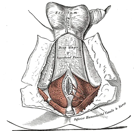
The perineum. The integument and superficial layer of superficial fascia reflected.

Figure 5: The perineum.
The perineum is the diamond-shaped region inferior to the pelvic diaphragm, bounded by the pubic symphysis anteriorly, the coccyx posteriorly, and the ischial tuberosities laterally. Its deep fascia is continuous with the deep fascia of the thigh and the pelvic fascia, and it splits to enclose the two main compartments of the perineum: the urogenital triangle (anterior) and the anal triangle (posterior). [2]
Deep to Colles' fascia lies the superficial perineal pouch, a closed space containing the bulbospongiosus, ischiocavernosus, and superficial transverse perineal muscles, plus the crura and bulb of the penis or clitoris.
The obturator internus fascia lines the lateral wall of the ischioanal fossa, a space on each side of the anal canal; the fossa is bounded medially by the external anal sphincter and laterally by the obturator internus, and it contains the pudendal canal (Alcock's canal) with the pudendal nerve and internal pudendal vessels. The deep fascia of the thigh (fascia lata) is continuous with the perineal fascia at the margins of the perineum, and it separates the thigh into med
Clinical pearlClinical pearl
A straddle injury can rupture the bulbous urethra; urine then extravasates into the superficial perineal pouch and tracks along Colles' fascia into the scrotum and penis, but is limited posteriorly by the attachment of Colles' fascia to the perineal membrane and laterally by the ischiopubic rami.
Figure 61116 Muscle of the Male Perineum
Long description
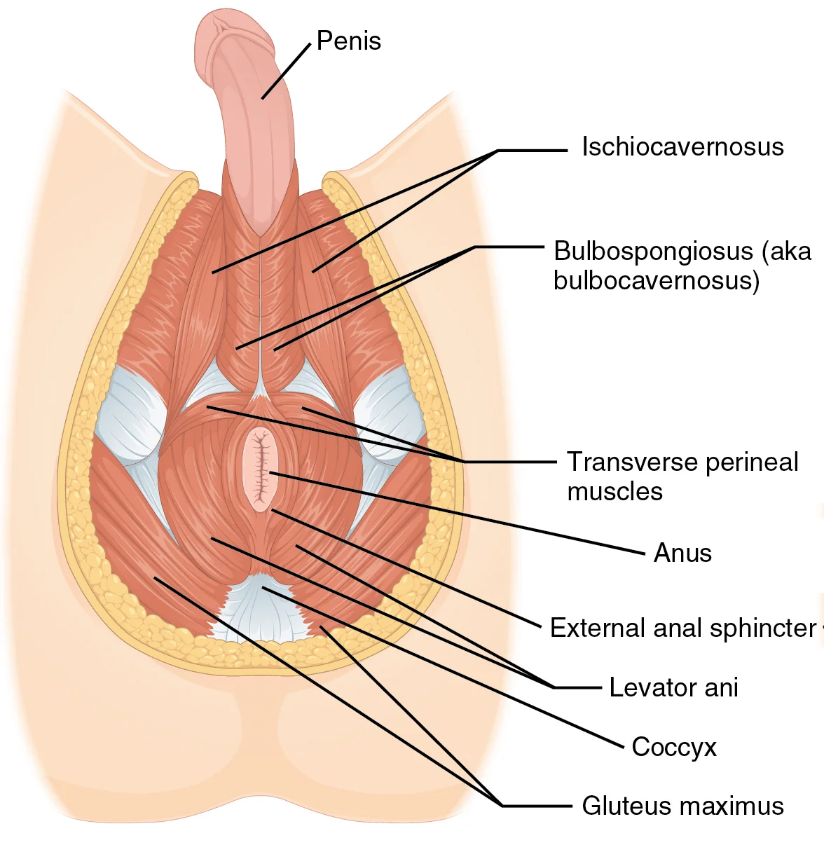
Caption: The perineum muscles play roles in urination in both sexes, ejaculation in men, and vaginal contraction in women. URL: https://cnx.org/contents/FPtK1zmh@8.108:b3YG6PIp@6/Axial-Muscles-of-the-Abdominal Version 8.

Figure 6: Caption: The perineum muscles play roles in urination in both sexes, ejaculation in men, and vaginal contraction in women (url: https://cnx.org/contents/fptk1zmh@8.108:b3yg6pip@6/axial-muscles-of-the-abdominal version 8).
Figure 71116 Muscle of the Perineum
Long description
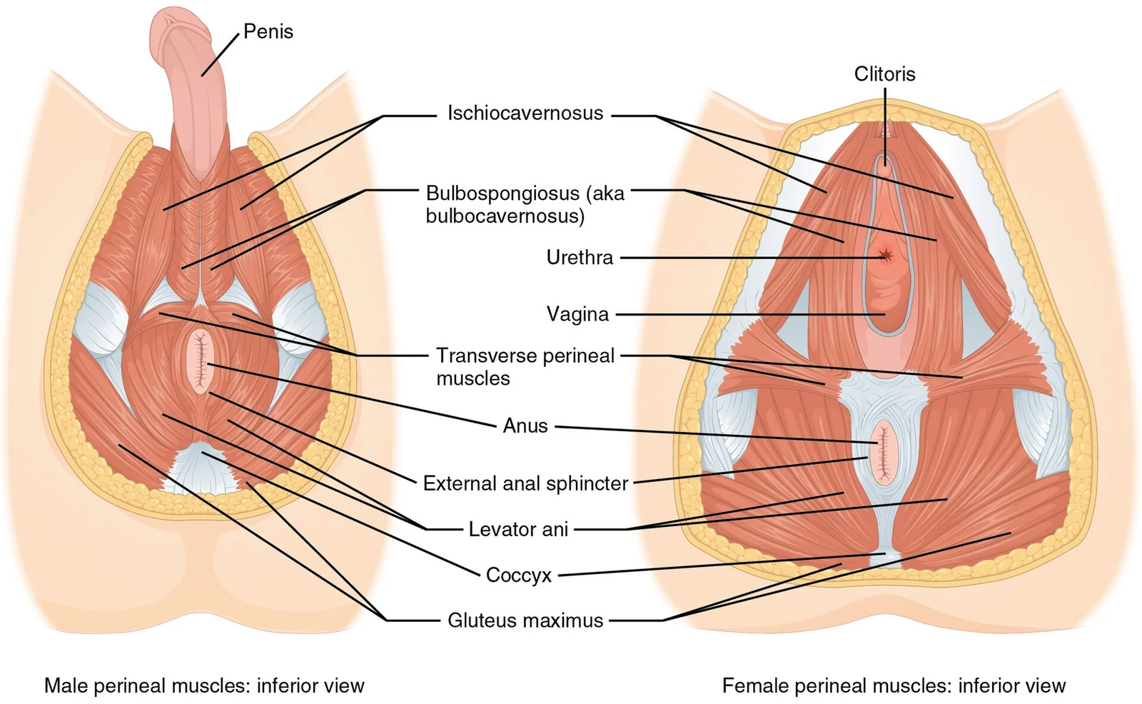
Caption: The perineum muscles play roles in urination in both sexes, ejaculation in men, and vaginal contraction in women. URL: https://cnx.org/contents/FPtK1zmh@8.108:b3YG6PIp@6/Axial-Muscles-of-the-Abdominal Version 8.

Figure 7: Caption: The perineum muscles play roles in urination in both sexes, ejaculation in men, and vaginal contraction in women (url: https://cnx.org/contents/fptk1zmh@8.108:b3yg6pip@6/axial-muscles-of-the-abdominal version 8).
The perineum is the diamond-shaped region inferior to the pelvic diaphragm, between the thighs. Its muscles are arranged in two main compartments: the urogenital triangle (anterior) and the anal triangle (posterior). These muscles support the pelvic viscera, control micturition and defecation, and are active in sexual function.
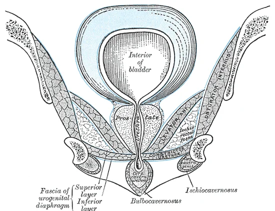
This triangle is bounded by the pubic symphysis anteriorly and the ischial tuberosities laterally. It contains the superficial and deep perineal pouches. The superficial pouch muscles include the bulbospongiosus, ischiocavernosus, and superficial transverse perineal muscles. The deep pouch contains the deep transverse perineal muscle and the external urethral sphincter.
Figure 8Gray407
Long description
Coronal section of anterior part of pelvis, through the pubic arch. Seen from in front. (Diagrammatic.)

Figure 8: Coronal section of anterior part of pelvis, through the pubic arch (seen from in front).
| Muscle | Origin | Insertion | Action | Nerve Supply |
|---|---|---|---|---|
| Bulbospongiosus | Central perineal tendon (male); central tendon and external anal sphincter (female) | Male: penile raphe and corpora cavernosa; Female: clitoris and corpus cavernosum | Male: expels semen/urine, assists erection; Female: constricts vaginal orifice, assists clitoral erection | Perineal branch of pudendal nerve (S2–S4) |
| Ischiocavernosus | Ischial tuberosity and ramus | Corpus cavernosum (clitoris or penis) | Compresses erectile tissue, maintains erection | Perineal branch of pudendal nerve (S2–S4) [1] |
| Superficial transverse perineal | Ischial tuberosity | Central perineal tendon | Fixes central tendon, supports pelvic floor | Perineal branch of pudendal nerve (S2–S4) |
| Deep transverse perineal | Ischial ramus | Central perineal tendon | Supports pelvic floor, fixes perineal body | Deep branch of pudendal nerve (S2–S4) |
| External urethral sphincter | Ischial ramus and pubic ramus | Encircles urethra (and vagina in female) | Compresses urethra, voluntary control of micturition | Pudendal nerve (S2–S4) |
This triangle contains the anal canal and the external anal sphincter, a striated voluntary muscle encircling the anus. It is supplied by the inferior rectal nerve, a branch of the pudendal nerve.
| Muscle | Origin | Insertion | Action | Nerve Supply |
|---|---|---|---|---|
| External anal sphincter | Coccyx and anococcygeal ligament | Central perineal tendon (anteriorly) | Voluntary closure of the anal canal | Inferior rectal nerve (S2–S4) |
Structures in the anterior perineum, including the scrotum in males or the labia majora in females, drain to the superficial inguinal lymph nodes. These nodes are located in the thigh, just inferior to the inguinal ligament. The v [1]
The perineal nerve supplies the urogenital muscles and skin; the dorsal nerve supplies the erectile tissue. [1]
- Perineal tear (obstetric): The bulbospongiosus and superficial transverse perineal muscles are commonly torn during vaginal delivery; repair requires suturing the central perineal tendon.
- Pudendal nerve block: Anaesthetic is injected near the ischial spine, where the nerve is most accessible as it crosses the sacrospinous ligament. [1]
- Ischioanal fossa abscess: Pus collects lateral to the anal canal; the external anal sphincter is spared unless infection tracks through it.
- Vasectomy: The ductus deferens is cut within the spermatic cord in the scrotum; this interrupts sperm transport without affecting the perineal muscles. [3]
The bulbospongiosus lies in the midline, the ischiocavernosus along the ischial rami, and the external anal sphincter encircles the anus posteriorly.
and the perineal artery, which contributes to the blood supply of the perineum. [1]
Within the pudendal canal, the internal pudendal artery gives rise to several important branches. [1]
Additionally, the perineum receives some contribution from the external pudendal artery, a branch of the femoral artery. [1]
| Artery | Origin | Course | Branches | Termination |
|---|---|---|---|---|
| Internal pudendal | Internal iliac artery | Through greater sciatic notch, lesser sciatic foramen, pudendal canal | Inferior rectal, perineal, posterior scrotal/labial | Deep artery of penis/clitoris |
| External pudendal | Femoral artery | Superficial to perineum | - | Anastomoses with other perineal vessels |
The venous drainage of the perineum follows a pattern that mirrors the arterial supply, ensuring efficient return of deoxygenated blood from the superficial and deep structures to the systemic circulation. Understanding this network is crucial for appreciating how infections or haemorrhages may spread within the pelvic region.
The central structure is the perineal vein, which drains the superficial perineal pouch.
The superficial perineal veins drain the skin and subcutaneous tissues of the perineum. These veins accompany the superficial perineal arteries and empty into the external pudendal veins. The external pudendal veins then drain into the great saphenous vein, which ultimately joins the femoral vein. This pathway provides a direct route for blood from the superficial perineal region to the lower limb venous system. [1]
The deep perineal veins are more complex. They drain the deep perineal pouch, including the bulb of the penis or vestibule, and the associated muscles. These veins form the internal pudendal veins, which run alongside the internal pudendal arteries. The internal pudendal veins receive tributaries from the deep dorsal vein of the penis or clitoris. In males, the prostatic venous plexus is a significant component, draining the prostate gland and bladder neck. In females, the vaginal venous plexus serves a similar role for the vagina.
This connection is vital because it links the perineal venous drainage to the major pelvic venous system. The internal iliac veins then join the common iliac veins, which converge to form the inferior vena cava. This anatomical arrangement ensures that blood from the perineum is efficiently returned to the heart.
Clinical relevance arises from the valveless nature of many of these veins. This characteristic allows for retrograde flow, which can facilitate the spread of infection or tumour cells from the perineum to the pelvic cavity or even the vertebral venous plexus. For instance, prostatic venous plexus connections to the vertebral veins explain the propensity for prostate cancer to metastasise to the spine. [3] [1]
Clinical pearlKey Point
The valveless nature of perineal veins allows for bidirectional flow, facilitating the spread of infection or malignancy to distant sites such as the vertebral column.
In summary, the venous drainage of the perineum involves a coordinated network of superficial and deep veins that ultimately empty into the internal iliac veins. This system ensures effective blood return while also presenting potential pathways for pathological spread.
The perineum is innervated by branches of the sacral plexus, pudendal nerve, and perineal branches of posterior femoral cutaneous nerve.
It exits the pelvis through the greater sciatic foramen [1]
It also provides cutaneous sensation to the perineum, genitalia, and surrounding areas. [1]
The perineal branch of the posterior femoral cutaneous nerve provides cutaneous sensation to the skin of the posterior scrotum or labium majus and the adjacent medial thigh. The ilioinguinal and genitofemoral nerves provide sensory innervation to the anterior scrotum/labium majus and upper medial thigh. [1]
| Nerve | Muscles Supplied |
|---|---|
| Pudendal nerve | External anal sphincter |
| Bulbospongiosus | |
| Ischiocavernosus | |
| Superficial transverse perineal |
| Nerve | Skin Area Supplied |
|---|---|
| Pudendal nerve | Perineum, genitalia, surrounding areas |
| Perineal branch of posterior femoral cutaneous nerve | Posterior scrotum/labium majus, adjacent medial thigh |
| Ilioinguinal nerve | Anterior scrotum/labium majus, upper medial thigh |
| Genitofemoral nerve | Anterior scrotum/labium majus, upper medial thigh |
The lymphatic drainage of the perineum is divided into structures anterior to the urogenital diaphragm and those posterior to it, with distinct terminal nodes for each region. [1]
Structures in the anterior perineum, including the scrotum in males or the labia majora in females, drain to the superficial inguinal lymph nodes. These nodes are located in the thigh, just inferior to the inguinal ligament. The v
DefinitionSuperficial Inguinal Nodes
These nodes receive lymph from the skin of the lower abdominal wall, buttocks, external genitalia, and lower limb, serving as the primary filter for anterior perineal structures.
The efferent vessels from the superficial inguinal nodes join the deep inguinal lymph nodes. The deep inguinal nodes receive lymph from the glans penis or clitoris, as well as from the superficial inguinal nodes. From the deep inguinal nodes, lymph flows into the external iliac lymph nodes. This pathway ensures that any pathology in the anterior perineum must pas [1]
This distinction is vital for understanding the spread of anal carcinoma. [1]
Exam trapAnal Canal Drainage
Remember that the anal canal has dual lymphatic drainage. The upper part drains to internal iliac nodes, while the lower part drains to superficial inguinal nodes. This is a common examination trap. [1]
| Structure | Primary Drainage | Secondary Drainage |
|---|---|---|
| Scrotum / Labia Majora | Superficial Inguinal Nodes | Deep Inguinal Nodes |
| Glans Penis / Clitoris | Deep Inguinal Nodes | External Iliac Nodes |
| Anal Canal (below pectinate line) | Superficial Inguinal Nodes | Internal Iliac Nodes |
| Anal Canal (above pectinate line) | Internal Iliac Nodes | Superior Rectal Nodes |
Understanding these pathways allows clinicians to predict the spread of disease and plan appropriate surgical interventions or radiation fields. The segmentation is not absolute, and collateral drainage may occur, but the primary pathways remain consistent and clinically significant.
The perineum is the diamond-shaped region between the thighs, inferior to the pelvic diaphragm. Its boundaries are the pubic symphysis anteriorly, the coccyx posteriorly, and the ischial tuberosities laterally. A transverse line joining the ischial tuberosities divides it into a urogenital triangle anteriorly and an anal triangle posteriorly. The urogenital triangle contains the external genitalia and the urethra; the anal triangle contains the anal canal and the ischioanal fossa. [3]
In the female, it contains the bulbs of the vestibule, the greater vestibular glands, and the clitoral crura. [3]
In the female, it also contains the urethra and the vagina as they pass through.
The deep perineal space lies superior to the membrane, with the sphincter urethrae surrounding the membranous urethra. The ischioanal fossa is lateral, filled with fat, and the pudendal canal lies on its lateral wall.
The muscles of the perineum are arranged in two layers. The superficial layer lies in the superficial perineal space and includes three paired muscles. The bulbospongiosus covers the bulb of the penis in the male and surrounds the vaginal orifice in the female; it compresses the bulb and aids erection and ejaculation. The ischiocavernosus covers the crura of the penis or clitoris and compresses them to maintain erection. The superficial transverse perineal muscle runs from the ischial tuberosity to the perineal body, stabilising it.
Figure 9Gray408
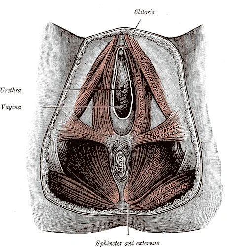
Long description
Muscles of the female perineum. (Modified from a drawing by Peter Thompson.)

Figure 9: Muscles of the female perineum.
The deep layer lies in the deep perineal space. The deep transverse perineal muscle runs from the ischial ramus to the perineal body. The perineal body is a node at the midpoint of the perineum, where the bulbospongiosus, superficial and deep transverse perineal muscles, and the external anal sphincter converge. [3]
| Muscle | Origin | Insertion | Action | Nerve supply |
|---|---|---|---|---|
| Bulbospongiosus | Perineal body, median raphe | Bulb of penis / clitoris, perineal membrane | Compresses bulb, aids erection and ejaculation | Perineal branch of pudendal nerve |
| Ischiocavernosus | Ischial tuberosity and ramus | Crus of penis / clitoris | Compresses crus, maintains erection | Perineal branch of pudendal nerve |
| Superficial transverse perineal | Ischial tuberosity | Perineal body | Stabilises perineal body | Perineal branch of pudendal nerve |
| Deep transverse perineal | Ischial ramus | Perineal body | Supports pelvic organs | Perineal branch of pudendal nerve |
| Sphincter urethrae | Ischial ramus | Median raphe around urethra | Voluntary urinary continence | Perineal branch of pudendal nerve |
It then runs in the pudendal canal on the lateral wall of the ischioanal fossa, giving off the inferior rectal nerve, the perineal nerve, and the dorsal nerve of the penis or clitoris. The perineal nerve supplies the muscles and skin of the urogenital triangle; the dorsal nerve supplies the erectile tissue. [1]
The ischioanal fossa is a wedge-shaped fat-filled space on each side of the anal canal, bounded laterally by the obturator internus fascia and medially by the levator ani and external anal sphincter. Its base is the skin of the anal triangle, and its apex is the junction of the obturator fascia and levator ani. It contains fat, the pudendal canal with the pudendal nerve and internal pudendal vessels, and the inferior rectal nerve and vessels crossing to the anal canal. Abscesses here can spread widely because of the fat and the communication across the midline. [3]
The perineum is best assessed radiologically with MRI, which gives superb soft-tissue contrast for the pelvic floor and ischiorectal fossae. On axial T1-weighted images, the fat of the ischioanal fossa appears bright, outlining the darker levator ani and obturator internus muscles. The anal canal is seen as a midline structure with its internal sphincter as a low-signal ring and the external sphincter as a slightly higher-signal ring around it. The urethra in males is visible as a thin, fluid-filled line passing through the perineal membrane; in females, the shorter urethra lies anterior to the vagina, which appears as a flattened, low-signal slit between the urethra and the rectum.
On CT, the perineum is less well defined, but the ischioanal fossae are visible as low-attenuation fat, and the anal canal can be identified by the gas within it. The obturator internus muscles are seen on each side, and the pudendal canal (Alcock's canal) is not directly visible but its position is inferred on the lateral wall of the ischioanal fossa. Ultrasound is used mainly for the anal sphincters: endoanal ultrasound shows the internal sphincter as a hypoechoic ring and the external sphincter as a mixed-echogenicity ring, useful for assessing obstetric tears.
For the radiological anatomy of the perineum, remember the key landmarks: the pubic symphysis anteriorly, the coccyx posteriorly, and the ischial tuberosities laterally.
The perineum is the diamond-shaped region between the thighs, bounded anteriorly by the pubic symphysis, posteriorly by the tip of the coccyx, and laterally by the ischial tuberosities. A transverse line joining the ischial tuberosities divides it into the urogenital triangle anteriorly and the anal triangle posteriorly. In clinical practice, the perineum is examined for injuries, infections, masses and obstetric trauma, and its surface landmarks guide every procedure from catheterisation to episiotomy.
Surface landmarks and clinical palpation. The ischial tuberosities are palpable when the patient sits; they mark the widest point of the pelvic outlet and the lateral border of the ischioanal fossae. The pubic symphysis is felt anteriorly, and the coccyx posteriorly. In the male, the scrotum and penis occupy the urogenital triangle; in the female, the labia majora, labia minora and clitoris. The anal canal opens in the anal triangle, surrounded by the external anal sphincter.
Injuries and trauma. Perineal trauma is common in vaginal delivery. A first-degree tear involves the fourchette and perineal skin only; a second-degree tear extends into the perineal muscles but not the anal sphincter; a third-degree tear involves the external anal sphincter; a fourth-degree tear extends through the sphincter into the rectal mucosa. Episiotomy is a deliberate incision of the perineum, usually mediolateral, to enlarge the vaginal outlet; it is performed to shorten the second stage of labour or to avoid a ragged tear. Blunt trauma to the perineum, as from a straddle injury, can cause a urethral injury in the male, often producing blood at the meatus and an inability to void.
Nerve palsies and lesions. The pudendal nerve (S2–S4) supplies most of the perineum. It passes through the greater sciatic foramen, hooks around the ischial spine, and enters the lesser sciatic foramen to run in the pudendal canal (Alcock’s canal) on the lateral wall of the ischioanal fossa. Compression of the nerve, as during prolonged cycling or childbirth, causes perineal numbness and, if the dorsal nerve of the penis or clitoris is affected, loss of sensation.
Imaging and procedures. Ultrasound is used to assess the anal sphincter integrity after obstetric injury. MRI defines the perineal muscles and the levator ani in cases of pelvic floor dysfunction. Abscesses in this fossa cause severe throbbing pain, worse on sitting, and require surgical drainage.
| Muscle | Origin | Insertion | Action | Nerve supply |
|---|---|---|---|---|
| Bulbospongiosus | Central tendon (male) / perineal body (female) | Penile raphe / clitoris | Ejects semen / compresses erectile tissue | Pudendal nerve (S2–S4) |
| Ischiocavernosus | Ischial tuberosity | Corpus cavernosum | Maintains erection | Pudendal nerve (S2–S4) |
| Superficial transverse perineal | Ischial tuberosity | Central tendon | Fixes perineal body | Pudendal nerve (S2–S4) |
| External anal sphincter | Coccyx | Central tendon | Keeps anal canal closed | Inferior rectal nerve (S2–S4) |
Exam trapConfusing pelvic boundaries
Students often mistake the pelvic brim for the pelvic outlet. The pelvic brim (inlet) is formed by the upper margin of the pubic symphysis anteriorly and the pectineal line, arcuate line, and sacral promontory posteriorly. The pelvic outlet is the inferior opening defined by the inferior margin of the pubic symphysis anteriorly and the ischiopubic ramus, ischial tuberosity, sacrotuberous ligament, and inferior tip of the coccyx posteriorly. [1]
Exam trapMisidentifying pelvic structures
The ilium is the fan-like superior region forming the largest part of the hip bone, united to the sacrum at the sacroiliac joint. The ischium forms the posteroinferior region and supports the body when sitting. The pubis forms the anterior portion and curves medially to join the opposite pubis at the pubic symphysis. These three bones must be correctly identified as distinct components of the hip bone.
Exam trapOverlooking pelvic sexual dimorphism
The female pelvis differs from the male pelvis in several ways: it is wider to accommodate childbirth, has a larger subpubic angle, and a broader greater sciatic notch. These anatomical differences are crucial for understanding obstetric considerations and cannot be generalized to both sexes. [1]
Exam trapConfusing testicular cell types
Spermatogonia are the stem cells lining the basement membrane of the seminiferous tubules and can differentiate into various cell types[1]. They divide to produce primary and secondary spermatocytes, then spermatids, which finally form sperm[1]. This process, spermatogenesis, must be understood in its proper sequence. [3]
Exam trapMisunderstanding semen composition
NoteClinical examination
At the bedside, knowledge of pelvic anatomy is essential for performing rectal examinations, where the examiner palpates the prostate gland anterior to the rectum at the base of the bladder[1]. The size and consistency of the prostate can be assessed to detect enlargement, which may indicate benign prostatic hyperplasia (BPH) or prostate cancer. [3]
NoteSurgical applications
In the operating theatre, understanding the pelvic brim and outlet is critical for pelvic surgeries, including prostatectomies and hysterectomies. The surgeon must navigate around structures like the sacrospinous and sacrotuberous ligaments, which contribute to the formation of the greater and lesser sciatic foramina. During prostate surgery, a portion of prostate tissue is removed to relieve symptoms of urinary obstruction[1].
NoteImaging interpretation
On imaging, the pelvic anatomy is evaluated in X-rays, CT scans, and MRIs. The sacroiliac joints, formed by the union of the ilium and sacrum, are assessed for conditions like sacroiliitis. The acetabulum, the cup-like structure forming part of the hip joint, is examined for fractures or degenerative changes. In males, the testes within the scrotum can be imaged to detect abnormalities like torsion or tumors.
NoteExamination preparation
In medical examinations, students must demonstrate knowledge of pelvic anatomy through questions about boundaries, contents, and clinical correlations. For example, students may be asked to identify the structures forming the pelvic brim or describe the differences between male and female pelves. Understanding spermatogenesis and the role of testosterone in male reproduction[1] is also frequently tested.
Testosterone Regulation
- Hypothalamus releases GnRHGonadotropin-releasing hormone stimulates pituitary
- Question: Pituitary responds?
Yes
- Pituitary releases LH and FSHLeydig cells produce testosterone
- Testosterone feedbackInhibits GnRH and LH release
No
- Low testosteroneResults in infertility
- The pelvic brim (inlet) and pelvic outlet are distinct boundaries formed by different bony structures
- The hip bone consists of three parts: ilium (superior), ischium (posteroinferior), and pubis (anterior)
- Female pelvis is wider with larger subpubic angle and broader sciatic notch for childbirth
- Spermatogenesis occurs in seminiferous tubules, starting with spermatogonia stem cells[1]
- Sperm constitute only 5% of semen volume; most is produced by accessory glands[1]
- Testosterone is produced by Leydig cells at 6-7 mg daily, crucial for spermatogenesis[1] [3]
Answer each one in your head first, then open the answer.
What structures form the pelvic brim?
Show answerHide answer
The upper margin of the pubic symphysis anteriorly, and the pectineal line of the pubis, the arcuate line of the ilium, and the sacral promontory posteriorlyWhat percentage of semen volume is composed of sperm?
Show answerHide answer
5 percent[1]Which cells produce testosterone in the testes?
Show answerHide answer
Leydig cells[1]What is the function of the prostate fluid in semen?
Show answerHide answer
To first coagulate and then decoagulate the semen following ejaculation[1]How much testosterone do Leydig cells produce per day?
Show answerHide answer
6 to 7 mg[1]
- Betts JG, Young KA, Wise JA, et al. The Pelvic Girdle and Pelvis. OpenStax, Anatomy and Physiology 2e (CC BY-NC-SA 4.0). Accessed .
- Betts JG, Young KA, Wise JA, et al. Appendicular Muscles of the Pelvic Girdle and Lower Limbs. OpenStax, Anatomy and Physiology 2e (CC BY-NC-SA 4.0). Accessed .
- Betts JG, Young KA, Wise JA, et al. Anatomy and Physiology of the Testicular Reproductive System. OpenStax, Anatomy and Physiology 2e (CC BY-NC-SA 4.0). Accessed .
Figure credits
- Figure 1: Henry Vandyke Carter, via Wikimedia Commons, Public domain, Public domain.
- Figure 2: Henry Vandyke Carter, via Wikimedia Commons, Public domain, Public domain.
- Figure 3: Henry Vandyke Carter, via Wikimedia Commons, Public domain, Public domain.
- Figure 4: Henry Vandyke Carter, via Wikimedia Commons, Public domain, Public domain.
- Figure 5: Henry Vandyke Carter, via Wikimedia Commons, Public domain, Public domain.
- Figure 6: Openstax, via Wikimedia Commons, CC BY 3.0, CC BY 3.0.
- Figure 7: OpenStax, via Wikimedia Commons, CC BY 4.0, CC BY 4.0. resized.
- Figure 8: Henry Vandyke Carter, via Wikimedia Commons, Public domain, Public domain.
- Figure 9: Henry Vandyke Carter, via Wikimedia Commons, Public domain, Public domain.
Educational content for medical students and doctors. It is not a substitute for clinical judgement or local guidelines.