Autonomic innervation of the abdomen
Detailed anatomy of sympathetic and parasympathetic pathways supplying abdominal viscera.
The nervous system is functionally divided into the somatic and autonomic systems. The somatic system causes contraction of skeletal muscles, whereas the autonomic system controls cardiac and smooth muscle, as well as glandular tissue[1]. The autonomic nervous system regulates many of the internal organs through a balance of two aspects, or divisions[1]. These two divisions are the sympathetic division and the parasympathetic division. The sympathetic system is associated with the fight-or-flight response, and parasympathetic activity is referred to by the epithet of rest and digest[1]. [2][3]
DefinitionAutonomic Innervation
Dual innervation refers to the simultaneous supply of an organ by both sympathetic and parasympathetic nerves, allowing for fine-tuned control of visceral function.
At each target effector, dual innervation determines activity[1]. For example, the heart receives connections from both the sympathetic and parasympathetic divisions[1]. One causes heart rate to increase, whereas the other causes heart rate to decrease[1]. This balance is critical; contradictory signals do not just cancel each other out, they alter the regularity of the heart rate and can cause arrhythmias. Both hypertension and arrhythmias are risk factors for heart disease. [2][3]
Synapses in the autonomic system are classified by the neurotransmitter released. Synapses of the autonomic system are classified as either cholinergic, meaning that acetylcholine (ACh) is released, or adrenergic, meaning that norepinephrine is released[1]. The terms cholinergic and adrenergic refer not only to the signaling molecule that is released but also to the class of receptors that each binds[1]. The autonomic system regulates organs through reflex circuits similar to somatic reflexes, but the effectors are visceral tissues rather than skeletal muscle. [2]
NoteClinical Relevance
Understanding the chemical basis of autonomic control is essential, as many drugs act by mimicking or interfering with endogenous neurotransmitters at these receptors.
The effector organs that are the targets of the autonomic system range from the iris and ciliary body of the eye to the urinary bladder and reproductive organs. The thoracolumbar output, through the various sympathetic ganglia, reaches all of these organs. The parasympathetic system has two components: the cranial component projects from the eye to part of the intestines, while the sacral component picks up with the majority of the large intestine and the pelvic organs of the urinary and reproductive systems. [2]
The autonomic innervation of the abdomen arises from two distinct embryological and functional sources: the sympathetic division, which originates from the thoracolumbar spinal cord, and the parasympathetic division, which is craniosacral in origin. Understanding this formation requires tracing the pathways from their central nuclei to the peripheral effector organs.
The sympathetic outflow begins with preganglionic neurons located in the lateral horn of the spinal cord segments T5 through L2. An axon leaving a central neuron of the lateral horn in the thoracolumbar spinal cord will pass through the white ramus communicans and enter the sympathetic chain, where it will branch toward a variety of targets. At the level of the spinal cord at which the preganglionic sympathetic fiber exits the spinal cord, a branch will synapse on a neuron in the adjacent chain ganglion. However, for abdominal viscera, these fibres typically do not synapse in the paravertebral chain. Instead, they pass through the chain ganglia and project through one of the splanchnic nerves to a collateral ganglion. [1]
The three collateral ganglia are the celiac ganglion, the superior mesenteric ganglion, and the inferior mesenteric ganglion. The preganglionic sympathetic fibres synapse within these ganglia. The postganglionic fibres then travel along the major arteries supplying the gut to reach their targets. This arrangement allows one preganglionic neuron to influence different regions of the sympathetic system very broadly, by acting on widely distributed organs. [1]
The parasympathetic supply to the abdomen is provided primarily by the vagus nerve (cranial nerve X). The cranial component of the parasympathetic system is based in particular nuclei of the brain stem. Specifically, the dorsal motor nucleus of the vagus and the nucleus ambiguus send preganglionic fibres that travel within the vagus nerve. These fibres descend through the thorax and enter the abdomen, forming the anterior and posterior vagal trunks. [1]
Unlike the sympathetic pathway, the parasympathetic preganglionic fibres travel a long distance before synapsing. That neuron, connected to the smooth muscle, is a postganglionic parasympathetic neuron that can be controlled by a fiber found in the vagus nerve. The synapse occurs in terminal ganglia located within or very close to the wall of the target organ. This short postganglionic distance allows for precise, localized control of gastrointestinal motility and secretion. [2]
The splanchnic nerves carry both sympathetic efferent fibres and visceral afferent (sensory) fibres. Visceral pain fibres travel retrograde along the sympathetic pathways to enter the spinal cord. When particularly strong visceral sensations rise to the level of conscious perception, the sensations are often felt in unexpected places. This irregular pattern of projection of conscious perception of visceral sensations is called referred pain. The most broadly accepted theory for this phenomenon is that the visceral sensory fibers enter into the same level of the spinal cord as the somatosensory fibers of the referred pain location. For example, pain from the foregut (supplied by the celiac plexus) may be referred to the epigastric region, while midgut pain (supplied by the superior mesenteric plexus) may refer to the periumbilical region. [2]
Figure 1Gray847
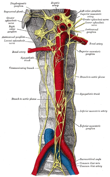
Long description
Abdominal portion of the sympathetic trunk, with the celiac and hypogastric plexuses. (Henle.)

Figure 1: Abdominal portion of the sympathetic trunk, with the celiac and hypogastric plexuses (Henle.).
Figure 2Gray848
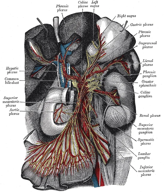
Long description
The celiac ganglia with the sympathetic plexuses of the abdominal viscera radiating from the ganglia. (Toldt.)

Figure 2: The celiac ganglia with the sympathetic plexuses of the abdominal viscera radiating from the ganglia (Toldt.).
The superior mesenteric ganglion lies inferior to it, associated with the superior mesenteric artery.
DefinitionCollateral Ganglia
Collateral ganglia, also called prevertebral ganglia, are situated anterior to the vertebral column and receive inputs from splanchnic nerves as well as central sympathetic neurons.
Clinical pearlReferred Pain
The most broadly accepted theory for this phenomenon is that the visceral sensory fibers enter into the same level of the spinal cord as the somatosensory fibers of the referred pain location.
The sympathetic division of the autonomic nervous system influences the various organ systems of the body through connections emerging from the thoracic and upper lumbar spinal cord. It is referred to as the thoracolumbar system to reflect this anatomical basis. A central neuron in the lateral horn of any of these spinal regions projects to ganglia adjacent to the vertebral column through the ventral spinal roots. The majority of ganglia of the sympathetic system belong to a network of sympathetic chain ganglia that runs alongside the vertebral column. The ganglia appear as a series of clusters of neurons linked by axonal bridges. There are typically 23 ganglia in the chain on either side of the spinal column. Three correspond to the cervical region, 12 are in the thoracic region, four are in the lumbar region, and four correspond to the sacral region. [1]
The prevertebral ganglia, including the coeliac, superior mesenteric, and inferior mesenteric ganglia, are situated anterior to the aorta.
The projections of the sympathetic division of the autonomic nervous system diverge widely, resulting in a broad influence of the system throughout the body. As a response to a threat, the sympathetic system would increase heart rate and breathing rate and cause blood flow to the skeletal muscle to increase and blood flow to the digestive system to decrease. Sweat gland secretion should also increase as part of an integrated response. All of those physiological changes are going to occur together to run away from a threat. This divergence is seen in the branching patterns of preganglionic sympathetic neurons. [1]
Signaling molecules utilized by the autonomic nervous system are released from axons and can be considered as either neurotransmitters (when they directly interact with the effector) or as hormones (when they are released into the bloodstream). The same molecule, such as norepinephrine, could be considered either a neurotransmitter or a hormone on the basis of whether it is released from a postganglionic sympathetic axon or from the adrenal gland. The synapses in the autonomic system are not always the typical type of connection first described in the neuromuscular junction. Instead of having synaptic end bulbs at the very end of an axonal fiber, they may have swellings—called varicosities—along the length of a fiber so that it makes a network of connections within the target tissue. [1]
The postganglionic fibers of the sympathetic and parasympathetic divisions both release neurotransmitters that bind to receptors on their targets. Postganglionic sympathetic fibers release norepinephrine, with a minor exception, whereas postganglionic parasympathetic fibers release ACh. For any given target, the difference in which division of the autonomic nervous system is exerting control is just in what chemical binds to its receptors. The target cells will have adrenergic and muscarinic receptors. If norepinephrine is released, it will bind to the adrenergic receptors present on the target cell, and if ACh is released, it will bind to the muscarinic receptors on the target cell. [2]
DefinitionThoracolumbar Outflow
The sympathetic nervous system originates from the lateral horns of the spinal cord segments T1 to L2.
Clinical pearlVaricosities
Autonomic synapses often feature varicosities along the axon, creating a network of connections rather than discrete end bulbs.
| Pathway | Neurotransmitter | Receptor Type |
|---|---|---|
| Preganglionic (Sympathetic) | ACh | Nicotinic |
| Postganglionic (Sympathetic) | Norepinephrine | Adrenergic |
| Postganglionic (Parasympathetic) | ACh | Muscarinic |
All ganglionic neurons of the autonomic system, in both sympathetic and parasympathetic ganglia, are activated by ACh released from preganglionic fibers. The ACh receptors on these neurons are of the nicotinic type, meaning that they are ligand-gated ion channels. When the neurotransmitter released from the preganglionic fiber binds to the receptor protein, a channel opens to allow positive ions to cross the cell membrane. The result is depolarization of the ganglia. [3] [1]
In the sympathetic system, there are exceptions to this pattern of dual innervation. The postganglionic sympathetic fibers that contact the blood vessels within skeletal muscle and that contact sweat glands do not release norepinephrine, they release ACh. This does not create any problem because there is no parasympathetic input to the sweat glands. Sweat glands have muscarinic receptors and produce and secrete sweat in response to the presence of ACh. [2]
The autonomic innervation of the abdomen is established through a complex network of splanchnic nerves that convey both sympathetic and parasympathetic signals to the visceral organs. To understand the supply, one must distinguish between the motor (efferent) pathways that drive organ function and the sensory (afferent) pathways that convey visceral information to the central nervous system. The efferent branch of a visceral reflex is a two-step pathway starting with the preganglionic fiber emerging from a lateral horn neuron in the spinal cord, or a cranial nucleus neuron in the brain stem, to a ganglion—followed by the postganglionic fiber projecting to a target effector. This contrasts with somatic reflexes, where the output is the lower motor neuron in the ventral horn of the spinal cord that projects directly to a skeletal muscle to cause its contraction. [2]
Figure 3Gray849
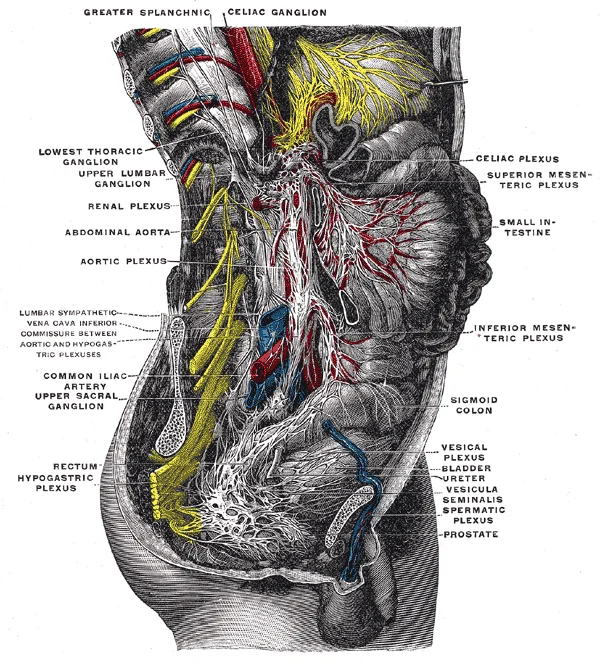
Long description
Lower half of right sympathetic cord. (Testut after Hirschfeld.)

Figure 3: Lower half of right sympathetic cord (Testut after Hirschfeld.).
The table below details the primary motor and sensory branches involved in abdominal autonomic innervation.
| Branch | Type | Origin | Target |
|---|---|---|---|
| Greater splanchnic nerve | Motor (Sympathetic) | Thoracic ganglia T5-T9 | Celiac ganglion |
| Lesser splanchnic nerve | Motor (Sympathetic) | Thoracic ganglia T10-T11 | Aorticorenal ganglion |
| Least splanchnic nerve | Motor (Sympathetic) | Thoracic ganglion T12 | Renal plexus |
| Vagus nerve (Abdominal part) | Motor (Parasympathetic) | Medulla oblongata | Foregut derivatives |
| Pelvic splanchnic nerves | Motor (Parasympathetic) | Sacral spinal cord S2-S4 | Hindgut and pelvic organs |
| Visceral afferents | Sensory | Various organs | Spinal cord / Brain stem |
Sensory neurons receiving input from the periphery—with cell bodies in the sensory ganglia, either of a cranial nerve or a dorsal root ganglion adjacent to the spinal cord—project into the CNS to initiate the reflex. These afferent fibres travel alongside the sympathetic efferent fibres, which is why visceral pain is often referred to somatic regions. The Latin root “effere” means “to carry.” Adding the prefix “ef-” suggests the meaning “to carry away,” whereas adding the prefix “af-” suggests “to carry toward or inward”. [2]
At most of the other targets of the autonomic system, the effector response is based on which neurotransmitter is released and what receptor is present. For the sympathetic supply, postganglionic sympathetic fibers release norepinephrine, which can be considered a neurotransmitter. These tissues have adrenergic receptors that respond to the release of norepinephrine from postganglionic sympathetic fibers by constricting and increasing blood pressure. The parasympathetic system has no significant input to the systemic blood vessels, so the sympathetic system determines their tone. However, for visceral organs such as the gut and heart, parasympathetic input is crucial. If ACh is released onto those cells, it binds to a muscarinic receptor that causes the cells to hyperpolarize so that they cannot reach threshold as easily, and the heart rate slows. [2] [1]
DefinitionVisceral Reflex
A reflex arc involving a two-step efferent pathway from the CNS to a ganglion and then to the effector, distinct from the single-neuron somatic reflex.
Understanding these branches allows for the prediction of symptoms in conditions such as splanchnic nerve block or surgical injury. The sensory component is vital for the perception of visceral pain, which travels via the same pathways as the sympathetic motor fibres.
The sympathetic division originates from the thoracolumbar region of the spinal cord. Preganglionic neurons exit via ventral roots and synapse in sympathetic ganglia. Postganglionic fibres then travel to abdominal viscera.
| Structure | Origin | Course | Termination |
|---|---|---|---|
| Greater Splanchnic Nerve | T5-T9 | Pierces crus of diaphragm | Celiac ganglion |
| Lesser Splanchnic Nerve | T10-T11 | Pierces crus of diaphragm | Aorticorenal ganglion |
| Least Splanchnic Nerve | T12 | Pierces crus of diaphragm | Renal plexus |
These nerves form plexuses around major abdominal arteries. The celiac plexus supplies foregut derivatives. The superior mesenteric plexus supplies midgut derivatives. The inferior mesenteric plexus supplies hindgut derivatives.
Exam trapSympathetic Effect
Sympathetic stimulation generally inhibits gastrointestinal motility and secretion while constricting sphincters, diverting blood flow to skeletal muscles during stress.
The parasympathetic division originates from cranial and sacral regions. The vagus nerve (CN X) provides the majority of parasympathetic innervation to the abdomen.
| Nerve | Origin | Target Region |
|---|---|---|
| Vagus Nerve | Medulla | Foregut and Midgut (up to splenic flexure) |
| Pelvic Splanchnic Nerves | S2-S4 | Hindgut and Pelvic Organs |
The vagus nerve travels through the oesophageal hiatus and forms anterior and posterior vagal trunks. These trunks contribute to the celiac and superior mesenteric plexuses. The pelvic splanchnic nerves arise from S2-S4 and supply the descending colon, rectum, and pelvic viscera.
Clinical pearlVagal Tone
Parasympathetic tone maintains baseline digestive activity and promotes nutrient absorption and waste elimination.
The sacral component picks up with the majority of the large intestine and the pelvic organs of the urinary and reproductive systems. This division promotes digestion and conservation of energy, contrasting with the sympathetic fight-or-flight response[1].
The clinical presentation of autonomic dysfunction in the abdomen is dictated by the specific spinal levels involved, as these determine which visceral organs lose their sympathetic or parasympathetic input. Understanding these deficits requires mapping the anatomical course of the splanchnic nerves to their vertebral origins.
Lesions at specific levels disrupt these pathways, leading to predictable deficits in blood flow, motility, and secretion.
The sympathetic innervation of the abdomen originates from the thoracic spinal cord. Preganglionic sympathetic fibres exit via the ventral roots and enter the sympathetic chain. Some synapse at the same level, while others pass through to collateral ganglia. For instance, the greater splanchnic nerve at the level of T5 synapses with a collateral ganglion outside the chain before making the connection to the postganglionic nerves that innervate the stomach. This pathway is critical for inhibiting gastric motility and constricting blood vessels during stress. [1]
Parasympathetic innervation is provided by the vagus nerve (cranial nerve X) and the pelvic splanchnic nerves (S2-S4). The vagus nerve supplies the foregut and midgut, extending to the splenic flexure of the colon. It promotes digestion, increasing motility and secretion. The pelvic splanchnic nerves supply the hindgut and pelvic organs, facilitating defecation and urination.
Lesions affecting the sympathetic chain or splanchnic nerves result in loss of inhibitory control. This can lead to increased motility, vasodilation, and secretion in the affected regions. Conversely, lesions affecting the parasympathetic supply result in reduced motility and secretion, potentially leading to constipation or urinary retention.
The enteric nervous system operates independently within the gut wall. It contains myenteric plexuses that directly influence digestive function. If stretch receptors in the stomach are activated by the filling and distension of the stomach, a short reflex will directly activate the smooth muscle fibers of the stomach wall to increase motility to digest the excessive food in the stomach. This local control is modulated by the autonomic nervous system but can function without central nervous system involvement. [2]
Visceral sensory fibres travel alongside autonomic fibres. This convergence explains referred pain, where visceral pathology is perceived as somatic pain in a distant dermatome. [2]
| Structure | Origin | Pathway | Target |
|---|---|---|---|
| Greater Splanchnic Nerve | T5-T9 | Collateral ganglion (Celiac) | Stomach, Liver, Spleen |
| Lesser Splanchnic Nerve | T10-T11 | Collateral ganglion (Superior Mesenteric) | Small Intestine, Ascending Colon |
| Vagus Nerve | Medulla | Direct to organ wall | Foregut, Midgut (to splenic flexure) |
| Pelvic Splanchnic Nerves | S2-S4 | Direct to organ wall | Hindgut, Pelvic Organs |
Deficits vary by level. High thoracic lesions may affect upper abdominal organs, while lower lesions impact the intestines and pelvic organs. Recognition of these patterns is essential for localising neurological lesions.
The autonomic nervous system regulates visceral function through two primary divisions: the sympathetic and parasympathetic systems. Understanding their anatomical pathways is essential for localising lesions. The sympathetic output originates from the lateral horn of the thoracolumbar spinal cord. Preganglionic neurons send axons via the ventral spinal nerve root to sympathetic ganglia. These ganglia are part of the sympathetic chain, also known as paravertebral ganglia. Synapses occur at chain ganglia superior or inferior to the central neuron’s level. Here, it synapses with a postganglionic neuron. The postganglionic fiber then projects to the target effector. It releases norepinephrine to bind adrenergic receptors. This causes physiological changes consistent with the sympathetic response. [2] [1]
Parasympathetic innervation of the abdomen relies heavily on the vagus nerve. Neurons in the dorsal nucleus of the vagus and the nucleus ambiguus project through cranial nerve X. These fibers reach terminal ganglia in the thoracic and abdominal cavities. In the abdomen, they influence the stomach, liver, pancreas, gall bladder, and small intestine. Postganglionic fibers are often incorporated into the organ structure. They form the mesenteric plexus of the digestive tract. Intramural ganglia are also common sites for these synapses. [1]
Clinical examination often assesses reflex arcs involving autonomic components. The baroreceptor apparatus is part of a unipolar neuron ending. Its cell body resides in a sensory ganglion. Baroreceptors from the carotid arteries have axons in the glossopharyngeal nerve. Those from the aorta have axons in the vagus nerve. Disruption of these pathways can alter blood pressure regulation. [2]
Another example involves pupillary size control, illustrating sympathetic pathways. If light levels are low, the sympathetic system activates. A signal travels from the upper thoracic spinal cord. It reaches the superior cervical ganglion of the sympathetic chain. The postganglionic fiber projects to the iris. It releases norepinephrine onto radial fibers of the iris. These are smooth muscles that contract to dilate the pupil. This increases light hitting the retina. Conversely, high light levels trigger parasympathetic signals. Signals originate from the Edinger–Westphal nucleus. They travel via the oculomotor nerve. Synapses occur in the ciliary ganglion in the posterior orbit. [2] [1]
DefinitionSympathetic Chain
A series of ganglia located alongside the vertebral column, also called paravertebral ganglia, where preganglionic fibers may synapse.
DefinitionParasympathetic Terminal Ganglia
Ganglia located near or within the target organs, such as the mesenteric plexus, where vagal preganglionic fibers synapse.
| Division | Origin | Ganglion Type | Target Organs |
|---|---|---|---|
| Sympathetic | Lateral horn (T1-L2) | Paravertebral or Collateral | Stomach, Liver, Pancreas, Intestines |
| Parasympathetic | Dorsal Vagus Nucleus | Terminal (Intramural) | Stomach, Liver, Pancreas, Gall Bladder, Small Intestine |
Examination of abdominal autonomic function requires understanding these distinct routes. Sympathetic lesions may affect blood flow and motility via norepinephrine release. Parasympathetic lesions disrupt digestive secretions and motility via the vagus nerve. Recognising the origin and course of these nerves aids in diagnosing visceral dysfunction.
The adrenal medulla functions as a modified sympathetic ganglion. Preganglionic sympathetic fibres project directly to chromaffin cells within the adrenal medulla, bypassing traditional ganglia. These chromaffin cells are neurosecretory cells derived from the neural crest, releasing catecholamines directly into the bloodstream rather than via axonal terminals. [1]
Cholinergic receptors are divided into two distinct classes: nicotinic and muscarinic. Both bind acetylcholine but differ in structure and mechanism. The nicotinic receptor is a ligand-gated cation channel, whereas the muscarinic receptor is a G protein–coupled receptor. [1]
Adrenergic receptors are exclusively G protein–coupled receptors, unlike nicotinic cholinergic receptors. They are classified into alpha (α) and beta (β) types. There are two α-adrenergic subtypes (α1 and α2) and three β-adrenergic subtypes (β1, β2, and β3). [1]
Epinephrine and adrenaline are synonymous terms for the same molecule. The chemical distinction between norepinephrine and epinephrine is the presence of a methyl group in epinephrine. In British scientific usage, 'adrenaline' is preferred, while 'epinephrine' is standard in the United States due to historical proprietary naming conventions. Similarly, norepinephrine and noradrenaline refer to the identical molecule. [1]
Phenylephrine is a selective α1-adrenergic agonist used as a sympathomimetic. It acts as a decongestant by causing vasoconstriction and can dilate the pupil. It is frequently combined with analgesics in over-the-counter sinus medications. [1]
β-blockers are sympatholytic drugs that treat cardiovascular disease by blocking β-receptors. They prevent vasoconstriction and cardioacceleration, thereby improving cardiac function in conditions such as congestive heart failure or post-myocardial infarction. Metoprolol is a selective β1-blocker, whereas propranolol is a non-selective β-blocker. [3]
Pilocarpine is a non-specific muscarinic agonist used in ophthalmology. It reverses mydriasis induced by agents like phenylephrine.
Atropine and scopolamine are muscarinic antagonists derived from Atropa belladonna (deadly nightshade). These compounds block muscarinic receptors in the iris, resulting in pupil dilation (mydriasis). Historically, this effect was exploited cosmetically to enlarge the appearance of the eyes. [3]
Clinical pearlAdrenal Medulla
The adrenal medulla is functionally a sympathetic ganglion where preganglionic fibres release ACh directly onto chromaffin cells.
DefinitionAdrenergic Receptors
All adrenergic receptors are G protein–coupled, subdivided into α1, α2, β1, β2, and β3 types.
NoteNomenclature
'Adrenaline' is the British term; 'epinephrine' is the US term for the same molecule.
Exam trapPhenylephrine Action
Phenylephrine is an α1 agonist, not a β agonist; it causes vasoconstriction, not bronchodilation via β2.
Clinical pearlβ-Blockers
Propranolol is non-selective; metoprolol is selective for β1 receptors in the heart. [3]
DefinitionPilocarpine
Pilocarpine is a muscarinic agonist that reduces intraocular pressure by increasing aqueous outflow. [3]
Exam trapBelladonna Effect
Anticholinergic drugs block muscarinic receptors, suppressing parasympathetic interaction with the organ. [3]
Students frequently confuse the anatomical and functional distinctions between the sympathetic and parasympathetic divisions, leading to errors in clinical reasoning. A primary error is assuming all postganglionic sympathetic fibres release noradrenaline. While this is true for most effectors, the postganglionic connections to sweat glands in the skin and blood vessels supplying skeletal muscle are exceptions; those fibres release ACh onto muscarinic receptors. Another common mistake is misidentifying the length of autonomic pathways. Students often assume preganglionic sympathetic fibres are long because they originate in the brain or spinal cord. In reality, because the sympathetic ganglia are adjacent to the vertebral column, preganglionic sympathetic fibers are relatively short, and they are myelinated. Conversely, postganglionic sympathetic fibers are long because of the relatively greater distance from the ganglion to the target effector. [1]
A further pitfall involves the terminology of neuronal structures. Students may incorrectly refer to the entire cell body and axon as a "postganglionic neuron." The problem with that usage is that the cell body is in the ganglion, and only the fiber is postganglionic. Typically, the term neuron applies to the entire cell. It is crucial to distinguish that an axon from the central neuron that projects to a sympathetic ganglion is referred to as a preganglionic fiber or neuron. [1]
Exam trapSweat Glands
Sympathetic innervation of sweat glands is cholinergic, not adrenergic. The postganglionic connections to sweat glands in the skin and blood vessels supplying skeletal muscle are, however, exceptions; those fibers release ACh onto muscarinic receptors.
Students also struggle with the concept of divergence in the sympathetic system. They may expect a one-to-one mapping of nerves to organs. To coordinate all these responses, the connections in the sympathetic system diverge from a limited region of the central nervous system (CNS) to a wide array of ganglia that project to the many effector organs simultaneously. This divergent architecture allows the sympathetic system to cause divergent effects as many different effector organs are activated together for a common purpose. [1]
DefinitionPreganglionic vs Postganglionic
Preganglionic fibers are short, myelinated, and originate from the CNS. Postganglionic fibers are long, unmyelinated, and originate from the ganglion.
Finally, students often overlook the hormonal component of sympathetic activation. They may think sympathetic effects are only local. The sympathetic system has a specialized preganglionic connection to the adrenal medulla that causes epinephrine and norepinephrine to be released into the bloodstream rather than exciting a neuron that contacts an organ directly. This hormonal component means that the sympathetic chemical signal can spread throughout the body very quickly and affect many organ systems at once.
Knowledge of autonomic innervation is applied at the bedside to interpret physiological responses to stress. To respond to a threat—to fight or to run away—the sympathetic system causes divergent effects as many different effector organs are activated together for a common purpose. Clinicians observe that more oxygen needs to be inhaled and delivered to skeletal muscle. The respiratory, cardiovascular, and musculoskeletal systems are all activated together. Additionally, sweating keeps the excess heat that comes from muscle contraction from causing the body to overheat. Understanding this helps explain why a patient in shock may have cool, clammy skin despite being hyperdynamic; the digestive system shuts down so that blood is not absorbing nutrients when it should be delivering oxygen to skeletal muscles. [1]
In the operating theatre, anaesthetists manage the complex set of structures that compose the output of the sympathetic system make it possible for these disparate effectors to come together in a coordinated, systemic change. They must anticipate the effects of drugs that enhance adrenergic function. A drug that enhances adrenergic function is known as a sympathomimetic drug[1]. Conversely, they may administer drugs that interrupt adrenergic function, known as sympatholytic drugs[1]. The neurochemistry of the sympathetic system is based on the adrenergic system[1]. Norepinephrine and epinephrine influence target effectors by binding to the α-adrenergic or β-adrenergic receptors[1]. [3]
On imaging, radiologists recognise that the sympathetic chain lies adjacent to the vertebral column. This anatomical relationship is critical when interpreting masses or trauma. The preganglionic sympathetic fibers are relatively short because the ganglia are adjacent to the vertebral column. This proximity means that pathology in the thoracic or lumbar spine can directly affect sympathetic outflow.
In examinations, students must distinguish visceral reflexes from somatic ones. The afferent branch of a reflex arc does differ between somatic and visceral reflexes in some instances[1]. Many of the inputs to visceral reflexes are from special or somatic senses, but particular senses are associated with the viscera that are not part of the conscious perception of the environment through the somatic nervous system[1]. For example, there is a specific type of mechanoreceptor, called a baroreceptor, in the walls of the aorta and carotid sinuses that senses the stretch of those organs when blood volume or pressure increases[1]. You do not have a conscious perception of having high blood pressure, but that is an important afferent branch of the cardiovascular and, particularly, vasomotor reflexes[1]. [2]
Sympathetic Response Pathway
- Threat DetectedFight or flight initiation
- Question: Local or Systemic?
Local
- Preganglionic Fiber (Short, Myelinated)Projects to sympathetic ganglion
- Postganglionic Fiber (Long, Unmyelinated)Releases NE or ACh to effector
Systemic
- Preganglionic Fiber to Adrenal MedullaSpecialized connection
- Hormone ReleaseEpinephrine/Norepinephrine into bloodstream
The efferent branch of the visceral reflex arc begins with the projection from the central neuron along the preganglionic fiber[1]. This fiber then makes a synapse on the ganglionic neuron that projects to the target effector[1]. This pathway underpins the autonomic control of abdominal organs, ensuring that the body can rapidly adjust blood flow and metabolic activity in response to internal and external stimuli. [2]
Sympathetic preganglionic fibers are short and myelinated. Sympathetic postganglionic fibers are long and unmyelinated. Sweat glands receive cholinergic sympathetic innervation. The adrenal medulla releases hormones into the bloodstream. Baroreceptors in the aorta and carotid sinuses are visceral mechanoreceptors. Visceral reflexes often operate without conscious perception. Sympathomimetics enhance adrenergic function. Sympatholytics interrupt adrenergic function.
Answer each one in your head first, then open the answer.
What neurotransmitter do postganglionic sympathetic fibers to sweat glands release?
Show answerHide answer
AChAre preganglionic sympathetic fibers myelinated or unmyelinated?
Show answerHide answer
MyelinatedWhat type of receptors do norepinephrine and epinephrine bind to in the sympathetic system?
Show answerHide answer
α-adrenergic or β-adrenergic receptorsWhere are sympathetic ganglia located relative to the vertebral column?
Show answerHide answer
Adjacent to the vertebral columnWhat is the term for a drug that interrupts adrenergic function?
Show answerHide answer
Sympatholytic drug
- Betts JG, Young KA, Wise JA, et al. Divisions of the Autonomic Nervous System. OpenStax, Anatomy and Physiology 2e (CC BY-NC-SA 4.0). Accessed .
- Betts JG, Young KA, Wise JA, et al. Autonomic Reflexes and Homeostasis. OpenStax, Anatomy and Physiology 2e (CC BY-NC-SA 4.0). Accessed .
- Betts JG, Young KA, Wise JA, et al. Drugs that Affect the Autonomic System. OpenStax, Anatomy and Physiology 2e (CC BY-NC-SA 4.0). Accessed .
Figure credits
Educational content for medical students and doctors. It is not a substitute for clinical judgement or local guidelines.