Abdominal lymphatics
Anatomical pathways of abdominal lymphatic drainage and autonomic innervation.
The lymphatic system serves as a critical return route for interstitial fluid and immune cells. Lymphatic capillaries are blind-ended vessels that collect fluid, which then travels through progressively larger vessels. These vessels pass through lymph nodes, which act as filters for pathogens and debris. Humans possess approximately 500–600 lymph nodes distributed throughout the body. [1]
DefinitionLymph Node Function
Lymph nodes remove debris and pathogens from the lymph, functioning as the “filters of the lymph”.
Lymph enters a node via afferent vessels and flows through the subcapsular sinus. Within the cortex, lymphoid follicles contain germinal centres of rapidly dividing B cells surrounded by T cells. The lymph then moves to the medulla, where it collects in medullary sinuses before exiting via efferent lymphatic vessels.
It receives lymph from the intestinal trunk and two lumbar trunks. The intestinal trunk drains the abdominal viscera, while the lumbar trunks drain the lower limbs and pelvic organs. The para-aortic nodes, also known as lateral aortic nodes, lie alongside the abdominal aorta. They receive lymph from the kidneys, adrenal glands, and gonads. These nodes drain into the lumbar trunks, which then feed into the cisterna chyli.
Figure 1Gray612
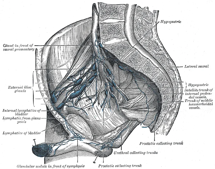
Long description
Iliopelvic glands (lateral view). (Cunéo and Marcille.)

Figure 1: Iliopelvic glands (lateral view) (Cunéo and Marcille.).
Figure 2Gray611
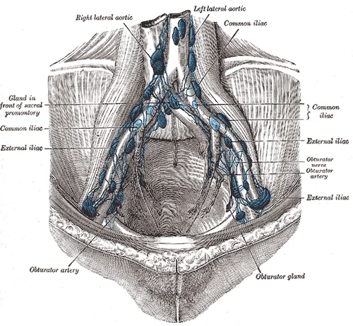
Long description
The parietal lymph glands of the pelvis. (Cunéo and Marcille.)

Figure 2: The parietal lymph glands of the pelvis (Cunéo and Marcille.).
The foregut, midgut, and hindgut receive sympathetic and parasympathetic innervation via specific plexuses. The coeliac plexus supplies the foregut. It is located around the coeliac trunk and receives greater splanchnic nerves from the T5–T9 spinal segments. The superior mesenteric plexus supplies the midgut. It surrounds the superior mesenteric artery and receives lesser splanchnic nerves from T10–T11.
NoteSplanchnic Nerves
Splanchnic nerves carry preganglionic sympathetic fibres and visceral afferent fibres to the prevertebral plexuses.
Visceral pain arises from the distension or ischaemia of hollow organs. Because visceral afferents enter the spinal cord at specific levels, pain is often referred to the corresponding dermatomes. Foregut pain is referred to the epigastric region (T5–T9). Midgut pain is referred to the periumbilical region (T10–T11). Hindgut pain is referred to the suprapubic or inguinal regions (T12–L1).
Lymphatic drainage follows arterial supply. The stomach and spleen drain to the coeliac nodes. The small intestine and ascending colon drain to the superior mesenteric nodes. The descending colon and rectum drain to the inferior mesenteric nodes.
Figure 3Gray617
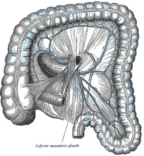
Long description
Lymphatics of colon. (Jamieson and Dobson.)

Figure 3: Lymphatics of colon (Jamieson and Dobson.).
Figure 4Gray616
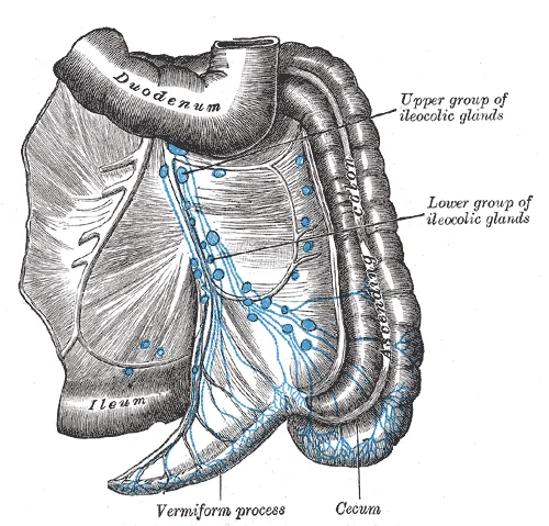
Long description
The lymphatics of cecum and vermiform process from behind. (Jamieson and Dobson.)

Figure 4: The lymphatics of cecum and vermiform process from behind (Jamieson and Dobson.).
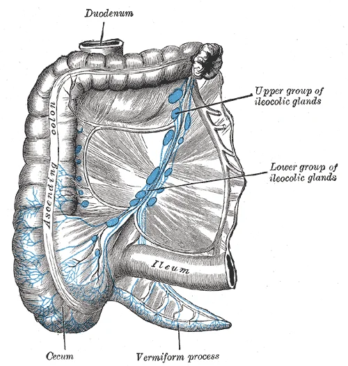
Figure 5Gray615
Long description
The lymphatics of cecum and vermiform process from the front. (Jamieson and Dobson.)

Figure 5: The lymphatics of cecum and vermiform process from the front (Jamieson and Dobson.).
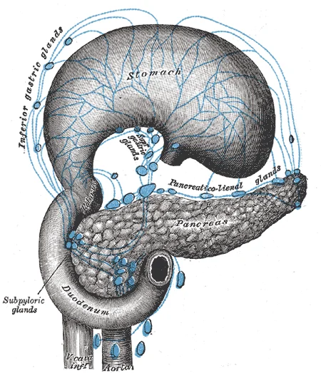
Figure 6Gray614
Long description
Lymphatics of stomach, etc. The stomach has been turned upward. (Jamieson and Dobson.)

Figure 6: Lymphatics of stomach, etc.
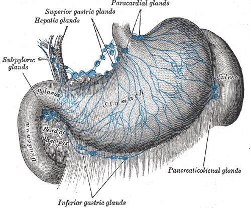
Figure 7Gray613
Long description
Lymphatics of stomach, etc. (Jamieson and Dobson.)

Figure 7: Lymphatics of stomach, etc (Jamieson and Dobson.).
The enteric nervous system consists of the myenteric (Auerbach’s) and submucosal (Meissner’s) plexuses. The myenteric plexus lies between the longitudinal and circular muscle layers and controls motility. The submucosal plexus lies in the submucosa and regulates secretion and blood flow. These plexuses operate independently but are modulated by the autonomic nervous system.
The abdominal lymphatic system originates from extensive networks of blind-ended capillaries within the submucosa and mesenteries of the gastrointestinal tract. These initial vessels converge to form larger collecting trunks that follow the arterial supply of the abdominal viscera. The primary route for lymph from the intestines is through the mesentery, where vessels accompany the superior mesenteric artery and its branches. These vessels transport chyle, a milky fluid rich in triglycerides absorbed from the diet, towards the central lymphatic ducts. [2]
From this origin, the thoracic duct ascends through the aortic hiatus of the diaphragm. Along its course, it receives lymph from the lumbar trunks, which drain the lower limbs and pelvic organs, and the intestinal trunk, which drains the stomach, intestines, liver, spleen, and pancreas. The duct terminates by emptying into the junction of the left internal jugular and left subclavian veins. This termination point ensures that lymph returns to the systemic circulation. The surrounding structures, including the aorta and inferior vena cava, provide important anatomical landmarks for identifying the path of the duct. [1]
The termination of the major abdominal lymphatic trunks is critical for understanding fluid balance and immune surveillance. The right lymphatic duct, which drains the right upper quadrant, terminates at the junction of the right internal jugular and right subclavian veins. In contrast, the thoracic duct, receiving the majority of abdominal lymph, terminates on the left side. This asymmetry is a key anatomical feature. The lymph nodes along these trunks, such as the celiac, superior mesenteric, and inferior mesenteric nodes, filter the lymph before it enters the main ducts. These nodes are arranged in chains parallel to the major arteries.
DefinitionCisterna Chyli
A dilated sac at the lower end of the thoracic duct, located anterior to the L1-L2 vertebrae, receiving lymph from the lumbar and intestinal trunks.
The course of these vessels is closely tied to the vascular supply. Lymphatics from the hindgut follow the inferior mesenteric artery, while those from the midgut follow the superior mesenteric artery. This relationship allows for predictable drainage patterns that are essential for surgical planning and understanding the spread of disease. The termination into the venous system at the venous angles completes the circuit, allowing for the return of proteins and fluids to the blood.
| Trunk | Origin | Termination |
|---|---|---|
| Thoracic Duct | Cisterna Chyli | Left venous angle |
| Right Lymphatic Duct | Right upper quadrant | Right venous angle |
| Lumbar Trunks | Lower limbs/pelvis | Cisterna Chyli |
| Intestinal Trunk | GI tract | Cisterna Chyli |
Understanding these pathways is fundamental for medical students, as they form the basis for diagnosing conditions such as lymphoedema and metastatic spread. The clear distinction between the right and left termination points prevents confusion in clinical practice. The anatomical relations to the aorta and vertebral column provide stable reference points for imaging and surgery.
The abdominal lymphatic system functions as a hierarchical drainage network, collecting interstitial fluid from visceral organs and the abdominal wall before conveying it to the central venous circulation. Understanding its branches requires tracing the flow from microscopic capillaries to major collecting trunks.
DefinitionLymphatic Capillaries
Lymphatic capillaries, also called the terminal lymphatics, are vessels where interstitial fluid enters the lymphatic system to become lymph fluid. [1]
These initial vessels are blind-ended tubes located in almost every tissue in the body, interlaced among the arterioles and venules of the circulatory system within soft connective tissues. Notably, exceptions to this widespread distribution include the central nervous system, bone marrow, bones, teeth, and the cornea of the eye, which do not contain lymph vessels. In the abdomen, these capillaries form dense plexuses around the organs they serve. [1]
| Structure | Origin | Course | Termination |
|---|---|---|---|
| Intestinal Trunk | Mesenteric nodes | Ascends anterior to the aorta | Cisterna chyli |
| Lumbar Trunks | Para-aortic nodes | Ascends lateral to the aorta | Cisterna chyli |
| Cisterna Chyli | Convergence of intestinal and lumbar trunks | Located at L1-L2 level | Thoracic duct |
The convergence of these tributaries forms the cisterna chyli, a dilated sac located typically at the level of the first or second lumbar vertebra. It receives lymph from the intestinal trunk and the paired lumbar trunks. The intestinal trunk drains the digestive organs, while the lumbar trunks drain the lower limbs, pelvic organs, and kidneys.
The thoracic duct ascends from the cisterna chyli, passing through the aortic hiatus of the diaphragm. The relationship is hierarchical: capillaries merge into pre-collecting vessels, which drain into regional nodes, and efferents from these nodes form the major trunks.
NoteClinical Relevance
Obstruction of the lumbar trunks can lead to chylous ascites due to the backflow of lymph into the peritoneal cavity.
It terminates by draining into the junction of the left internal jugular and left subclavian veins. This anatomical arrangement ensures that lymph from the abdominal viscera eventually returns to the systemic circulation, maintaining fluid balance and facilitating immune surveillance.
The abdominal lymphatic system is defined by its role in collecting fluid from the viscera and abdominal wall, ultimately merging into larger trunks that feed into the central venous system. The superficial and deep lymphatics eventually merge to form larger lymphatic vessels known as lymphatic trunks. These trunks serve as the primary conduits for lymph derived from the abdominal organs, the lower limbs, and the pelvis. [1]
The overall drainage system of the body is asymmetrical. This asymmetry is critical when considering the abdominal territory. On the right side of the body, the right sides of the head, thorax, and right upper limb drain lymph fluid into the right subclavian vein via the right lymphatic duct. However, the abdomen does not drain into this right-sided duct. Instead, the lymph from the rest of the body enters the bloodstream through the thoracic duct via all the remaining lymphatic trunks. [1]
The cisterna chyli is therefore the foundational collecting reservoir for the entire abdominal lymphatic output. It sits anterior to the vertebral column, typically at the level of the first or second lumbar vertebra, receiving input from the paired lumbar trunks which drain the lower limbs and pelvic walls, and the single intestinal trunk which drains the gastrointestinal viscera. [1]
In general, lymphatic vessels of the subcutaneous tissues of the skin, that is, the superficial lymphatics, follow the same routes as veins. Conversely, the deep lymphatic vessels of the viscera generally follow the paths of arteries. This anatomical rule applies strictly to the abdominal viscera. For instance, the lymphatics draining the stomach and intestines accompany the branches of the celiac trunk and the superior and inferior mesenteric arteries. As these arteries branch and anastomose, their accompanying lymphatic vessels do the same, forming plexuses around the vessels before converging into larger trunks that eventually empty into the intestinal trunk or directly into the cisterna chyli. [1]
From this sac, the thoracic duct ascends. The viewer should note the relationship between the aorta and the thoracic duct, with the duct lying to the right of the aorta as it ascends, crossing to the left as it passes the diaphragm.
DefinitionCisterna Chyli
A dilated sac at the lower end of the thoracic duct that receives lymph from the lumbar and intestinal trunks.
The intestinal trunk is a major collector for the abdominal viscera. It receives lymph from the stomach, intestines, pancreas, spleen, and liver. The lymph from these organs travels along the arterial supply. For example, lymph from the small intestine follows the superior mesenteric artery. The vessels form a complex network before coalescing into the intestinal trunk. This trunk then empties into the cisterna chyli. The lumbar trunks, meanwhile, collect lymph from the lower limbs and pelvic organs. They ascend along the psoas major muscles and enter the cisterna chyli from either side.
| Trunk | Primary Drainage Territory | Termination |
|---|---|---|
| Intestinal Trunk | Gastrointestinal viscera | Cisterna Chyli |
| Left Lumbar Trunk | Lower limbs, pelvic walls | Cisterna Chyli |
| Right Lumbar Trunk | Lower limbs, pelvic walls | Cisterna Chyli |
This arrangement ensures that all lymph from the abdomen, except for the small portion that might drain via right-sided collateral pathways (which are minor), is directed into the thoracic duct. This single large vessel handles the vast majority of the body's lymphatic return, including all of the abdominal content.
The lymphatic system functions as a series of vessels, ducts, and trunks that remove interstitial fluid from the tissues and return it to the blood. This system is also utilized to transport dietary lipids and cells of the immune system. Cells of the immune system all come from the hematopoietic system of the bone marrow. Primary lymphoid organs, the bone marrow and thymus gland, are the locations where lymphocytes of the adaptive immune system proliferate and mature. Secondary lymphoid organs are sites in which mature lymphocytes congregate to mount immune responses. Many immune system cells use the lymphatic and circulatory systems for transport throughout the body to search for and then protect against pathogens. [1]
DefinitionLymphatic Drainage
The lymphatic system removes interstitial fluid and returns it to the blood, while also transporting dietary lipids and immune cells.
Abdominal lymphatic collateral pathways form extensive anastomotic networks that ensure drainage continuity when primary routes are obstructed. The retroaortic (lateral aortic) lymph nodes, positioned on the third and fourth lumbar vertebral bodies below the cisterna chyli, serve as a major convergence zone receiving afferents from both the lateral aortic and preaortic groups before draining into the cisterna chyli. The duodenum demonstrates a critical bidirectional collateral arrangement: pancreaticoduodenal lymph nodes on the pancreaticoduodenal groove send efferents superiorly to the hepatic nodes at the porta hepatis and inferiorly to the superior mesenteric (preaortic) nodes at the origin of the superior mesenteric artery. Hepatic superficial lymphatics from the convex surface distribute through three distinct collateral routes: a middle group traverses the vena caval foramen to nodes around the terminal inferior vena cava; left-sided vessels course posteriorly through the esophageal hiatus to paracardial gastric nodes; and right-sided vessels cross the right diaphragmatic crus to reach phrenic nodes. The suprarenal gland drains via multiple parallel channels — following the renal vein to lateral aortic nodes, joining renal efferents, descending to common iliac nodes, or connecting with vesical and hypogastric pathways. Superficial abdominal wall lymphatics converge on the superficial inguinal nodes via the superficial epigastric and superficial circumflex iliac vessels, while deep parietal vessels accompany the deep epigastric and lumbar arteries to reach deep inguinal and lateral aortic nodes, creating overlapping superficial and deep drainage territories.
NoteCollateral Pathways
While cranial nerve lymphatics are debated, the abdominal system relies on extensive anastomoses to ensure fluid return, though specific abdominal collateral criteria are not listed in the provided texts.
The intestinal trunk is located anterior to the vertebral column, receiving lymph from the digestive organs. The lumbar trunks lie on either side of the vertebral column, draining the lower limbs and pelvic organs. These structures anastomose to form the cisterna chyli, which then continues as the thoracic duct.
These investigators had succeeded in establishing a method through which the cranial and spinal subarachnoid spaces were separated from each other in living dogs using rings of muscle tissue wrapped around the spinal cord. It was only possible to fill the lymph nodes along the abdominal wall when introducing the ink into the caudal region of the isolated spinal space. Ink introduced in small volumes in the intact SAS either into the cisterna magna or at the caudal spine did not demonstrate significant outflow from the spine. Two rather illuminating studies on spinal outflow were performed in this lab by Galkin. [2]
Importantly, this procedure led to significantly higher spikes in ICP and increased CSF outflow resistance in response to fluid infusions into the ventricles and, remarkably, even increased ICP during homeostatic conditions when a ligation of the SAS of the spine was performed to block flow into this compartment. [2]
Clinical utility was short-lived, however, as it was soon concluded from studies in cats, dogs and monkeys that intracranial administration of this contrast agent was harmful. [2]
The lymphatic drainage of the abdomen exhibits significant anatomical variability, which is crucial for understanding the spread of malignancy and the distribution of infectious agents. While standard textbooks describe a predictable pattern, individual differences frequently alter the expected pathways. These variations are not merely academic curiosities; they directly impact surgical planning and the interpretation of imaging studies.
The lumbar trunks are formed by the convergence of lymphatic vessels from the lower limbs, pelvic walls, and abdominal walls. They typically drain into the cisterna chyli, but their termination points can vary. The left lumbar trunk often joins the thoracic duct directly, bypassing the cisterna chyli. The right lumbar trunk may also drain directly into the thoracic duct or into the right lymphatic duct. In some individuals, multiple small lumbar trunks drain into the cisterna chyli rather than a single large trunk. These variations affect the volume of lymph entering the venous system and can influence the spread of abdominal cancers. For instance, tumours of the testis or ovary may metastasise to para-aortic nodes via these variable pathways. Surgeons must be aware that ligation of a single lumbar trunk may not prevent lymph leakage if collateral channels exist.
It usually arises from the celiac, superior mesenteric, and inferior mesenteric lymph nodes. The trunk typically enters the cisterna chyli, but it may drain directly into the thoracic duct. In some cases, the intestinal trunk is absent, with its tributaries draining separately into the cisterna chyli or the thoracic duct. The size and number of intestinal trunks can vary, with some individuals having multiple small trunks. These variations are important in the context of lymph node dissection during cancer surgery. Incomplete removal of lymphatic tissue due to unexpected branching patterns can lead to local recurrence. Additionally, the intestinal trunk plays a key role in the absorption of dietary fats, and its obstruction can result in fat malabsorption.
However, this crossing can occur at different levels, ranging from the fourth to the seventh thoracic vertebra. The duct may also have multiple origins or terminate in unusual veins, such as the left subclavian vein or the junction of the left internal jugular and subclavian veins. In some cases, the thoracic duct is duplicated or forms a complex network of anastomoses. These variations are critical during thoracic surgery to avoid injury, which can cause chylothorax. Radiologists must also consider these anatomical differences when interpreting lymphoscintigraphy or MR lymphangiography.
The clinical anatomy of abdominal lymphatics is defined by the drainage patterns of the gastrointestinal tract and retroperitoneal organs, which converge into specific nodal groups before entering the thoracic duct. Understanding these pathways is critical for staging malignancies and identifying sources of infection.
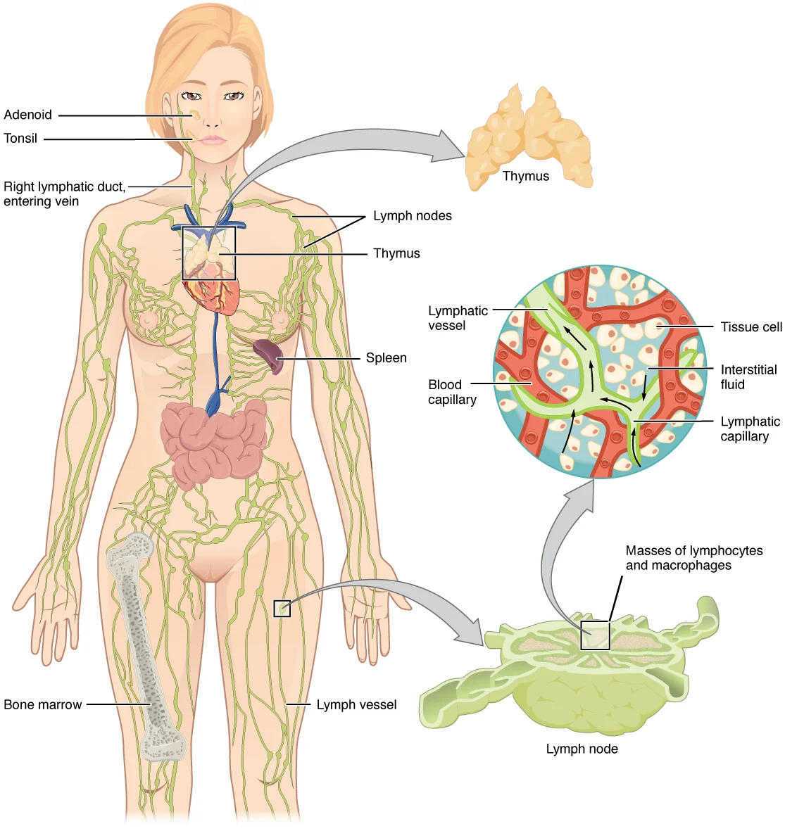
Figure 82201 Anatomy of the Lymphatic System
Long description
Lymphatic vessels in the arms and legs convey lymph to the larger lymphatic vessels in the torso.

Figure 8: Lymphatic vessels in the arms and legs convey lymph to the larger lymphatic vessels in the torso.
Lateral to these are the iliac nodes, which receive drainage from the lower limbs and pelvic viscera. The superior mesenteric nodes are located around the superior mesenteric artery, draining the midgut. The inferior mesenteric nodes are found near the inferior mesenteric artery, draining the hindgut.
The primary access points for surgical assessment or biopsy are the para-aortic and iliac regions. The para-aortic nodes are accessible via a midline laparotomy or retroperitoneal approach. They lie between the aorta and the inferior vena cava. The iliac nodes are accessed via the inguinal or pelvic approaches. The external iliac nodes lie along the external iliac vein. The internal iliac nodes surround the internal iliac artery.
Ligation of lymphatic vessels is rarely performed as a primary treatment due to the risk of lymphoedema. However, in cases of traumatic injury or severe chylous ascites, ligation may be necessary. The thoracic duct can be ligated above the diaphragm if a leak is identified. This is typically done via a left thoracotomy. The cisterna chyli may be ligated in rare cases of congenital malformation.
Aneurysms of the abdominal aorta can compress adjacent lymphatic structures. The abdominal aorta lies anterior to the vertebral column. The inferior vena cava lies to its right. The lymphatic vessels run alongside these major vessels. Compression can lead to impaired drainage and subsequent oedema.
Pulse points are not directly associated with lymphatic vessels. However, the femoral pulse is a landmark for accessing the external iliac lymph nodes. The abdominal aortic pulse can be palpated in the epigastrium, indicating the location of the para-aortic nodes.
The following table summarises the key nodal groups and their drainage territories.
| Node Group | Location | Primary Drainage Territory |
|---|---|---|
| Celiac | Around celiac trunk | Foregut (stomach, liver, spleen, pancreas) |
| Superior Mesenteric | Around superior mesenteric artery | Midgut (small intestine, ascending colon) |
| Inferior Mesenteric | Around inferior mesenteric artery | Hindgut (descending colon, rectum) |
| Para-aortic | Anterior to aorta and IVC | Gonads, kidneys, uterus |
| Iliac | Along iliac vessels | Lower limbs, pelvic viscera |
Lesions in these nodes often present as palpable masses. Virchow's node, a left supraclavicular node, may indicate abdominal malignancy via the thoracic duct. This is known as Troisier's sign. The presence of such nodes suggests advanced disease.
Clinical pearlTroisier's Sign
Palpable left supraclavicular lymphadenopathy (Virchow's node) often indicates intra-abdominal malignancy, particularly gastric cancer, due to drainage via the thoracic duct.
Exam trapLymphatic Congruence
Do not assume lymphatic drainage follows arterial supply exactly. Venous drainage patterns are more consistent. Lymphatics may cross compartmental boundaries.
The lymphatic system functions as a unidirectional drainage network, returning excess interstitial fluid to the bloodstream while filtering pathogens. It is structurally distinct from the venous system despite similarities in vessel wall composition.
DefinitionLymphatic Capillaries
The initial blind-ended vessels that collect interstitial fluid, which then drain into larger collecting vessels.
Lymphatic capillaries empty into larger lymphatic vessels. These larger vessels possess a three-tunic structure similar to veins. They contain one-way valves located fairly close to one another. Each valve causes a bulge in the vessel wall. This creates a characteristic beaded appearance of the lymphatic vessels. [1]
Exam trapValve Density
Do not confuse lymphatic valves with venous valves; lymphatic valves are more closely spaced, creating the 'beaded' look.
Lymphocytes are the primary cellular components of this system. They develop and mature in primary lymphoid organs. They mount immune responses from secondary lymphoid organs. A naïve lymphocyte has left the primary organ but has not yet encountered an antigen. These cells concentrate in secondary lymphoid organs. These organs include the lymph nodes, spleen, and lymphoid nodules. [1]
Clinical pearlNaïve Lymphocytes
Naïve lymphocytes are fully functional immunologically but have yet to encounter an antigen to respond to. [1]
Natural killer cells are a fourth important lymphocyte type. They participate in the innate immune response. They are circulating blood cells with cytotoxic granules. They share this mechanism with cytotoxic T cells. NK cells are among the body’s first lines of defense against viruses. They also defend against certain types of cancer. [1]
NoteNK Cells
Natural killer cells are innate immune cells, distinct from the adaptive T and B cell lineages.
The immune system is a complex collection of cells and organs. It destroys or neutralizes pathogens that would otherwise cause disease or death. The lymphatic system carries excess fluids to the bloodstream. It also filters pathogens from the blood. Swelling of lymph nodes during infection is a key clinical sign. Lymphocytes are transported via lymphatic vessels. [1]
DefinitionLymph Node Swelling
Swelling indicates active immune response and pathogen filtration within the lymphatic system.
Arachnoid projections within the skull are the main CSF outflow site. These are outcroppings of arachnoid tissue extending through the dura. They enter major venous sinuses and associated lacunae laterales. There are two types of arachnoid projections. The only major criterion differentiating them is size. Villi are described as microscopic. Granulations are grossly visible to the naked eye. [2]
Exam trapVilli vs Granulations
Differentiation is purely size-based: villi are microscopic, granulations are visible to the naked eye.
Examination of human tissue gives evidence of a continuous arachnoid cell layer. This layer is at the cap of the arachnoid villi. [2]
Clinical pearlCSF Outflow Barrier
Fluid exits CSF through two thin membranes: the villus surface and the dural sheath.
A functional arachnoid barrier is also present in these regions. In the subarachnoid angle region, the arachnoid layer is reduced. It is two or three layers thick. The dura mater is thus closely associated with the subarachnoid space. Tight junctions were not always found in this region. [2]
NoteCervical Lymphatics
Dural lymphatic vessels are abundant in the cervical spine, where the arachnoid barrier is thinnest.
Flow down the spine may act as an alternate efflux site. This occurs when flow from the nasal region is reduced. [2]
Exam trapCribriform Plate
Obstruction here reduces tracer recovery to deep cervical lymph, but spinal flow may compensate.
Students frequently confuse the systemic lymphatic drainage of the abdomen with the specific drainage of the central nervous system, particularly cerebrospinal fluid (CSF). A major error is assuming that the lymphatic system drains CSF directly into the venous sinuses via simple filtration. The correct principle is that CSF drainage involves complex mechanisms, including the arachnoid villi, where a valve-like mechanism allows fluid to pass into the dural venous sinuses. Furthermore, historical concepts suggested bulk flow, but modern understanding indicates that tight junctions in the endothelial lining create a barrier, requiring active processes like micropinocytosis for high molecular weight substances.
Another frequent mistake is misidentifying the spleen’s role in lymphatic function. Students often overlook that the spleen is a major secondary lymphoid organ, distinct from the abdominal lymph nodes[1]. It is approximately 12 cm long and attached to the stomach via the gastrosplenic ligament[1]. Its function is not merely structural but physiological: it acts as a filter for blood-borne pathogens and removes dying red blood cells via macrophages and dendritic cells[1]. Confusing splenic filtration with lymph node filtration leads to errors in understanding immune responses to blood-borne versus tissue-borne antigens.
Students also struggle with the origin of lymph. It is crucial to remember that lymph is defined as interstitial fluid once it has entered the lymphatic system[1]. The daily volume of plasma filtered into the interstitial space is 20 liters, of which 17 liters is reabsorbed by blood vessels, leaving 3 liters to be drained by the lymphatics[1]. Failing to appreciate this balance explains why lymphatic obstruction leads to oedema.
A specific anatomical pitfall involves the cribriform plate. Some students believe CSF drains primarily through the nasal cavity via this structure as a normal physiological route. While Galen described this pathway, it is not the primary drainage route for CSF in health; rather, it is a site where CSF outflow can occur, but the main drainage is via the arachnoid villi into the venous sinuses.
Finally, students may misinterpret tracer studies. For instance, methylene blue can cross the blood-brain barrier due to lipid solubility, not because it is a lymphatic marker. Similarly, phenolsulfonphthalein was once thought to indicate CSF drainage but is actually subject to active transport into the blood vascular system. Understanding these distinctions prevents confusion between true lymphatic/CSF pathways and non-specific diffusion or active transport mechanisms.
In clinical practice, understanding abdominal lymphatics is vital for assessing fluid balance and oedema. The knowledge that 3 liters of daily interstitial fluid relies on lymphatic drainage[1] helps clinicians understand the pathophysiology of conditions like lymphoedema or chylous ascites. When a patient presents with unexplained swelling, recognising that the lymphatic system returns excess fluid to the bloodstream[1] guides the differential diagnosis towards lymphatic obstruction or insufficiency.
In the operating theatre, awareness of the spleen’s anatomy is critical. The spleen’s attachment via the gastrosplenic ligament and its fragile nature without a strong capsule[1] necessitate careful handling during abdominal surgeries, such as gastrectomies or splenectomies. Surgeons must respect its vascularization and position relative to the stomach to avoid iatrogenic injury, which can lead to significant haemorrhage due to its extensive blood supply[1].
On imaging, particularly MRI or CT, identifying the spleen as a secondary lymphoid organ[1] helps in staging lymphomas or detecting metastatic disease. The presence of macrophages and dendritic cells[1] means the spleen is a site of immune activity, which can manifest as enlargement in response to blood-borne infections. Radiologists look for changes in size and density, correlating with its role in filtering microbes and dying red blood cells[1].
In examinations, students are often tested on the distinction between lymph and blood filtration. Questions may ask about the volume of fluid handled by the lymphatic system. Remembering that 20 liters of plasma is filtered daily, with 17 liters reabsorbed and 3 liters drained by lymphatics[1], provides the numerical basis for answering questions on fluid dynamics. Similarly, knowing the spleen’s length of 12 cm[1] is a common factual recall point.
Furthermore, understanding the barriers to CSF drainage, such as the tight junctions in the dural sinus endothelium, is relevant in neurology. It explains why certain substances do not easily enter the blood from the CSF, impacting the interpretation of lumbar puncture results or the administration of intrathecal drugs. The concept that active processes like micropinocytosis are needed for high molecular weight substances is a key physiological principle tested in advanced exams.
DefinitionLymph
Interstitial fluid that has entered the lymphatic system, primarily draining the 3 liters of daily excess fluid not reabsorbed by blood capillaries.
Clinical pearlSpleen Anatomy
The spleen is a fragile, dark red secondary lymphoid organ, about 12 cm long, attached to the stomach via the gastrosplenic ligament.
CSF Drainage Pathways
- CSF in Subarachnoid SpaceFluid surrounding brain and spinal cord
- Question: Primary Drainage Route?
Yes
- Arachnoid VilliProject into dural venous sinuses
- Question: Mechanism?
Bulk Flow
- Valve MechanismAllows fluid entry to venous blood
Macromolecules
- MicropinocytosisActive transport across endothelial barrier
Alternative/Pathological
- Cribriform PlateOutflow to nasal cavity (historical concept)
- Lymphatic ConnectionsSacral/diaphragmatic lymph nodes (emerging evidence)
| Structure | Key Feature | Clinical/Anatomical Relevance |
|---|---|---|
| Spleen | Secondary lymphoid organ | Filters blood-borne pathogens; fragile capsule[1] |
| Gastrosplenic Ligament | Attachment site | Connects spleen to lateral border of stomach[1] |
| Arachnoid Villi | CSF drainage site | Valve mechanism allows fluid to dural sinuses |
| Dural Sinus Endothelium | Tight junctions | Barrier requiring micropinocytosis for macromolecules |
| Cribriform Plate | Ethmoid bone part | Historical route for CSF outflow to nasal cavity |
Lymph is interstitial fluid that has entered the lymphatic system[1]. The lymphatic system drains 3 liters of daily excess interstitial fluid[1]. The spleen is a major secondary lymphoid organ, approximately 12 cm long[1]. The spleen is attached to the stomach via the gastrosplenic ligament[1]. The spleen filters blood-borne pathogens and removes dying red blood cells[1]. CSF drainage occurs primarily via arachnoid villi into dural venous sinuses. [2] Tight junctions in dural sinus endothelium require micropinocytosis for macromolecules. [2] The cribriform plate is a historical but not primary route for CSF outflow. [2]
Answer each one in your head first, then open the answer.
What is the daily volume of plasma filtered into the interstitial space?
Show answerHide answer
20 litersHow much of the filtered plasma is reabsorbed directly by blood vessels?
Show answerHide answer
17 litersWhat is the approximate length of the spleen?
Show answerHide answer
12 cmWhich ligament attaches the spleen to the stomach?
Show answerHide answer
Gastrosplenic ligamentWhat cellular mechanism is proposed for transporting high molecular weight substances from CSF to blood?
Show answerHide answer
Micropinocytosis
- Betts JG, Young KA, Wise JA, et al. Anatomy of the Lymphatic and Immune Systems. OpenStax, Anatomy and Physiology 2e (CC BY-NC-SA 4.0). Accessed .
- Proulx ST. Cerebrospinal fluid outflow: a review of the historical and contemporary evidence for arachnoid villi, perineural routes, and dural lymphatics. Cellular and molecular life sciences : CMLS 2021;78(6):2429-2457. doi:10.1007/s00018-020-03706-5. Accessed .
Figure credits
- Figure 1: Henry Vandyke Carter, via Wikimedia Commons, Public domain, Public domain.
- Figure 2: Henry Vandyke Carter, via Wikimedia Commons, Public domain, Public domain.
- Figure 3: Henry Vandyke Carter, via Wikimedia Commons, Public domain, Public domain.
- Figure 4: Henry Vandyke Carter, via Wikimedia Commons, Public domain, Public domain.
- Figure 5: Henry Vandyke Carter, via Wikimedia Commons, Public domain, Public domain.
- Figure 6: Henry Vandyke Carter, via Wikimedia Commons, Public domain, Public domain.
- Figure 7: Henry Vandyke Carter, via Wikimedia Commons, Public domain, Public domain.
- Figure 8: OpenStax College, via Wikimedia Commons, CC BY 3.0, CC BY 3.0.
Educational content for medical students and doctors. It is not a substitute for clinical judgement or local guidelines.