Portal venous system
The portal venous system transports nutrient-rich blood from the gastrointestinal tract to the liver for metabolic processing.
The liver functions as a complex biochemical processing plant, packaging nutrients, producing plasma proteins, and disposing of waste. Instead of entering systemic circulation directly, absorbed nutrients and wastes travel via the hepatic portal system. Portal systems begin and end in capillaries. Initial capillaries from the stomach, small intestine, large intestine, and spleen lead to the hepatic portal vein. These vessels end in specialized capillaries within the liver known as hepatic sinusoids. [1]
The hepatic portal system consists of the hepatic portal vein and the veins draining into it. The hepatic portal vein is relatively short, beginning at the level of L2 with the confluence of the superior mesenteric and splenic veins. It also receives branches from the inferior mesenteric vein. The inferior mesenteric vein drains the distal third of the large intestine, including the descending colon, the sigmoid colon, and the rectum.
The hepatic portal vein delivers materials from these digestive and circulatory organs directly to the liver for processing. Because of the hepatic portal system, the liver receives its blood supply from two different sources: from normal systemic circulation via the hepatic artery and from the hepatic portal vein. The liver processes the blood from the portal system to remove certain wastes and excess nutrients, which are stored for later use. This processed blood, as well as the systemic blood that came from the hepatic artery, exits the liver via the right, left, and middle hepatic veins, and flows into the inferior vena cava. Overall systemic blood composition remains relatively stable, since the liver is able to metabolize the absorbed digestive components. [1]
The porta hepatis (“gate to the liver”) is where the hepatic artery and hepatic portal vein enter the liver. These two vessels, along with the common hepatic duct, run behind the lateral border of the lesser omentum on the way to their destinations. In addition to nutrients, drugs and toxins are also absorbed. After processing the bloodborne nutrients and toxins, the liver releases nutrients needed by other cells back into the blood, which drains into the central vein and then through the hepatic vein to the inferior vena cava. [3]
A hepatic sinusoid is an open, porous blood space formed by fenestrated capillaries from nutrient-rich hepatic portal veins and oxygen-rich hepatic arteries. Hepatocytes are tightly packed around the fenestrated endothelium of these spaces, giving them easy access to the blood. From their central position, hepatocytes process the nutrients, toxins, and waste materials carried by the blood. Materials such as bilirubin are processed and excreted into the bile canaliculi. Other materials including proteins, lipids, and carbohydrates are processed and secreted into the sinusoids or just stored in the cells until called upon. The hepatic sinusoids combine and send blood to a central vein. Blood then flows through a hepatic vein into the inferior vena cava. This means that blood and bile flow in opposite directions. [3]
The portal triad is a distinctive arrangement around the perimeter of hepatic lobules, consisting of three basic structures: a bile duct, a hepatic artery branch, and a hepatic portal vein branch. [3]
The portal venous system is a unique circuit that transports nutrient-rich blood from the gastrointestinal tract and spleen to the liver for processing before it enters the systemic circulation. This system originates at the confluence of the superior mesenteric vein and the splenic vein. These two major tributaries unite posterior to the neck of the pancreas to form the main portal vein. The splenic vein collects blood from the spleen, pancreas, and stomach.
DefinitionPortal Vein
The main trunk of the portal system, formed by the union of the superior mesenteric and splenic veins, which ascends to the liver.
The portal vein courses superiorly and to the right within the free edge of the lesser omentum. It lies anterior to the inferior vena cava and the common bile duct, with the hepatic artery proper positioned anteriorly and to the left. This anatomical arrangement is critical during surgical procedures to avoid inadvertent injury to these structures.
Understanding this spatial relationship helps in recognising the portal vein on cross-sectional imaging.
Portosystemic anastomoses are connections between the portal venous system and the systemic venous system. These sites are clinically significant because they provide collateral pathways when portal pressure increases. In portal hypertension, blood flow is diverted through these anastomoses, leading to the formation of varices. Common sites include the lower oesophagus, the anal canal, and the umbilical region. The lower oesophageal veins connect the left gastric vein (portal) with the azygos vein (systemic). The superior rectal vein (portal) anastomoses with the middle and inferior rectal veins (systemic). The paraumbilical veins (portal) connect with the superficial epigastric veins (systemic).
Exam trapPortal Hypertension
Increased pressure in the portal vein forces blood into portosystemic anastomoses, causing varices that are prone to life-threatening bleeding.
The clinical manifestation of these collaterals includes oesophageal varices, which can rupture and cause massive haematemesis. Rectal varices may present as haemorrhoids, although true haemorrhoids are dilated veins in the anal canal, not necessarily portal in origin. Caput medusae refers to the dilation of paraumbilical veins around the umbilicus, resembling the head of Medusa. These physical signs are direct consequences of the altered haemodynamics in portal hypertension.
| Site | Portal Vein | Systemic Vein | Clinical Sign |
|---|---|---|---|
| Lower Oesophagus | Left gastric vein | Azygos vein | Oesophageal varices |
| Anal Canal | Superior rectal vein | Middle/Inferior rectal veins | Rectal varices |
| Umbilicus | Paraumbilical veins | Superficial epigastric veins | Caput medusae |
The liver itself does not drain into the portal vein; rather, the portal vein supplies it. The hepatic veins then drain the liver into the inferior vena cava. This distinction is vital for understanding liver pathology and surgical anatomy. The portal triad, consisting of the portal vein, hepatic artery, and bile duct, is a key landmark in liver surgery.
The portal venous system is a unique anatomical arrangement where blood from the gastrointestinal tract, spleen, and pancreas is collected and directed to the liver for processing before entering the systemic circulation. This system originates from the convergence of major tributaries, primarily the superior mesenteric vein and the splenic vein. These vessels join posterior to the neck of the pancreas to form the main portal vein. The portal vein then ascends within the hepatoduodenal ligament, carrying nutrient-rich but oxygen-poor blood to the liver sinusoids.
The splenic vein runs horizontally across the posterior abdomen, receiving tributaries from the spleen and pancreas. It joins the superior mesenteric vein, which ascends from the mesentery of the small intestine. Together, they form the portal vein, which lies anterior to the inferior vena cava and posterior to the common bile duct and hepatic artery. The portal vein enters the liver at the porta hepatis, dividing into right and left branches that follow the distribution of the hepatic artery and bile ducts.
DefinitionPortal Vein
The main trunk formed by the union of the superior mesenteric and splenic veins, delivering blood to the liver.
The tributaries of the portal system can be categorised by their origin. The splenic vein drains the spleen, the tail of the pancreas, and the stomach via the short gastric and left gastro-omental veins. The superior mesenteric vein drains the small intestine, the caecum, the ascending colon, and the transverse colon. It receives the inferior pancreaticoduodenal vein and the right gastro-omental vein. The left gastric vein drains the lesser curvature of the stomach and the lower oesophagus. The cystic vein, draining the gallbladder, often joins the right branch of the portal vein or the superior mesenteric vein.
| Structure | Origin | Termination | Key Relations |
|---|---|---|---|
| Splenic Vein | Spleen | Joins SMV to form Portal Vein | Runs along pancreas |
| Superior Mesenteric Vein | Small Intestine | Joins Splenic Vein | Ascends in mesentery |
| Left Gastric Vein | Stomach | Portal Vein | Lesser curvature |
| Right Gastric Vein | Stomach | Portal Vein | Lesser curvature |
| Inferior Mesenteric Vein | Rectum/Colon | Splenic Vein or SMV | Descending colon |
This variability is clinically significant during surgical procedures.
NoteClinical Relevance
Obstruction of the portal vein leads to portal hypertension, causing collateral circulation through anastomoses with systemic veins.
The anatomical relationships are critical for understanding pathology. The portal vein lies anterior to the inferior vena cava and the common bile duct. The hepatic artery lies to the left of the portal vein. This triad is enclosed within the hepatoduodenal ligament, part of the lesser omentum. Compression of this ligament, known as the Pringle manoeuvre, can control bleeding from the liver by occluding the portal vein and hepatic artery.
Portal Venous Drainage
- StartBlood from GI tract
- Question: Which region?
Foregut
- Left Gastric Vein
Midgut
- Superior Mesenteric Vein
Hindgut
- Inferior Mesenteric Vein
- ConfluenceSplenic + SMV = Portal Vein
- EndLiver Sinusoids
This dual supply ensures adequate nutrition and oxygenation for hepatic metabolism. The blood mixes in the sinusoids, where hepatocytes perform detoxification, protein synthesis, and bile production. The processed blood then drains into the hepatic veins, which empty into the inferior vena cava.
Exam trapCommon Error
Do not confuse the portal vein with the hepatic veins. The portal vein brings blood TO the liver; hepatic veins take blood AWAY from the liver.
Understanding the branches is essential for interpreting imaging and planning surgery. Variations in the origin and termination of the tributaries are common. For example, the inferior mesenteric vein may drain into the splenic vein, the superior mesenteric vein, or the junction of the two. Awareness of these variations prevents iatrogenic injury during abdominal procedures. The portal system also contains numerous anastomoses with systemic veins, such as the oesophageal, rectal, and paraumbilical veins. These anastomoses become prominent in portal hypertension, leading to varices and caput medusae.
The anatomy of the portal venous system is a testament to the body's efficiency in nutrient processing. By routing blood from the gut directly to the liver, the body ensures that absorbed nutrients are immediately available for metabolic regulation. This arrangement highlights the liver's central role in homeostasis. The precise knowledge of these branches allows clinicians to diagnose and treat conditions affecting the liver and gastrointestinal tract effectively.
The portal venous system is a unique vascular arrangement where blood from the gastrointestinal tract and spleen is directed to the liver for processing before entering the systemic circulation. This pathway ensures that all blood from the alimentary canal passes through the liver. This anatomical feature largely explains why the liver is the most common site for the metastasis of cancers that originate in the alimentary canal. [3]
The central structure is the portal vein, formed by the confluence of the superior mesenteric vein and the splenic vein. To the right of the midline, the superior mesenteric vein ascends vertically, receiving tributaries from the small intestine and right colon. To the left, the splenic vein runs horizontally across the posterior abdomen, draining the spleen and pancreas. These two major veins join behind the neck of the pancreas to form the main portal trunk. This trunk ascends within the hepatoduodenal ligament, lying anterior to the inferior vena cava and to the right of the hepatic artery proper.
These breakdown products, including proteins, iron, and toxic bilirubin, are transported to the liver via the splenic vein of the hepatic portal system. In the liver, proteins and iron are recycled, whereas bilirubin is excreted in the bile. It accounts for the green color of bile. Bilirubin is eventually transformed by intestinal bacteria into stercobilin, a brown pigment that gives your stool its characteristic color. [3]
Bile salts act as emulsifying agents, so they are also important for the absorption of digested lipids. While most constituents of bile are eliminated in feces, bile salts are reclaimed by the enterohepatic circulation. Once bile salts reach the ileum, they are absorbed and returned to the liver in the hepatic portal blood. The hepatocytes then excrete the bile salts into newly formed bile. Thus, this precious resource is recycled. [3]
Clinical pearlClinical Relevance
The portal system acts as a filter, explaining why liver metastases are common from gastrointestinal cancers.
| Vein | Primary Drainage Area |
|---|---|
| Superior Mesenteric Vein | Small intestine, cecum, ascending colon |
| Splenic Vein | Spleen, pancreas, stomach |
| Inferior Mesenteric Vein | Descending colon, rectum (usually drains into splenic vein) |
Anastomoses are connections where two blood vessels that previously branched reconnect. They are especially common in veins, where they help maintain blood flow even when one vessel is blocked or narrowed. This redundancy is critical in the portal venous system, which lacks valves and relies on collateral pathways to bypass obstructions. [1]
DefinitionPortal System
A venous system where blood passes through two capillary beds before returning to the heart.
The portal venous system forms via the union of the superior mesenteric vein and the splenic vein. This confluence typically occurs posterior to the neck of the pancreas. The resulting portal vein then courses upwards behind the first part of the duodenum. It enters the hepatoduodenal ligament, lying anterior to the inferior vena cava and to the right of the hepatic artery proper. Within the liver, it divides into right and left branches.
The superior rectal vein (portal) connects with the middle and inferior rectal veins (systemic).
| Site | Portal Vein | Systemic Vein | Clinical Significance |
|---|---|---|---|
| Lower oesophagus | Left gastric vein | Azygos vein | Oesophageal varices |
| Rectum | Superior rectal vein | Middle/inferior rectal veins | Haemorrhoids |
| Umbilicus | Paraumbilical veins | Superficial epigastric veins | Caput medusae |
| Retroperitoneum | Colic veins | Retroperitoneal veins | Usually asymptomatic |
| Structure | Origin | Course | Termination |
|---|---|---|---|
| Portal Vein | Union of SMV and Splenic V | Anterior to IVC, in hepatoduodenal ligament | Divides into right/left branches in liver |
| Splenic Vein | Hilum of spleen | Behind pancreas | Joins SMV to form portal vein |
| Superior Mesenteric Vein | Mesenteric capillaries | Ascends in mesentery | Joins splenic vein behind pancreas |
When portal hypertension occurs, blood seeks alternative routes to the systemic circulation. These collaterals dilate and become tortuous. In the lower oesophagus, the left gastric vein connects with the azygos system. This leads to oesophageal varices, which are prone to rupture. In the rectum, the superior rectal vein connects with the middle and inferior rectal veins. This causes haemorrhoids. At the umbilicus, paraumbilical veins connect with superficial epigastric veins.
These are usually clinically silent because the collateral flow is insufficient to cause visible changes. However, they provide a vital safety net in severe portal obstruction. Understanding these pathways allows clinicians to predict complications of liver disease. The anatomical relationships ensure that blood can bypass the liver when resistance is high. This is a fundamental principle of vascular anatomy.
The portal venous system is a complex network that drains blood from the gastrointestinal tract and spleen to the liver. While the general architecture is consistent, individual variations in circulation patterns are not uncommon. Understanding these variations is crucial for interpreting imaging and surgical outcomes. The system originates from the confluence of the superior mesenteric vein and the splenic vein, typically occurring posterior to the neck of the pancreas. This confluence forms the main portal vein, which then courses superiorly and to the right within the hepatoduodenal ligament. [1]
The portal vein enters the liver at the porta hepatis and divides into right and left branches. These branches further subdivide to follow the lobular architecture of the liver. The right branch is generally larger and shorter than the left. The left branch crosses the falciform ligament to reach the left lobe. It is important to note that the portal vein has no valves, allowing for bidirectional flow in pathological states such as portal hypertension.
The tributaries of the portal system include the superior mesenteric vein, which drains the small intestine and large intestine up to the splenic flexure. The splenic vein drains the spleen, pancreas, and parts of the stomach. The inferior mesenteric vein usually drains into the splenic vein, although it may occasionally join the superior mesenteric vein or the confluence directly. The cystic vein, draining the gallbladder, typically joins the right branch of the portal vein or the superior mesenteric vein.
Variations in the origin and termination of these veins are frequent. For instance, the inferior mesenteric vein may drain into the superior mesenteric vein in a significant minority of individuals. The splenic vein may have multiple tributaries from the pancreas that vary in number and size. The right gastric vein may drain directly into the portal vein or into the superior mesenteric vein. These anatomical variations can complicate surgical procedures such as liver transplantation or portal vein embolization.
Figure 1Gray591
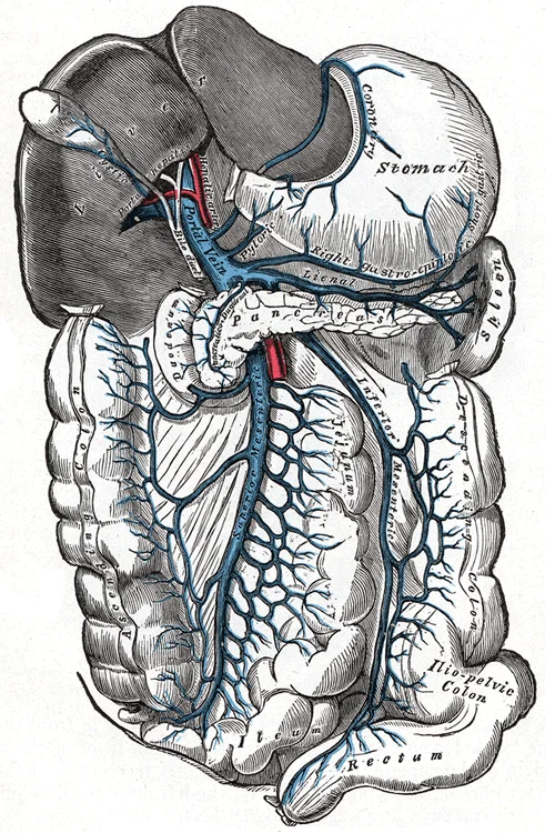
Long description
The portal vein and its tributaries. Labelled illustration from Wikimedia Commons, used under its open licence.

Figure 1: The portal vein and its tributaries.
The portal system also communicates with the systemic circulation via portosystemic anastomoses. These connections exist at the oesophagus, rectum, umbilicus, and retroperitoneum. In portal hypertension, these anastomoses become engorged, leading to complications such as oesophageal varices and caput medusae. The presence and size of these anastomoses can vary between individuals, influencing the clinical presentation of portal hypertension.
The hepatic veins, which drain the liver into the inferior vena cava, are distinct from the portal system. However, in cases of Budd-Chiari syndrome or severe cirrhosis, the relationship between the portal and hepatic venous systems becomes clinically significant. The pressure gradient between the portal vein and the hepatic vein is a key determinant of portal hypertension. Normal values are typically low, but this can increase significantly in liver disease.
The portal system is also influenced by the autonomic nervous system. The nervi vasorum regulate the tone of the vessels, although their role in the portal system is less pronounced than in the systemic circulation. Hormonal factors, such as vasopressin and angiotensin II, can also affect portal pressure. Understanding these regulatory mechanisms is essential for managing conditions like variceal bleeding.
The portal system's anatomy is critical for drug metabolism. Drugs absorbed from the gut pass through the liver before reaching the systemic circulation, a process known as first-pass metabolism. Variations in portal anatomy can alter this process, affecting drug bioavailability. For example, a portosystemic shunt can bypass the liver, reducing first-pass metabolism and increasing systemic drug levels.
In summary, the portal venous system exhibits considerable individual variation. These variations are not merely academic curiosities but have significant clinical implications. Surgeons and radiologists must be aware of these variations to avoid complications and to accurately interpret diagnostic images. The portal system's unique anatomy and physiology make it a vital component of human circulation.
The portal venous system is a unique high-pressure circuit that connects the gastrointestinal tract to the liver, serving as the primary route for nutrient processing and detoxification. Understanding its anatomy is critical for managing conditions such as portal hypertension, where increased resistance to blood flow leads to the development of collateral circulation.
When portal pressure rises, blood seeks alternative pathways to bypass the liver and return to the systemic circulation. These portosystemic anastomoses are clinically significant because their dilation leads to specific pathological signs.
- Lower oesophagus: The left gastric vein (portal) anastomoses with the oesophageal veins draining into the azygos vein (systemic). Dilation here causes oesophageal varices, which are prone to life-threatening haemorrhage.
- Rectum and anal canal: The superior rectal vein (portal) anastomoses with the middle and inferior rectal veins (systemic). Umbilical region: The paraumbilical veins (portal) anastomose with the superficial epigastric veins (systemic). In severe portal hypertension, these dilate and radiate from the umbilicus, creating the classic "caput medusae" appearance.
- Retroperitoneum: Small veins connecting the colon and kidneys (portal) with the retroperitoneal veins (systemic). These are often less clinically visible but contribute to collateral flow.
Clinical pearlClinical Pearl
The presence of caput medusae indicates significant portal hypertension and suggests that the paraumbilical veins have recanalised to bypass the obstructed portal flow.
While true aneurysms are rare in the portal system due to its low-pressure nature compared to arteries, venous ectasia can occur. The portal vein itself is the main trunk, formed by the union of the superior mesenteric vein and the splenic vein behind the neck of the pancreas.
Access points for diagnostic or therapeutic procedures often involve the umbilical region or percutaneous transhepatic routes. The umbilical vein, which is normally obliterated after birth as the ligamentum teres hepatis, can sometimes be cannulated in cases of severe portal hypertension to establish a shunt.
The portal system does not have pulse points, as it carries venous blood under low pressure. However, understanding the vascular supply is vital for surgical ligation. The splenic vein is a major tributary, draining the spleen, pancreas, and parts of the stomach. Ligation of the splenic artery or vein may be performed in cases of trauma or severe pancreatitis to control bleeding or reduce pressure.
The superior mesenteric vein drains the small intestine and large intestine. Injury to this vessel requires careful repair to prevent intestinal ischaemia. The inferior mesenteric vein drains the distal colon and rectum, joining the splenic vein or the superior mesenteric vein.
| Vein | Drainage Area | Clinical Significance |
|---|---|---|
| Superior Mesenteric Vein | Small intestine, right colon | Major source of portal flow; injury causes significant bleeding. |
| Splenic Vein | Spleen, pancreas, stomach | Ligation may be used in trauma; thrombosis can cause isolated gastric varices. |
| Inferior Mesenteric Vein | Left colon, rectum | Contributes to portosystemic anastomoses in the rectum. |
| Left Gastric Vein | Stomach, lower oesophagus | Primary cause of oesophageal varices when dilated. |
Understanding these structures allows for precise surgical intervention and effective management of complications arising from portal hypertension. The interplay between these veins and their systemic counterparts is central to the pathophysiology of liver disease.
The portal venous system is a unique circuit where blood flows through two capillary beds before returning to the heart. Unlike most systemic veins, portal veins do not drain directly into the heart but instead connect two capillary networks: those of the gastrointestinal tract and those of the liver. This arrangement allows the liver to process nutrients and detoxify substances absorbed from the gut before they reach the general circulation.
DefinitionPortal Venous System
A venous network that drains blood from the gastrointestinal tract, spleen, and pancreas into the liver for processing before returning to the systemic circulation.
The primary vessel is the portal vein, formed by the union of the superior mesenteric vein and the splenic vein. This junction typically occurs posterior to the neck of the pancreas. The portal vein then ascends within the hepatoduodenal ligament, part of the lesser omentum, to enter the liver at the porta hepatis.
Exam trapCommon Trap
Do not confuse the portal vein with the hepatic veins. The portal vein brings blood to the liver, while hepatic veins carry blood away from the liver to the inferior vena cava.
Key tributaries include the left and right gastric veins, which drain the stomach, and the cystic vein, which drains the gallbladder. The superior mesenteric vein drains the small intestine and the majority of the large intestine. The splenic vein drains the spleen and receives the inferior mesenteric vein in some anatomical variations, though the inferior mesenteric vein often drains directly into the splenic vein or the junction of the splenic and superior mesenteric veins.
Clinical pearlClinical Pearl
Portal hypertension, often caused by liver cirrhosis, leads to the formation of portosystemic anastomoses. These collateral vessels can become dilated, resulting in esophageal varices, which are prone to life-threatening bleeding.
The portal system lacks valves, allowing for bidirectional flow under pathological conditions. This is critical in understanding the spread of metastatic cancer from the gut to the liver. The liver receives approximately 75% of its blood supply from the portal vein and 25% from the hepatic artery.
| Vessel | Primary Drainage Area |
|---|---|
| Splenic Vein | Spleen, pancreas, part of stomach |
| Superior Mesenteric Vein | Small intestine, cecum, ascending colon |
| Inferior Mesenteric Vein | Descending colon, sigmoid colon, rectum |
| Left Gastric Vein | Lesser curvature of stomach |
| Right Gastric Vein | Lesser curvature of stomach |
| Cystic Vein | Gallbladder |
Anatomical relations are crucial for surgical safety. The portal vein lies posterior to the common bile duct and the hepatic artery proper within the hepatoduodenal ligament. This triad is enclosed in the free edge of the lesser omentum. Compression of this ligament (Pringle manoeuvre) can control bleeding from the liver by occluding the portal vein and hepatic artery.
NoteDevelopmental Note
The portal system develops from the vitelline veins. Abnormalities in this development can lead to congenital portosystemic shunts, bypassing the liver entirely.
Understanding the portal system is essential for interpreting liver function tests and managing conditions like portal hypertension. The liver's dual blood supply ensures it receives both oxygenated blood from the hepatic artery and nutrient-rich, deoxygenated blood from the portal vein. This unique physiology supports the liver's metabolic, detoxification, and synthetic functions.
Students frequently confuse the arterial and venous systems, particularly when describing the path of blood from the gut to the liver. A critical error is assuming the portal vein carries oxygenated blood from the heart. In reality, the portal system is a venous network that transports nutrient-rich, deoxygenated blood from the gastrointestinal tract to the liver for processing. This distinction is vital because the liver acts as a metabolic filter before blood returns to the general circulation via the hepatic veins and inferior vena cava. Confusing the direction of flow or the oxygenation status leads to fundamental misunderstandings of hepatic physiology and portal hypertension.
Another frequent mistake involves the anatomical landmarks of the aorta, which is often conflated with the portal vein in student recall. The aorta is the largest artery in the body, arising from the left ventricle and descending to bifurcate at the level of the fourth lumbar vertebra into the two common iliac arteries. It is essential to remember that the aorta distributes blood to virtually all tissues, whereas the portal vein specifically serves the digestive organs. The aortic arch ends at the level of the intervertebral disk between the fourth and fifth thoracic vertebrae, after which it becomes the descending aorta. Mixing up these vertebral levels can lead to errors in localizing pathology on imaging. [1]
Students also often misidentify the layers of the heart wall when discussing coronary circulation, which is closely related to cardiac output and thus portal pressure. The middle and thickest layer is the myocardium, made largely of cardiac muscle cells. This layer is responsible for pumping blood through the heart and into the major arteries. The contraction of the myocardium drives the systemic circulation, including the splanchnic circulation that feeds the portal system. Understanding that coronary circulation cycles, reaching a peak when the heart muscle is relaxed, helps explain why cardiac function directly influences venous return and portal pressure. [2]
A common trap is ignoring the valve structures that ensure one-way flow. The septum between the atria and ventricles is marked by four openings that allow blood to move from the atria into the ventricles and from the ventricles into the pulmonary trunk and aorta. The valves between the atria and ventricles are known as atrioventricular valves, while those leading to the pulmonary trunk and aorta are semilunar valves. While these are cardiac structures, their dysfunction can lead to backpressure that affects the entire venous system, including the portal vein. Forgetting that the aortic semilunar valve prevents backflow into the left ventricle is a basic anatomical oversight. [2]
Finally, students may confuse the gallbladder's anatomy with the portal vein's course. The gallbladder is nested in a shallow area on the posterior aspect of the right lobe of the liver. It stores and concentrates bile, propelling it into the duodenum via the common bile duct. While the gallbladder is a liver accessory organ, it is not part of the portal venous drainage itself. Confus [1]
Knowledge of the portal venous system is indispensable at the bedside for assessing patients with liver disease. Clinicians use this anatomical understanding to interpret signs of portal hypertension, such as ascites and caput medusae. The portal vein is formed by the union of the superior mesenteric and splenic veins, carrying blood from the intestines and spleen. When resistance increases in the liver, blood seeks collateral pathways, leading to varices in the oesophagus and stomach. Recognising the origin and termination of these vessels allows doctors to predict the location of bleeding and plan endoscopic interventions.
In the operating theatre, precise knowledge of vascular anatomy is crucial during hepatic surgeries. Surgeons must identify the portal triad, which contains the portal vein, hepatic artery, and bile duct. The abdominal aorta terminates when it bifurcates into the two common iliac arteries at the level of the fourth lumbar vertebra. While this is an arterial landmark, it helps orient the surgeon in the retroperitoneum. Understanding that the abdominal aorta remains to the left of the vertebral column and is embedded in adipose tissue behind the peritoneal cavity helps in navigating deep abdominal structures. This spatial awareness prevents accidental injury to major vessels during liver resections or transplants. [1]
On imaging, such as CT or MRI, the portal vein is a key landmark for liver segmentation. Radiologists assess the diameter and flow direction of the portal vein to diagnose thrombosis or stenosis. The internal carotid arteries along with the vertebral arteries are the two primary suppliers of blood to the human brain. While this refers to cerebral circulation, the principle of dual supply and collateral flow is analogous in the liver. Blood flow to the brain is remarkably constant, with approximately 20 percent of blood flow directed to this organ at any given time. Similarly, the liver receives dual blood supply from the portal vein and hepatic artery, and imaging must evaluate both to assess hepatic viability. [1]
In examinations, students are often tested on the relationships between the portal vein and surrounding structures. The gallbladder is nested in a shallow area on the posterior aspect of the right lobe of the liver. This close anatomical relationship means that gallbladder disease can sometimes mimic or complicate portal vein pathology. Questions may ask about the course of the cystic duct, which is 1–2 cm lon
Clinical pearlClinical Pearl
Portal hypertension leads to portosystemic anastomoses, causing varices in the oesophagus, stomach, and rectum. Always consider the liver's role as a filter when evaluating gastrointestinal bleeding.
| Structure | Landmark / Relation | Clinical Significance |
|---|---|---|
| Abdominal Aorta | Bifurcates at L4 into common iliac arteries | Landmark for surgical orientation |
| Portal Vein | Formed by SMV and splenic vein | Carries nutrient-rich blood to liver |
| Gallbladder | Nested in right lobe of liver | Accessory organ, not part of portal vein |
| Cystic Duct | 1–2 cm long, turns inferiorly | Connects gallbladder to hepatic duct [1] |
Portal Hypertension Assessment
- Patient presents with ascitesSuspect portal hypertension
- Question: Check for varices?
Yes
- Endoscopy to locate bleeding source
No
- Ultrasound to assess portal vein flow
The portal vein carries deoxygenated, nutrient-rich blood from the gut to the liver. The abdominal aorta bifurcates at the level of the fourth lumbar vertebra. The myocardium is the thickest layer of the heart wall, responsible for pumping. Coronary circulation peaks when the heart muscle is relaxed. The gallbladder is 8–10 cm long and stores bile. The cystic duct is 1–2 cm long and connects the gallbladder to the hepatic duct. Portal hypertension leads to collateral circulation and varices. The aortic arch ends at the level of the intervertebral disk between T4 and T5.
Answer each one in your head first, then open the answer.
At which vertebral level does the abdominal aorta bifurcate into the common iliac arteries?
Show answerHide answer
Fourth lumbar vertebra (L4)What is the length of the cystic duct?
Show answerHide answer
1–2 cmWhich layer of the heart wall is the thickest and made of cardiac muscle cells?
Show answerHide answer
MyocardiumWhat percentage of blood flow is directed to the brain at any given time?
Show answerHide answer
20 percentWhere is the gallbladder located relative to the liver?
Show answerHide answer
Nested in a shallow area on the posterior aspect of the right lobe
- Betts JG, Young KA, Wise JA, et al. Circulatory Pathways. OpenStax, Anatomy and Physiology 2e (CC BY-NC-SA 4.0). Accessed .
- Betts JG, Young KA, Wise JA, et al. Heart Anatomy. OpenStax, Anatomy and Physiology 2e (CC BY-NC-SA 4.0). Accessed .
- Betts JG, Young KA, Wise JA, et al. Accessory Organs in Digestion: The Liver, Pancreas, and Gallbladder. OpenStax, Anatomy and Physiology 2e (CC BY-NC-SA 4.0). Accessed .
Figure credits
Educational content for medical students and doctors. It is not a substitute for clinical judgement or local guidelines.