Abdominal aorta and branches
Anatomical overview of the abdominal aorta and its major visceral branches.
Figure 1Gray539
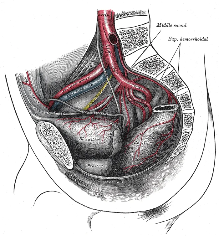
Long description
The arteries of the pelvis. Labelled illustration from Wikimedia Commons, used under its open licence.

Figure 1: The arteries of the pelvis.
The abdominal aorta is the continuation of the thoracic aorta, descending anterior to the vertebral column. It bifurcates into the common iliac arteries at the level of the fourth lumbar vertebra. Its primary function is to supply oxygenated blood to the abdominal viscera, the posterior abdominal wall, and the lower limbs. Understanding its branching pattern is fundamental to interpreting abdominal pathology and planning surgical interventions.
The coeliac trunk is the first major anterior branch of the abdominal aorta, typically arising just below the aortic hiatus of the diaphragm at the level of T12. It is a short, wide trunk that divides almost immediately into three major branches: the left gastric artery, the splenic artery, and the common hepatic artery. These vessels supply the foregut structures, including the distal oesophagus, stomach, proximal duodenum, liver, gallbladder, pancreas, and spleen.
It supplies the midgut, which includes the distal half of the duodenum, jejunum, ileum, caecum, ascending colon, and the proximal two-thirds of the transverse colon. The SMA passes anterior to the left renal vein and the uncinate process of the pancreas. Compression of the duodenum between the aorta and the SMA can lead to superior mesente [1]
Figure 2Gray536
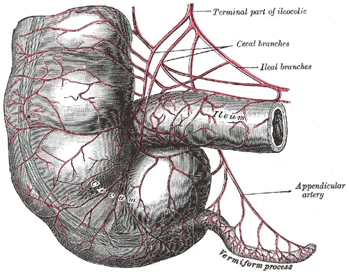
Long description
Arteries of cecum and vermiform process.

Figure 2: Arteries of cecum and vermiform process.
It supplies the hindgut, comprising the distal third of the transverse colon, the descending colon, the sigmoid colon, and the upper part of the rectum. The IMA gives rise to the left colic artery, the sigmoid arteries, and the superior rectal artery. Anastomoses exist between the SMA and IMA via the marginal artery of Drummond and the arc of Riolan, providing collateral circulation.
Figure 3Gray538
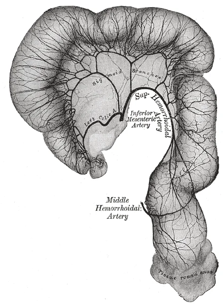
Long description
Sigmoid colon and rectum, showing distribution of branches of inferior mesenteric artery and their anastomoses. (From a preparation by Mr. Hamilton Drummond.) Prepared in same manner as Fig. 535.

Figure 3: Sigmoid colon and rectum, showing distribution of branches of inferior mesenteric artery and their anastomoses (From a preparation by Mr.
Nutcracker syndrome, or left renal vein entrapment, occurs when the left renal vein is compressed between the abdominal aorta and the superior mesenteric artery. This anatomical relationship can lead to venous hypertension, resulting in haematuria, varicocele, and pelvic congestion.
| Structure | Level of Origin | Primary Supply |
|---|---|---|
| Coeliac Trunk | T12 | Foregut |
| Superior Mesenteric Artery | L1 | Midgut |
| Renal Arteries | L1-L2 | Kidneys |
| Inferior Mesenteric Artery | L3 | Hindgut |
The abdominal aorta is the continuation of the thoracic aorta, descending through the diaphragm at the level of the twelfth thoracic vertebra. It travels anterior to the vertebral column, slightly to the left of the midline, until it bifurcates into the common iliac arteries at the level of the fourth lumbar vertebra. This termination point is a critical landmark for surgical and radiological procedures. The vessel maintains a relatively consistent diameter in its proximal segment, though variations exist across the population.
Figure 4Gray531
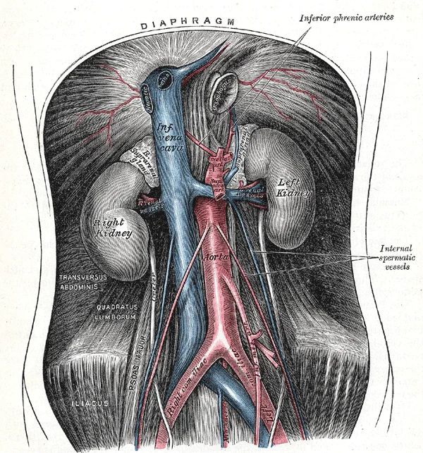
Long description
The abdominal aorta and its branches. Labelled illustration from Wikimedia Commons, used under its open licence.

Figure 4: The abdominal aorta and its branches.
The diameter of the abdominal aorta is a significant factor in vascular health. The aortic diameter tends to change with age and sex, although the specific influence of these factors on branching angles remains less clearly defined in some literature. Histological changes also occur over time; young individuals possess thick, concentric elastin fibers, whereas elderly individuals have thinner, fragmented fibers. [3] [1]
Aneurysms are a notable pathology associated with the abdominal aorta. They are approximately four times more frequent in the abdominal segment than in the thoracic aorta, which has a larger diameter. This increased incidence in smaller diameter segments suggests that factors beyond simple size play a role in aneurysm formation. The origins of such complex diseases are multifactorial. [1]
Understanding the precise anatomy is essential for interventions. In smaller diameter aortas, less backup force is generated for guiding catheters, making intervention more challenging. Therefore, accurate knowledge of the origin, course, and termination of the abdominal aorta, along with its branching patterns, is fundamental for clinical practice. [3]
Figure 5Gray532
Long description
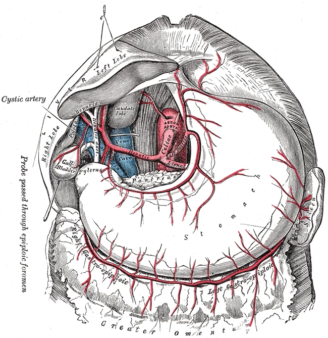
The celiac artery and its branches; the liver has been raised, and the lesser omentum and anterior layer of the greater omentum removed.

Figure 5: The celiac artery and its branches; the liver has been raised, and the lesser omentum and anterior layer of the greater omentum removed.
The abdominal aorta is the major arterial trunk supplying the abdominal viscera, pelvic walls, and lower limbs. It descends anterior to the vertebral column, slightly to the left of the midline, terminating at the level of the fourth lumbar vertebra by dividing into the common iliac arteries. Understanding its branching pattern is fundamental to interpreting cross-sectional imaging and surgical anatomy.
The Abdominal aorta and branches are clearly delineated. The inferior vena cava lies to the right of the aorta. The coeliac trunk arises anteriorly. The superior mesenteric artery (SMA) arises just below the coeliac trunk. The inferior mesenteric artery (IMA) arises lower down. The renal arteries emerge laterally. The gonadal arteries arise inferior to the renal arteries. Anastomoses connect these vessels.
The coeliac trunk is the first major anterior branch of the abdominal aorta. It typically arises at the level of the twelfth thoracic vertebra. It is short and wide, dividing almost immediately into three terminal branches: the left gastric artery, the splenic artery, and the common hepatic artery. These vessels supply the foregut derivatives, including the stomach, liver, spleen, and proximal duodenum.
The superior mesenteric artery arises from the anterior aspect of the abdominal aorta, just inferior to the coeliac trunk, usually at the level of the first lumbar vertebra. It descends anterior to the uncinate process of the pancreas and the left renal vein. Its branches supply the midgut, including the distal duodenum, jejunum, ileum, caecum, ascending colon, and proximal two-thirds of the transverse colon.
Figure 6Gray537
Long description
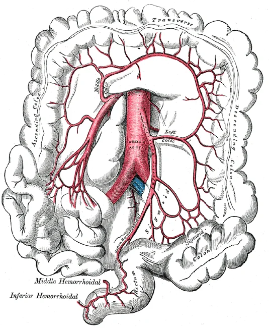
The inferior mesenteric artery and its branches.

Figure 6: The inferior mesenteric artery and its branches.
The abdominal aorta terminates by bifurcating into the common iliac arteries. Extensive anastomoses exist between the coeliac, superior mesenteric, and inferior mesenteric arteries, providing collateral circulation to the gut. These include the marginal artery of Drummond and the arc of Riolan. The inferior vena cava runs parallel to the aorta on its right side, receiving blood from the lower body.
Figure 7Gray535
Long description
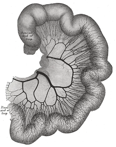
Loop of small intestine showing distribution of intestinal arteries. (From a preparation by Mr. Hamilton Drummond.) The vessels were injected while the gut was in situ; the gut was then removed, and an x -ray photograph taken.

Figure 7: Loop of small intestine showing distribution of intestinal arteries (From a preparation by Mr.
| Structure | Level of Origin | Primary Supply |
|---|---|---|
| Coeliac Trunk | T12 | Foregut |
| Superior Mesenteric Artery | L1 | Midgut |
| Renal Arteries | L1-L2 | Kidneys |
| Inferior Mesenteric Artery | L3 | Hindgut |
Figure 8Gray533
Long description
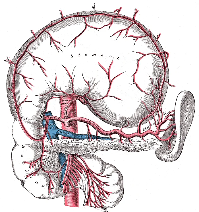
The celiac artery and its branches; the stomach has been raised and the peritoneum removed.

Figure 8: The celiac artery and its branches; the stomach has been raised and the peritoneum removed.
The abdominal aorta is the principal arterial trunk supplying the posterior abdominal wall and the intraperitoneal and retroperitoneal viscera. It terminates at the level of the fourth lumbar vertebra by dividing into the common iliac arteries. Understanding its territories requires tracing the origin, course, and anastomoses of its branches.
These three arteries arise from the anterior aspect of the aorta. The coeliac trunk arises at the level of T12. It divides into the left gastric, splenic, and common hepatic arteries. The left gastric artery supplies the lesser curvature of the stomach. The splenic artery supplies the spleen, pancreas, and greater curvature of the stomach via the short gastric and left gastro-omental arteries. The common hepatic artery gives rise to the proper hepatic artery, which supplies the liver, and the gastroduodenal artery. The gastroduodenal artery further divides into the right gastro-omental and superior pancreaticoduodenal arteries.
The superior mesenteric artery arises at the level of L1. It supplies the midgut, including the distal duodenum, jejunum, ileum, caecum, ascending colon, and proximal two-thirds of the transverse colon. Its branches include the inferior pancreaticoduodenal artery, jejunal and ileal arteries, ileocolic artery, right colic artery, and middle colic artery.
The inferior mesenteric artery arises at the level of L3. It supplies the hindgut, including the distal one-third of the transverse colon, descending colon, sigmoid colon, and upper rectum. Its branches are the left colic artery, sigmoid arteries, and superior rectal artery.
They supply the posterior abdominal wall muscles and the spinal cord via spinal branches.
Rich anastomotic networks ensure collateral circulation. Between the foregut and midgut, the left gastro-omental artery (from the splenic artery) anastomoses with the right gastro-omental artery (from the gastroduodenal artery) along the greater curvature of the stomach. Between the midgut and hindgut, the middle colic artery (from the superior mesenteric artery) anastomoses with the left colic artery (from the inferior mesenteric artery) along the transverse colon. This connection is known as the marginal artery of Drummond. Additionally, the superior rectal artery (from the inferior mesenteric artery) anastomoses with the middle and inferior rectal arteries (from the internal iliac system) in the rectum.
The aorta is central, running vertically. The superior mesenteric artery arises below it. The inferior mesenteric artery arises lower down.
DefinitionMarginal Artery
A continuous arterial channel formed by anastomoses between the branches of the superior and inferior mesenteric arteries along the inner border of the colon.
Figure 9Gray534
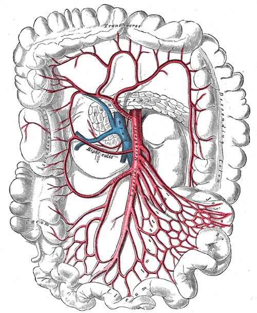
Long description
The superior mesenteric artery and its branches.

Figure 9: The superior mesenteric artery and its branches.
Understanding these anatomical variations and age-related changes is useful when planning invasive procedures. This includes choosing approaches, the curvature of catheters, and the diameters of angioplasty balloons and stents in endovascular procedures. Future device manufacturing may also benefit from tailoring diameters and curvatures to fit patients better on the basis of their sex and age group. [1]
Anatomical variations of arterial branches may involve their origin, course, or branching pattern. These variations can lead to unfamiliar surgical landmarks, making appreciation of the underlying anatomy essential. This understanding minimizes complications and refines the interpretation of angiography.
Clinical pearlAge-Related Changes
Arteries with developed elastic layers become dilated, elongated, and tortuous with age, influencing catheter curvature and stent diameter choices. [1]
The abdominal aorta is a critical landmark for vascular access and surgical intervention. Clinicians must understand its course, branching, and common pathologies to manage emergencies and plan interventions effectively.
Abdominal aortic aneurysms (AAA) typically occur infrarenally, below the origin of the renal arteries. This location is significant because the infrarenal segment lacks supportive branches, making it prone to dilation. When planning endovascular aneurysm repair (EVAR), precise anatomical knowledge is essential. Noninvasive examinations provide vital anatomic information for intervention planning. For example, when attempting selective catheterization of the left renal artery (LRA) in a 68-year-o [1]
The abdominal aorta bifurcates into the common iliac arteries at the level of the fourth lumbar vertebra. The femoral arteries, derived from the external iliacs, are primary pulse points for assessing peripheral circulation. In emergency situations, such as severe hemorrhage, ligation of specific arteries may be necessary.
The aorta descends anterior to the vertebral column. The renal arteries branch laterally at the L1/L2 level. The inferior mesenteric artery arises anteriorly at L3. The common iliac arteries bifurcate at L4.
Clinical pearlKey Access Point
Femoral artery puncture is a common access point for vascular interventions, but anatomical variations, such as those seen in female patients, can increase procedural difficulty.
| Feature | Description | Clinical Significance |
|---|---|---|
| Main RA | Major target for balloons/stents | Treats stenosis |
| Branch RAs | Variable number and distribution | RDN targeting both main and branch RAs had greater efficacy than RDN in the main RA alone in animals and humans [3] |
| Imaging | Catheter angiography | Gold standard for morphological diagnosis |
Clinical pearlBifurcation Level
The aortic bifurcation occurs at the level of L4, not L5. Confusing this with the sacral promontory (S1) is a common error.
Exam trapRenal Vein Course
The left renal vein crosses anterior to the aorta. The right renal vein is short and enters directly. Do not confuse this with the inferior vena cava, which lies to the right.
DefinitionVisceral Branches
Unpaired branches supply the gastrointestinal tract (coeliac, SMA, IMA). Paired branches supply retroperitoneal organs (suprarenal, renal, gonadal).
Clinical pearlCoeliac Trunk
The coeliac trunk arises at T12. It divides into the left gastric, splenic, and common hepatic arteries. This is the first major visceral branch.
Clinical pearlMedian Sacral Artery
This small artery arises from the posterior aspect of the aortic bifurcation. It is often small and may be absent. It supplies the sacrum and coccyx.
Exam trapAortic Aneurysm
Abdominal aortic aneurysms typically occur infrarenally. The lack of adventitia and muscular support in this segment predisposes to dilation. Screening is recommended for men aged 65-75 who have ever smoked.
Students frequently confuse the anatomical territories and clinical presentations of the major abdominal aortic branches, leading to diagnostic errors. A primary error is attributing acute abdominal pain solely to gastrointestinal causes without considering vascular origins. The superior mesenteric artery (SMA) supplies the midgut, extending from the distal duodenum to the proximal two-thirds of the transverse colon. Ischaemia here presents with pain out of proportion to physical findings, where [1]
A second frequent pitfall involves the interpretation of imaging regarding age-related changes. Students may assume arterial dimensions are static, but anatomical studies show that the SMA diameter and angle of origin vary significantly with age and sex. For instance, in older men, the SMA tends to emerge at an angle that is around 26° larger than in younger men, specifically measuring 81.27° in the seventh decade compared to 54.64° in the third decade. Conversely, among female patients, this an [1]
A third error is the incorrect identification of collateral pathways. The arc of Riolan and the marginal artery of Drummond are critical anastomoses between the SMA and IMA. Students often forget that these vessels provide collateral circulation that can maintain bowel viability during acute occlusion of one major trunk. In clinical practice, appreciating this network is essential for surgical planning during aortic aneurysm repair to prevent colonic ischaemia. Additionally, confusion often arises regarding the renal arteries. The right renal artery passes posterior to the inferior vena cava, whereas the left renal artery passes anterior to it. Misidentifying this relationship can lead to vascular injury during retroperitoneal surgery.
Finally, students frequently neglect the detailed branching patterns of the SMA. The SMA gives off the inferior pancreaticoduodenal artery, jejunal and ileal arteries, ileocolic artery, right colic artery, and middle colic artery. Missing the specific territory of the middle colic artery, which supplies the transverse colon, can result in inadequate resection margins during oncological surgery. Understanding that the SMA is the primary blood supply to the small intestine and most of the large intestine is fundamental; any deviation from this rule requires careful anatomical verification.
This knowledge is applied at the bedside when evaluating patients with acute abdominal pain. Clinicians use the anatomical distribution of the SMA and IMA to localise ischaemic pain. For example, periumbilical pain suggests midgut ischaemia (SMA territory), while left lower quadrant pain suggests hindgut ischaemia (IMA territory). In the operating theatre, surgeons rely on precise anatomical landmarks to control haemorrhage. During a Whipple procedure or pancreaticoduodenectomy, the SMA is a critical landmark; the uncinate process hooks around the SMA and superior mesenteric vein. Surgeons must identify the SMA early to ensure clear margins in pancreatic cancer resection. Similarly, in colorectal surgery, ligation of the inferior mesenteric artery must be performed with care to preserve collateral flow via the marginal artery of Drummond, preventing left-sided colonic ischaemia.
On imaging, radiologists assess the angle and diameter of the SMA to diagnose vascular syndromes. As noted, the angle of origin can widen with age in men, potentially predisposing to duodenal compression. Radiologists also measure the aortic diameter to screen for abdominal aortic aneurysms. A dilation index can be calculated to assess age-related changes, using a formula that compares mean diameters across age groups. This quantitative approach helps distinguish normal ageing from pathological dilation. In in [1]
In examinations, students are tested on the origin, course, and relations of these vessels. Questions often require identifying the vertebral level of origin (L1 for SMA, L3 for IMA) and the structures crossed by the vessels. For example, the SMA crosses the left renal vein and the duodenum. Understanding these relations is crucial for answering questions on vascular compression syndromes. Furthermore, students must know the branches of the SMA to identify the blood supply to specific bowel segments, which is vital for surgical questions regarding resection boundaries.
The clinical relevance extends to understanding sex-specific differences in vascular anatomy. The lesser predisposition towards changes in arterial anatomy among women may be related to the role played by estrogen, which increases the bioavailability of nitric oxide without increasing expression and/or activity of endothelial nitric oxide-synthase. This biological insight explains why older men are more prone to significant anatomical changes in the SMA angle and diameter. Surgeons should strive to acquire the most detailed knowledge possible of the structures involved in each procedure performed, because this has positive impacts on the quality of the technique employed and on avoidance of iatrogenic injuries. This principle underpins all practical applications of abdominal aortic anatomy. [1]
- The SMA arises anteriorly from the aorta at L1, supplying the midgut.
- The IMA arises anteriorly at L3, supplying the hindgut.
- SMA angle of origin widens significantly in older men but remains stable in women.
- SMA diameter is similar in young sexes but larger in older men.
- The right renal artery passes posterior to the IVC; the left passes anterior.
- Collateral circulation via the marginal artery of Drummond protects the colon.
- Estrogen protects women from age-related SMA anatomical changes via nitric oxide.
- Precise anatomical knowledge prevents iatrogenic injury during abdominal surgery.
Answer each one in your head first, then open the answer.
At which vertebral level does the superior mesenteric artery typically originate?
Show answerHide answer
L1Which artery passes posterior to the inferior vena cava?
Show answerHide answer
Right renal arteryWhat is the primary blood supply to the midgut?
Show answerHide answer
Superior mesenteric arteryWhich hormone is linked to reduced age-related changes in SMA anatomy in women?
Show answerHide answer
EstrogenWhat structure does the SMA cross anterior to before branching?
Show answerHide answer
Left renal vein
- Góes AMO, de Albuquerque FBA, Beckmann FA, et al. Age and sex and their influence on the anatomy of the abdominal aorta and its branches. Jornal vascular brasileiro 2020;19:e20200073. doi:10.1590/1677-5449.200073. Accessed .
- Na J, Kakazu A, Muthana A, et al. Lingual artery: Angiographic anatomy and variations review for neurosurgeons. Surgical neurology international 2025;16:156. doi:10.25259/sni_282_2025. Accessed .
- Sakaoka A, Koshimizu M, Nakamura S, Matsumura K. Quantitative angiographic anatomy of the renal arteries and adjacent aorta in the swine for preclinical studies of intravascular catheterization devices. Experimental animals 2018;67(2):291-299. doi:10.1538/expanim.17-0125. Accessed .
Figure credits
- Figure 1: Henry Vandyke Carter, via Wikimedia Commons, Public domain, Public domain.
- Figure 2: Henry Vandyke Carter, via Wikimedia Commons, Public domain, Public domain.
- Figure 3: Henry Vandyke Carter, via Wikimedia Commons, Public domain, Public domain.
- Figure 4: Henry Vandyke Carter, via Wikimedia Commons, Public domain, Public domain.
- Figure 5: Henry Vandyke Carter, via Wikimedia Commons, Public domain, Public domain.
- Figure 6: Henry Vandyke Carter, via Wikimedia Commons, Public domain, Public domain.
- Figure 7: Henry Vandyke Carter, via Wikimedia Commons, Public domain, Public domain.
- Figure 8: Henry Vandyke Carter, via Wikimedia Commons, Public domain, Public domain.
- Figure 9: Henry Vandyke Carter, via Wikimedia Commons, Public domain, Public domain.
Educational content for medical students and doctors. It is not a substitute for clinical judgement or local guidelines.