Suprarenal glands
Paired endocrine organs situated superior to the kidneys, regulating metabolism and stress response via cortical and medullary hormones.
The suprarenal glands are paired endocrine structures that regulate a myriad of bodily functions such as metabolism, immune system, blood pressure, and response to stress via the hormones released. Historically termed the adrenal glands, the term adrenal was abolished by the Federative Committee on Anatomical Terminology in 1998, and the term suprarenal should be used based on the position of the glands superior to the kidneys. Each gland is related closely to the diaphragm superiorly, the kidney [2]
The gland is composed of two distinct functional parts: the outer cortex and the inner medulla. The cortical and medullary secretions of the adrenal are released into the sinuses receiving one of the highest arterial flow rates per gram of tissue (5–10 ml/g/min). This abundant blood supply is received through numerous branches of the three different arteries, namely, superior suprarenal artery (SSA), middle suprarenal artery (MSA), and inferior suprarenal artery (ISA). [2]
Figure 1Gray1186
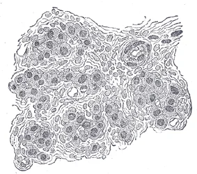
Long description
Section of part of human glomus caroticum. (Schaper.) Highly magnified. Numerous bloodvessels are seen in section among the gland cells.

Figure 1: Section of part of human glomus caroticum (Schaper.) Highly magnified.
Figure 2Gray1185
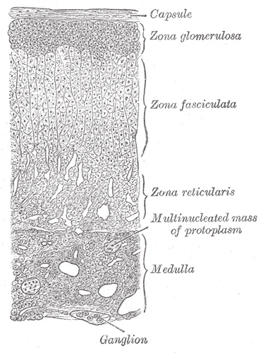
Long description
Section of a part of a suprarenal gland. (Magnified.)

Figure 2: Section of a part of a suprarenal gland (Magnified.).
The superior suprarenal artery usually arises from the ipsilateral inferior phrenic artery and passes to the gland as four or five small branches. The middle suprarenal artery is usually single and arises from the lateral aspect of the abdominal aorta at around the level of the superior mesenteric artery. Developmentally, the lateral splanchnic branches from the dorsal aorta are dedicated to supplying the kidneys, gonads, and suprarenal glands. Felix gave the Ladder Theory and proposed that there are nine lateral splanchnic branches that normally disappear in caudal direction thus leaving behind cranial, middle, and caudal groups of arteries. [2]
Figure 3Gray1184
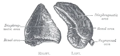
Long description
Suprarenal glands viewed from behind. Labelled illustration from Wikimedia Commons, used under its open licence.

Figure 3: Suprarenal glands viewed from behind.
DefinitionSuprarenal Gland
A paired endocrine organ situated superior to the kidney, comprising a cortex and medulla, with high arterial flow rates of 5–10 ml/g/min.
The surface marking of the suprarenal glands corresponds to the upper pole of the kidney. The right gland is pyramidal and lies anterior to the diaphragm and the IVC. The left gland is semilunar and lies anterior to the diaphragm and the upper pole of the left kidney. Awareness of variations in angioarchitecture of the suprarenal gland is beneficial while doing radiological and surgical procedures in the subphrenic region concerning the presence of extrahepatic collateral arteries formed, which often involves IPA and therefore the SSA. [2]
| Artery | Origin | Branches |
|---|---|---|
| Superior | Inferior phrenic | 4-5 small branches |
| Middle | Abdominal aorta | Single branch |
| Inferior | Renal artery | Variable |
NoteNomenclature
The term adrenal was abolished in 1998; vessels are now called suprarenal arteries and veins based on position superior to the kidneys. [2]
The cortex produces steroid hormones, while the medulla produces catecholamines. The glands are surrounded by a fibrous capsule. The blood supply is rich, with the arteries anastomosing within the gland. The veins drain directly into the IVC on the right and the left renal vein on the left. This anatomical arrangement is crucial for understanding the spread of pathology and the approach for surgical interventions such as suprarenal segmentectomy.
Arterial Supply
- StartIdentify suprarenal gland
- Question: Which artery?
Superior
- From inferior phrenic
Middle
- From aorta
Inferior
- From renal artery
Figure 4Gray1183
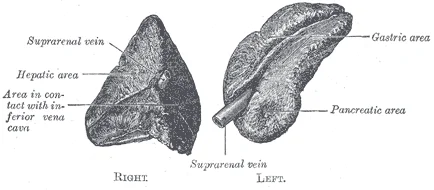
Long description
Suprarenal glands viewed from the front.

Figure 4: Suprarenal glands viewed from the front.
The left suprarenal gland is also situated superior to the left kidney but has a more crescentic or semilunar shape.
The suprarenal glands are endocrine organs located at the superior pole of each kidney. They are composed of two distinct functional parts: the outer cortex and the inner medulla. The cortex produces steroid hormones, while the medulla produces catecholamines. The glands are highly vascularised, receiving blood supply from multiple arterial sources. This rich vascular supply is critical for their endocrine function. This anatomical difference is important for surgical procedures. [3]
The external surface of the glands is smooth and covered by a thin capsule. The capsule sends septa into the gland, dividing it into lobes. Both glands are surrounded by adipose tissue, which helps to anchor them in place. The proximity to the kidneys and major vessels makes them accessible for surgical intervention when necessary. Understanding the external anatomy is essential for radiologists and surgeons to avoid injury during procedures.
Variation in the arterial supply of the suprarenal glands is quite common and very frequently reported. Previous studies have been done utilizing cadavers, angiographic data from human adult and fetuses, and corrosion casts as study samples. In the literature, the most common variation noted was in ISA followed by MSA, while the least common variations were observed in SSA. Variation in the multiplicity of arteries was observed more frequently in ISA and MSA of the right side. The absence of MSA was observed to be associated with increased number of ISA. Furthermore, the variation in suprarenal arteries was often correlated to variations in inferior phrenic and gonadal arteries. [2]
The variations of suprarenal arteries regarding their presence/absence, origin, and number were mostly reported as case presentations and very few as original studies. The scattered data required an overall presentation concerning the prevalence and clinical effects of such a common variation in human anatomy. Therefore, this review aims to condense all the data on variant anatomy of suprarenal arteries regarding its origin, number, and branching pattern, while emphasizing the possible etiology behind such variations making use of the literature available, such that this information can be used by nephrologists, radiologists to reduce inadvertent mortality in neonates as well as adults undergoing adrenal procedures. [2]
Paitre et al. did a study to distinguish between main and accessory suprarenal arteries though it was not accepted. The variations in suprarenal arteries are commonly reported, but an embryological theory explaining these variations is yet to be established. The authors did not attempt to provide any specific clinical advice in this aspect. Hence, analytical interpretation from a clinical perspective is suggested in this regard. Future direction on this topic of research is that the research should be multicentric and must include a large sample size with real-time clinical data to comprehend the horizon of variations of arterial supply of suprarenal glands with a broader perspective. [2]
The lack of an established embryological model explaining the variations provide further scope of research on the topic. The present review tabulates and analyses the available data on the variations of suprarenal arteries. This strengthens the database which provides essential information to instill awareness among the concerned clinicians regarding the possibilities of vascular variation related to suprarenal arteries. [2]
NoteClinical Relevance
Variations in arterial supply are common and can impact surgical outcomes.
| Feature | Description |
|---|---|
| Common Variation | ISA followed by MSA |
| Least Common | SSA |
| Right Side | Multiplicity more frequent in ISA and MSA |
| Left Side | Absence of MSA associated with increased ISA |
The suprarenal glands are retroperitoneal organs situated in the upper abdomen, lying superior to the kidneys. They are covered by peritoneum anteriorly and are closely related to various visceral structures.
It is related anteriorly to the liver and the inferior vena cava. The left suprarenal gland is semilunar in shape and lies anterior to the upper part of the left kidney. It is related anteriorly to the stomach, pancreas, and splenic artery. Both glands are enclosed in a thin layer of connective tissue that extends into the gland, dividing it into lobules.
The cortex of the suprarenal gland is the outer layer and is responsible for producing steroid hormones. It is divided into three zones: the zona glomerulosa, zona fasciculata, and zona reticularis. The medulla is the inner part of the gland and is responsible for producing catecholamines. It is derived from neural crest cells and is innervated by preganglionic sympathetic fibres. [3]
The arterial supply to the suprarenal glands is derived from three sources: the superior suprarenal artery, the middle suprarenal artery, and the inferior suprarenal artery. The superior suprarenal artery arises from the inferior phrenic artery. The middle suprarenal artery arises from the abdominal aorta. The venous drainage is via the suprarenal veins.
The suprarenal glands are closely related to the diaphragm posteriorly and are separated from the kidneys by a layer of fat. They are also related to the liver on the right side and the spleen on the left side. The glands are supplied by the sympathetic nervous system, which plays a key role in the regulation of their function.
The suprarenal glands are important organs in the body, playing a key role in the regulation of blood pressure, electrolyte balance, and stress response. Understanding their anatomy and relations is essential for medical students to appreciate their function and pathology.
The suprarenal gland exhibits a distinct architectural division into an outer cortex and an inner medulla, each with unique histological and vascular characteristics. The cortex is responsible for steroid hormone production and is organized into three concentric zones. The zona glomerulosa, located immediately beneath the capsule, consists of cells arranged in small clusters or arches. These cells produce mineralocorticoids, primarily aldosterone, which regulate sodium and potassium balance. Beneath this lies the zona fasciculata, the thickest layer, composed of cells arranged in straight cords or columns. These cells are rich in lipid droplets, giving them a pale, foamy appearance, and they synthesize glucocorticoids such as cortisol. The innermost cortical layer is the zona reticularis, where cells form an interconnecting network. This zone produces weak androgens and contributes to the overall steroid output of the gland.
DefinitionCortical Zones
The adrenal cortex is divided into the zona glomerulosa, zona fasciculata, and zona reticularis, each producing distinct classes of steroid hormones.
The medulla occupies the central portion of the gland and is derived from neural crest cells. It consists of chromaffin cells arranged in clusters or cords, separated by a rich sinusoidal capillary network. These cells secrete catecholamines, primarily adrenaline and noradrenaline, directly into the bloodstream. The vascular supply is critical to this function. The superior suprarenal arteries, typically branching from the inferior phrenic arteries, supply the upper part of the gland. The middle suprarenal arteries arise directly from the abdominal aorta, supplying the central region. The inferior suprarenal arteries, branching from the renal arteries, supply the lower pole. This dual arterial supply ensures robust perfusion, while venous drainage occurs via a single suprarenal vein on each side, which empties into the inferior vena cava on the right and the left renal vein on the left. [1]
The outermost thin layer is the fibrous capsule. Just inside the capsule lies the zona glomerulosa, appearing as small, rounded clusters of cells. Deep to this is the broad, pale-staining zona fasciculata, characterized by columns of lipid-rich cells. At the center lies the medulla, distinguished by its darker staining chromaffin cells and prominent sinusoidal vessels. The medullary veins converge towards the center, preparing to exit via the central suprarenal vein.
| Artery | Typical Origin | Region Supplied |
|---|---|---|
| Superior suprarenal | Inferior phrenic artery | Upper pole |
| Middle suprarenal | Abdominal aorta | Central region |
| Inferior suprarenal | Renal artery | Lower pole |
Variations in arterial origin are common. In most cases, the superior suprarenal artery originates from the ipsilateral inferior phrenic artery. However, variant origins from the abdominal aorta or coeliac trunk are documented. Understanding these variations is crucial for surgical planning, as aberrant branching patterns can affect blood supply during procedures. The para-aortic ridge gives rise to the gland during development, and vascular variations may result from defects in vasculogenesis or signal molecule gradients. These anatomical details ensure the gland receives adequate blood flow for its endocrine functions.
The suprarenal glands receive a rich arterial supply from three distinct sources, ensuring robust perfusion to support their endocrine function. Understanding this vascular arrangement is critical for surgical procedures involving the adrenal region. The primary vessels are the superior suprarenal arteries, the middle suprarenal arteries, and the inferior suprarenal arteries.
These arteries arise from the inferior phrenic arteries. They are the most consistent in their origin and course. They descend to supply the superior aspect of the gland.
The middle suprarenal arteries originate directly from the abdominal aorta. Their origin is variable, but they typically arise just below the superior mesenteric artery. They run laterally to reach the medial aspect of the gland. [2]
These arteries branch from the renal arteries. They ascend to supply the inferior pole of the suprarenal gland. This arrangement ensures that even if one source is compromised, collateral flow may maintain perfusion.
Despite the triple arterial supply, venous drainage is singular. Each suprarenal gland drains via a single central vein. This asymmetry is a key anatomical landmark.
Clinical pearlVenous Asymmetry
Remember the right suprarenal vein is short and enters the IVC directly, while the left is longer and enters the left renal vein first.
| Artery | Origin | Course |
|---|---|---|
| Superior | Inferior phrenic artery | Descends to superior gland |
| Middle | Abdominal aorta | Lateral to medial |
| Inferior | Renal artery | Ascends to inferior gland |
The venous drainage of the suprarenal glands is distinct from most other abdominal organs, characterised by the absence of a common trunk on either side. This anatomical arrangement has significant clinical implications for surgical access and the spread of pathology. Understanding the specific pathways for the right and left glands is essential for mastering abdominal anatomy.
The right suprarenal vein is typically shorter and more direct than its left counterpart. It originates from the anterior aspect of the right suprarenal gland. The vessel courses inferiorly and medially. It lies anterior to the right crus of the diaphragm. It passes deep to the inferior vena cava. The right suprarenal vein terminates by draining directly into the inferior vena cava. This direct entry into a large, low-pressure vessel facilitates rapid return of corticosteroids to the systemic ci [1]
The left suprarenal vein is longer and more tortuous than the right. It originates from the medial aspect of the left suprarenal gland. The vessel courses inferiorly and medially. It lies anterior to the left crus of the diaphragm. The left suprarenal vein terminates by draining into the left renal vein. This pathway is longer and involves a right-angle turn. The left renal vein then crosses anterior to the aorta to join the inferior vena cava. The le [1]
The right suprarenal vein is short and direct. The left suprarenal vein is long and indirect. This asymmetry is a key anatomical feature. The labels are precise and accurate. The viewer can trace the path of blood flow.
The suprarenal glands, also known as the adrenal glands, are paired endocrine organs situated superior to the kidneys. Their lymphatic drainage is extensive and follows the vascular supply, reflecting their embryological origin and functional integration with the retroperitoneal space. Understanding this drainage is critical for tracing the spread of pathology, such as metastatic carcinoma or infectious processes, from the gland to regional lymph nodes.
These nodes are located around the origin of the renal arteries and the suprarenal arteries. This direct drainage pathway explains why primary tumours of the suprarenal gland or metastases from other sites often present with enlarged lateral aortic nodes.
DefinitionLateral Aortic Nodes
A group of lymph nodes situated along the abdominal aorta, receiving drainage from the suprarenal, renal, and gonadal organs.
The lymphatic drainage is bilateral but predominantly ipsilateral. The right suprarenal gland drains primarily into the right lateral aortic nodes, while the left suprarenal gland drains into the left lateral aortic nodes. However, there is some cross-drainage, particularly in the midline, where lymphatics may anastomose. This anatomical arrangement ensures that any obstruction in the main drainage pathway can potentially be compensated for by collateral circulation, although this is rare in clinical practice.
| Structure | Drainage Target | Clinical Relevance |
|---|---|---|
| Right Suprarenal Gland | Right Lateral Aortic Nodes | Metastasis from adrenal carcinoma |
| Left Suprarenal Gland | Left Lateral Aortic Nodes | Metastasis from adrenal carcinoma |
| Efferent Vessels | Lateral Aortic Nodes | Tracing spread of infection |
The nerves supplying the suprarenal glands are primarily sympathetic, originating from the greater splanchnic nerve. These nerves pass through the celiac ganglion without synapsing and directly innervate the chromaffin cells of the adrenal medulla. The parasympathetic supply is minimal and comes from the vagus nerve, contributing to the regulation of blood flow rather than hormone secretion. This neurovascular bundle travels with the lymphatic vessels, ensuring coordinated physiological and pathological responses.
Figure 5Gray1187
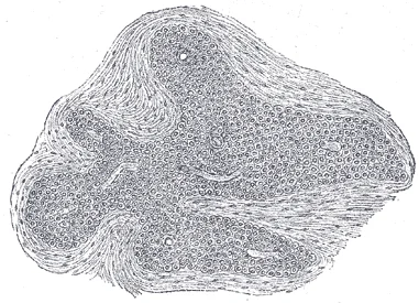
Long description
Section of an irregular nodule of the glomus coccygeum. (Sertoli.) X 85. The section shows the fibrous covering of the nodule, the bloodvessels within it, and the epithelial cells of which it is constituted.

Figure 5: Section of an irregular nodule of the glomus coccygeum (Sertoli.) X 85.
The suprarenal glands receive a complex innervation that integrates autonomic control with somatic sensory pathways. This dual supply is critical for understanding both the physiological response to stress and the clinical presentation of pathology in this region.
DefinitionAutonomic Supply
The sympathetic nervous system provides direct preganglionic innervation to the adrenal medulla, bypassing the typical ganglionic relay found in most autonomic pathways.
These nerves arise from the thoracic spinal segments T5 through T9. They descend through the diaphragm to reach the suprarenal glands. Unlike most sympathetic postganglionic fibres, these preganglionic fibres do not synapse in a peripheral ganglion. Instead, they pass directly through the adrenal cortex to reach the medulla. Here, they synapse with chromaffin cells. These cells function as modified postganglionic neurons. They release catecholamines, primarily adrenaline and noradrenaline, directly into the bloodstream. This mechanism allows for a rapid, systemic hormonal response to stress.
NoteClinical Pearl
Because the preganglionic fibres act directly on chromaffin cells, the adrenal medulla is essentially an extension of the sympathetic nervous system.
These fibres likely modulate blood flow and may have minor regulatory roles in secretion, though their exact physiological significance is less defined than the sympathetic drive.
These nerves supply the capsule and the surrounding connective tissue. They carry pain and proprioceptive information. Pain from the suprarenal glands is often perceived in the upper abdomen or the lower chest. This is due to the shared dermatomal origins of the somatic nerves supplying the gland and the overlying skin.
Exam trapReferred Pain
Pain from the suprarenal glands may be referred to the T5–T9 dermatomes, mimicking cardiac or pulmonary pathology.
| Nerve Type | Origin | Target | Function |
|---|---|---|---|
| Sympathetic | T5–T9 | Medulla | Catecholamine release |
| Parasympathetic | Vagus | Cortex/Medulla | Modulation |
| Somatic | Phrenic | Capsule | Pain sensation |
The suprarenal glands, or adrenal glands, are paired endocrine organs situated superior to the kidneys. They consist of two distinct functional components: the outer cortex and the inner medulla. This histological arrangement reflects their dual role in steroid hormone production and catecholamine secretion.
DefinitionAdrenal Cortex
The outer layer of the adrenal gland, responsible for synthesising steroid hormones including glucocorticoids, mineralocorticoids, and androgens. It is organised into three zones: zona glomerulosa, zona fasciculata, and zona reticularis.
The zona glomerulosa is the outermost layer, arranged in clusters. It produces mineralocorticoids, primarily aldosterone, under the influence of angiotensin II and potassium levels. Beneath this lies the zona fasciculata, the thickest layer, composed of cells arranged in straight cords. These cells produce glucocorticoids, mainly cortisol, regulated by adrenocorticotropic hormone (ACTH). The innermost cortical layer, the zona reticularis, consists of anastomosing cords and produces androgens such as dehydroepiandrosterone (DHEA).
Clinical pearlZonation
Remember the mnemonic 'GFR' for the adrenal cortex zones from outside to inside: Glomerulosa, Fasciculata, Reticularis. Each zone produces a different class of steroid hormone.
These cells are modified postganglionic sympathetic neurons that secrete catecholamines, primarily adrenaline and noradrenaline, directly into the bloodstream. They are stimulated by preganglionic sympathetic fibres releasing acetylcholine.
Exam trapBlood Supply
The adrenal glands have a rich blood supply. Cortical blood flows from the periphery towards the centre, bathing the medulla. This counter-current exchange allows cortical hormones to stimulate medullary catecholamine release.
In clinical practice, adrenalectomy may be required for conditions such as pheochromocytoma, adrenal cortical carcinoma, or Cushing’s syndrome. Preoperative management is critical. For pheochromocytoma, alpha-adrenergic blockade must precede beta-blockade to prevent hypertensive crisis. emergency care points for adrenal pathology include recognising the signs of hypercortisolism (Cushing’s syndrome) and hypoaldosteronism (Addison’s disease). Addison’s disease presents with hyperpigmentation, hypotension, and hyponatraemia due to autoimmune destruction of the cortex. In contrast, Conn’s syndrome involves aldosterone excess, leading to hypertension and hypokalaemia. Understanding the histological zonation is essential for correlating structural damage with specific hormonal deficits. The cortex’s ability to regenerate is limited, making surgical removal or autoimmune destruction permanent unless replaced by hormone therapy. The medulla’s neural origin means it responds rapidly to stress, unlike the slower-acting cortical hormones. This histological distinction underpins the physiological response to acute versus chronic stress.
The suprarenal glands, or adrenal glands, arise from two distinct embryological origins that fuse during development. This dual origin is critical for understanding their final anatomical position and vascular supply.
The cortex derives from the mesoderm, specifically the coelomic epithelium lining the posterior abdominal wall. These mesenchymal cells proliferate and form a capsule around the developing kidney. This mesodermal component eventually differentiates into the three layers of the adrenal cortex: the zona glomerulosa, zona fasciculata, and zona reticularis. The timing of this mesodermal migration coincides with the establishment of the renal system, ensuring the suprarenal gland sits superior to the kidney.
In contrast, the medulla originates from the neural crest. These ectodermal cells migrate ventrally and differentiate into chromaffin cells. These cells are essentially modified postganglionic sympathetic neurons. They cluster together to form the central medullary tissue. This neural crest origin explains why the medulla responds directly to sympathetic nervous system stimulation rather than hormonal trophic factors alone.
By the end of the first trimester, the mesodermal cortical tissue and the neural crest medullary tissue have fused. This fusion creates the characteristic triangular or Y-shaped gland. The right gland is pyramidal, while the left is more semilunar. They sit within the perirenal fat, enclosed by Gerota’s fascia along with the kidney. This anatomical relationship protects the gland and provides a consistent landmark for surgical approaches.
The blood supply reflects this complex development. The superior suprarenal arteries arise from the inferior phrenic arteries. The middle suprarenal arteries branch directly from the abdominal aorta. The inferior suprarenal arteries come from the renal arteries. This rich, multi-source arterial supply ensures adequate perfusion despite the gland's small size. This anatomical variation is crucial during adrenalectomy to avoid catastrophic haemorrhage.
DefinitionEmbryological Origin
The adrenal cortex is mesodermal, while the medulla is neural crest derived.
| Component | Embryological Origin | Key Derivative |
|---|---|---|
| Cortex | Mesoderm (Coelomic Epithelium) | Steroid-producing cells |
| Medulla | Neural Crest | Chromaffin cells |
Understanding these origins clarifies why the cortex responds to ACTH (a pituitary hormone) while the medulla responds to preganglionic sympathetic fibres. The fusion process is complete before birth, meaning the adult gland functions as a single organ with two distinct physiological roles. Any disruption in this migration or fusion can lead to congenital anomalies, though these are rare. The close proximity to the kidney allows for shared pathological processes, such as metastasis, but the distinct embryological roots maintain separate functional identities.
It is pyramidal in shape and rests on the diaphragm. In the image, it appears as a soft-tissue density structure adjacent to the inferior vena cava. The gland is separated from the liver by the hepatorenal recess. Its medial border relates to the crus of the diaphragm. The lateral border is in contact with the right kidney. The anterior surface is covered by peritoneum. The posterior surface is related to the diaphragm. The superior pole is near the right crus. The inferior pole rests on the kidney.
It is semilunar in shape. In the image, it is located medial to the spleen and anterior to the left kidney. The gland is separated from the pancreas by the splenic vessels. Its medial border relates to the aorta. The lateral border is in contact with the left kidney. The anterior surface is covered by peritoneum. The posterior surface is related to the diaphragm. The superior pole is near the left crus. The inferior pole rests on the kidney.
The suprarenal glands are supplied by three arteries. The superior suprarenal artery arises from the inferior phrenic artery. The middle suprarenal artery arises from the abdominal aorta. These arteries anastomose within the gland. The venous drainage differs between the two sides. This anatomical difference is crucial for surgical planning.
DefinitionSuprarenal Gland Anatomy
The suprarenal glands are endocrine organs located superior to the kidneys, with distinct vascular supplies and venous drainage patterns that are critical for radiological identification and surgical intervention.
The imaging appearance of the suprarenal glands is homogeneous on computed tomography. They are isodense to muscle. On magnetic resonance imaging, they have intermediate signal intensity on T1-weighted images. The glands are surrounded by perirenal fat, which provides contrast. The adrenal glands are often difficult to distinguish from lymph nodes if enlarged. Normal suprarenal glands are thin and flat. They are not easily visible on ultrasound. Computed tomography is the modality of choice for evaluating suprarenal pathology.
| Structure | Relation |
|---|---|
| Right Suprarenal Vein | Drains into Inferior Vena Cava |
| Left Suprarenal Vein | Drains into Left Renal Vein |
| Superior Suprarenal Artery | Arises from Inferior Phrenic Artery |
| Middle Suprarenal Artery | Arises from Abdominal Aorta |
| Inferior Suprarenal Artery | Arises from Renal Artery |
The radiological anatomy of the suprarenal glands is essential for diagnosing adrenal disorders. Pathologies such as adenomas, carcinomas, and metastases can alter the appearance of the glands. The size and shape of the glands are important diagnostic criteria. Normal suprarenal glands are less than 10 mm in thickness. Enlargement suggests pathology. The presence of calcification or fat within the gland is also significant. Radiologists must be familiar with the normal anatomy to detect abnormalities.
The suprarenal glands, also known as the adrenal glands, are paired endocrine organs situated in the retroperitoneum. They lie superior to the kidneys, nestled within the perirenal fat but separated from the renal capsule by a distinct layer of connective tissue. This anatomical positioning places them in close proximity to major vascular structures, which is critical for understanding both their blood supply and potential sites of injury during abdominal procedures.
The left suprarenal gland appears more crescent-shaped, located medial to the upper pole of the left kidney and posterior to the stomach. Both glands are surrounded by a thin fibrous capsule. Understanding this spatial arrangement is essential for identifying lesion sites during imaging.
The blood supply to the suprarenal glands is derived from three distinct sources, reflecting their embryological origin and functional demands. The superior suprarenal arteries arise from the inferior phrenic arteries. The middle suprarenal arteries branch directly from the abdominal aorta. The inferior suprarenal arteries are branches of the renal arteries. This rich vascular network ensures adequate perfusion for hormone synthesis. Venous drainage is asymmetric. This anatomical difference is clinically significant during surgical dissection.
Lymphatics from the right gland drain into the lateral aortic nodes. Lymphatics from the left gland drain into the left lateral aortic nodes. The innervation is provided by the splanchnic nerves. They terminate directly on the chromaffin cells of the adrenal medulla. This direct neural control allows for rapid release of catecholamines during stress.
Disorders of the suprarenal glands can arise from various causes. Congenital adrenal hyperplasia is a common genetic disorder affecting steroidogenesis. Tumours, such as adenomas or carcinomas, may develop within the gland. Metastatic disease is also frequent, given the gland's rich blood supply. Recognition of these conditions relies on understanding the normal anatomy and its variations.
| Vessel | Origin |
|---|---|
| Superior suprarenal artery | Inferior phrenic artery |
| Middle suprarenal artery | Abdominal aorta |
| Inferior suprarenal artery | Renal artery |
DefinitionSuprarenal Gland
A paired endocrine organ located superior to the kidney, responsible for producing steroid hormones and catecholamines.
The suprarenal glands, or adrenal glands, are paired endocrine organs situated superior to the kidneys. They are distinct from the kidneys themselves, separated by a layer of perirenal fat and the renal fascia.
The right suprarenal gland is pyramidal in shape. It sits on the superomedial aspect of the right kidney. Its anterior surface relates to the liver and the inferior vena cava. The posterior surface relates to the diaphragm. The medial border is closely related to the inferior vena cava. The gland is drained by a single suprarenal vein that empties directly into the inferior vena cava. This venous drainage is asymmetrical compared to the left side.
The left suprarenal gland is semilunar or crescent-shaped. It lies on the superomedial aspect of the left kidney. Its anterior surface relates to the stomach, pancreas, and splenic artery. The posterior surface relates to the diaphragm. The medial border is related to the abdominal aorta. The gland is drained by a single suprarenal vein that empties into the left renal vein. This difference in venous drainage is a key anatomical distinction. [1]
The arterial supply is rich and comes from three sources. The superior suprarenal arteries arise from the inferior phrenic arteries. The middle suprarenal arteries arise directly from the abdominal aorta. The inferior suprarenal arteries arise from the renal arteries. These arteries anastomose extensively within the capsule. This rich supply ensures adequate perfusion for hormone production.
As noted, the right suprarenal vein drains into the inferior vena cava. The left suprarenal vein drains into the left renal vein. This asymmetry is critical during surgical procedures to avoid venous injury. The veins are short and thin-walled.
The glands are innervated by sympathetic nerve fibres from the splanchnic nerves. These fibres are preganglionic and synapse directly with chromaffin cells in the medulla. This is unique as it is a direct neural control of an endocrine gland.
The gland has two distinct parts. The outer cortex produces steroid hormones. The inner medulla produces catecholamines. The cortex is divided into three zones. The zona glomerulosa produces mineralocorticoids. The zona fasciculata produces glucocorticoids. The zona reticularis produces androgens. The medulla produces adrenaline and noradrenaline.
Adrenal insufficiency can result from destruction of the cortex. This leads to a lack of cortisol and aldosterone. This is a life-threatening condition requiring immediate steroid replacement.
Students frequently confuse the endocrine function of the suprarenal glands with the reproductive physiology of the ovaries. A primary error is attributing the production of progesterone to the adrenal cortex. Progesterone is produced by the corpus luteum in the ovary, not the suprarenal glands. The corpus luteum forms from the collapsed follicle after ovulation and secretes progesterone to maintain the endometrium. The suprarenal glands produce corticosteroids such as cortisol and aldosterone, not sex steroids like progesterone in significant amounts for pregnancy maintenance. [1]
Another common mistake involves the anatomical relationship between the suprarenal glands and the kidneys. Students often assume the glands are embedded within the renal parenchyma. In reality, the suprarenal glands sit superior to the kidneys, separated by a distinct fascial plane. They are retroperitoneal structures but are not part of the kidney itself. Confusing this leads to errors in surgical approaches and imaging interpretation.
Students also misidentify the blood supply. The suprarenal glands have a rich, multi-source arterial supply. The superior suprarenal artery arises from the inferior phrenic artery. The middle suprarenal artery arises directly from the abdominal aorta. Assuming a single source or confusing it with the renal artery alone is incorrect. This rich supply is why haemorrhage is a significant risk during adrenalectomy.
Venous drainage is another frequent point of confusion. Assuming bilateral symmetry in venous drainage is a critical error. This asymmetry has implications for surgical ligation and the spread of metastatic disease.
A further pitfall is confusing the embryological origin of the adrenal cortex and medulla. The cortex derives from mesoderm. The medulla derives from neural crest cells. Treating them as a single embryological unit ignores their distinct developmental pathways and functional differences. The cortex responds to ACTH. The medulla responds to preganglionic sympathetic fibres.
Finally, students often overlook the clinical significance of the adrenal medulla's function. The medulla secretes catecholamines, primarily adrenaline and noradrenaline. It is not involved in the regulation of blood pressure via the renin-angiotensin-aldosterone system. That is the function of the zona glomerulosa in the cortex. Confusing these roles leads to incorrect management of hypertensive crises.
Knowledge of suprarenal anatomy is critical at the bedside for interpreting hormonal profiles. In cases of suspected Cushing’s syndrome, understanding the hypothalamic-pituitary-adrenal axis is essential. The adrenal cortex produces cortisol, which is regulated by ACTH. High cortisol levels suppress ACTH via negative feedback. If ACTH is high, the source is likely adrenal or ectopic. If ACTH is low, the source is likely exogenous or pituitary. This distinction guides further imaging and treatment.
In the operating theatre, precise anatomical knowledge prevents catastrophic haemorrhage. During an adrenalectomy, surgeons must identify the three arterial supplies. Ligation of the superior, middle, and inferior suprarenal arteries is required. The venous drainage must also be controlled carefully. The right suprarenal vein is short and drains directly into the vena cava. Injury here can cause massive bleeding. The left vein drains into the renal vein, which is longer and more accessible.
On imaging, recognizing the normal shape and position of the glands is vital. The right gland is triangular and sits posterior to the inferior vena cava. The left gland is crescentic and lies anterior to the left kidney. Masses such as adenomas or metastases can distort this shape. CT and MRI are used to characterize these lesions. Size and density help differentiate benign from malignant growths.
In examinations, students are tested on the embryological origins and vascular supply. Questions often ask about the nerve supply. The medulla is innervated by preganglionic sympathetic fibres from the splanchnic nerves. The cortex is not directly innervated but is regulated by ACTH. Understanding this distinction is key to answering questions on stress response and shock.
The suprarenal glands are also relevant in trauma. Blunt abdominal trauma can rupture the glands. This is rare but life-threatening. Imaging must include the adrenal regions. Surgical exploration may be required if haemodynamic instability persists. Knowledge of the close relationship to the kidneys helps in assessing associated injuries.
Finally, in endocrinology clinics, the distinction between primary and secondary adrenal insufficiency is crucial. Primary insufficiency (Addison’s disease) involves destruction of the cortex. This leads to low cortisol and high ACTH. Secondary insufficiency involves pituitary failure. This leads to low cortisol and low ACTH. Treatment differs significantly. Primary requires mineralocorticoid replacement. Secondary does not. Anatomical knowledge supports this physiological understanding.
The suprarenal glands are retroperitoneal structures superior to the kidneys. The cortex produces corticosteroids; the medulla produces catecholamines. The right suprarenal vein drains into the inferior vena cava. The gland has three arterial supplies: superior, middle, and inferior. [3] The medulla is innervated by preganglionic sympathetic fibres. The cortex is regulated by ACTH from the anterior pituitary. [1] Adrenal masses require careful imaging to assess for malignancy. [3]
Answer each one in your head first, then open the answer.
Which vein drains the right suprarenal gland directly into the inferior vena cava?
Show answerHide answer
Right suprarenal veinWhat is the embryological origin of the adrenal medulla?
Show answerHide answer
Neural crest cellsWhich artery arises directly from the abdominal aorta to supply the suprarenal gland?
Show answerHide answer
Middle suprarenal arteryWhat hormone is produced by the corpus luteum to maintain the endometrium?
Show answerHide answer
ProgesteroneWhat is the primary function of the adrenal cortex?
Show answerHide answer
Production of corticosteroids
- Betts JG, Young KA, Wise JA, et al. Anatomy and Physiology of the Ovarian Reproductive System. OpenStax, Anatomy and Physiology 2e (CC BY-NC-SA 4.0). Accessed .
- Priya A, Narayan RK, Ghosh SK. Prevalence and clinical relevance of the anatomical variations of suprarenal arteries: a review. Anatomy & cell biology 2022;55(1):28-39. doi:10.5115/acb.21.211. Accessed .
- Geragotellis A, Cox K, Yip HCA, et al. Renal outcomes of suprarenal <i>vs.</i> infrarenal endograft fixation in endovascular abdominal aortic aneurysm repair: a narrative review. Cardiovascular diagnosis and therapy 2022;12(4):531-544. doi:10.21037/cdt-22-196. Accessed .
Figure credits
- Figure 1: Henry Vandyke Carter, via Wikimedia Commons, Public domain, Public domain.
- Figure 2: Henry Vandyke Carter, via Wikimedia Commons, Public domain, Public domain.
- Figure 3: Henry Vandyke Carter, via Wikimedia Commons, Public domain, Public domain.
- Figure 4: Henry Vandyke Carter, via Wikimedia Commons, Public domain, Public domain.
- Figure 5: Henry Vandyke Carter, via Wikimedia Commons, Public domain, Public domain.
Educational content for medical students and doctors. It is not a substitute for clinical judgement or local guidelines.