Kidneys
Paired retroperitoneal organs filtering blood to produce urine.
The kidneys lie on either side of the spine in the retroperitoneal space between the parietal peritoneum and the posterior abdominal wall, well protected by muscle, fat, and ribs[1]. They are roughly the size of a fist, with the male kidney typically being slightly larger than the female kidney[1]. Each kidney weighs approximately 125–175 g in males and 115–155 g in females, and measures about 11–14 cm in length, 6 cm wide, and 4 cm thick[1].
Figure 12608 Kidney Position in Abdomen
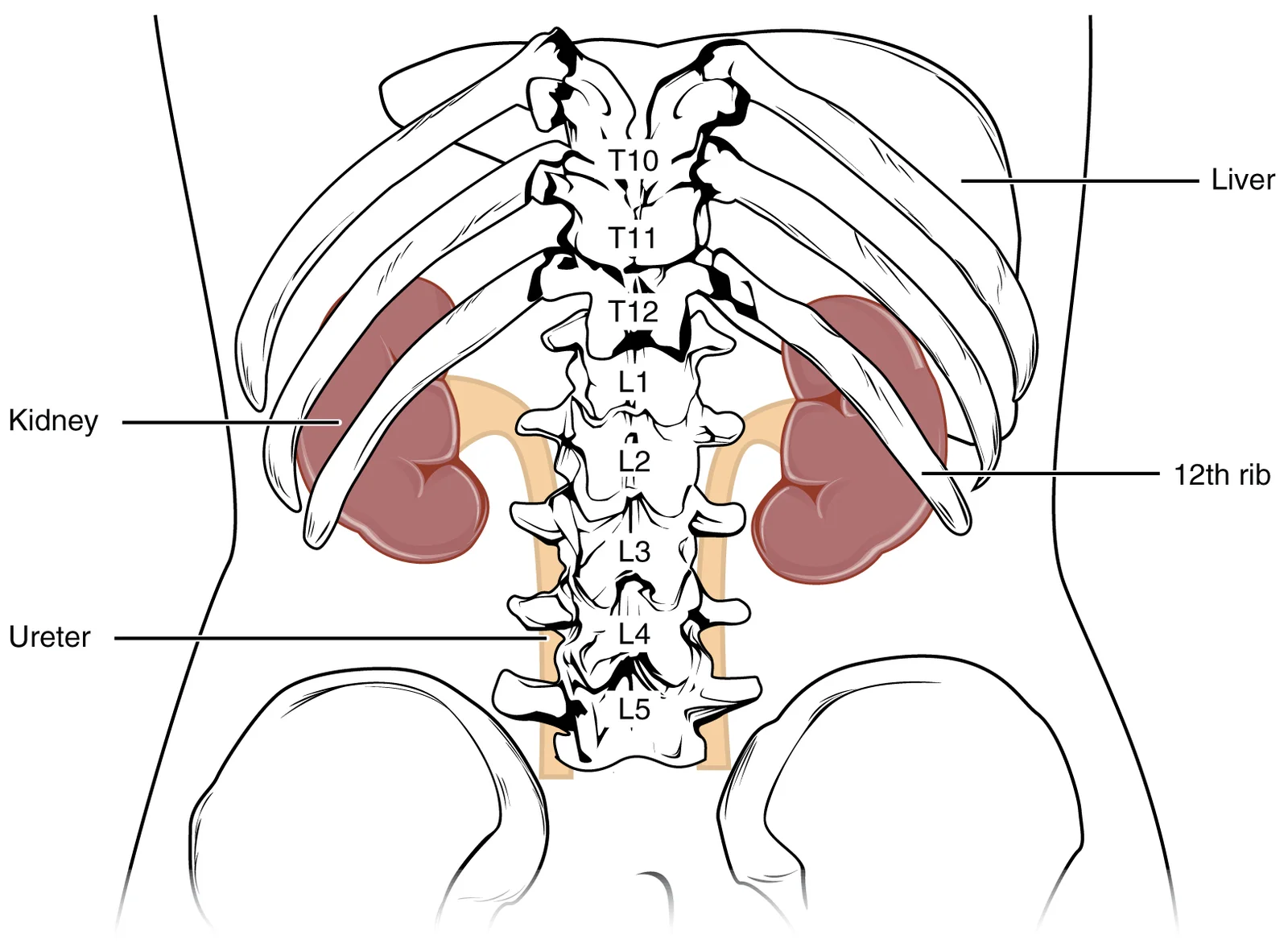
Long description
The kidneys are slightly protected by the ribs and are surrounded by fat for protection (not shown).

Figure 1: The kidneys are slightly protected by the ribs and are surrounded by fat for protection (not shown).
The left kidney is positioned at approximately the T12 to L3 vertebrae, while the right kidney sits slightly lower due to displacement by the liver[1]. The upper portions of the kidneys are somewhat protected by the eleventh and twelfth ribs[1]. Each kidney is directly covered by a fibrous capsule composed of dense, irregular connective tissue that helps maintain its shape and provide protection[1]. This capsule is enveloped by a shock-absorbing layer of adipose tissue known as the renal fat pad, which in turn is encompassed by a tough renal fascia that anchors the kidneys to the posterior abdominal wall[1].
On the superior aspect of each kidney lies the adrenal gland, with the adrenal cortex directly influencing renal function through the production of aldosterone, which stimulates sodium reabsorption[1]. The kidneys receive about 25 percent of cardiac output at rest, making them well vascularized organs[1].
A frontal section through the kidney reveals an outer region called the renal cortex and an inner region called the medulla[1]. The papillae consist of bundles of collecting ducts that transport urine to the calyces for excretion[1].
The renal hilum serves as the entry and exit site for vessels, nerves, lymphatics, and the ureter[1]. Structures emerging from the hilum include the renal pelvis, formed from the major and minor calyxes, which funnels urine via peristalsis into the ureter[1]. The renal arteries originate directly from the descending aorta, while the renal veins return blood directly to the inferior vena cava[1].
The renal artery first divides into segmental arteries, which further branch into interlobar arteries passing through the renal columns to reach the cortex[1]. These interlobar arteries then branch into arcuate arteries, cortical radiate arteries, and finally into afferent arterioles that service approximately 1.3 million nephrons in each kidney[1].
Figure 2Gray1121
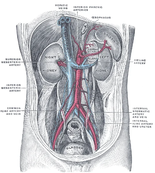
Long description
Posterior abdominal wall, after removal of the peritoneum, showing kidneys, suprarenal capsules, and great vessels. (Corning).

Figure 2: Posterior abdominal wall, after removal of the peritoneum, showing kidneys, suprarenal capsules, and great vessels (Corning).
The kidneys are paired, bean-shaped organs located in the retroperitoneal space of the abdominal cavity. Each kidney weighs approximately 150 grams and is about 11 cm long, 6 cm wide, and 4 cm thick. The superior pole of each kidney is capped by the adrenal gland, while the inferior pole tapers toward the renal pelvis. The kidneys lie at the level of the 12th thoracic to the 3rd lumbar vertebrae, with the right kidney typically positioned slightly lower than the left due to the overlying liver. [3] [1]
The external surface of each kidney is covered by a fibrous capsule that protects the underlying renal parenchyma. The kidney has two main regions: the outer cortex and the inner medulla. The medulla is divided into 8-12 renal pyramids, each with a base adjacent to the cortex and an apex (renal papilla) that projects into a minor calyx. The renal papillae contain openings that allow urine to drain into the minor calyces. [3]
The hilum is a medial indentation where the renal artery enters and the renal vein exits, along with the ureter. The ureter transports urine from the renal pelvis to the bladder. The renal pelvis is a funnel-shaped structure that collects urine from the major calyces and channels it into the ureter. The bladder is largely retroperitoneal and can hold up to 500-600 mL of urine. [3]
The adrenal gland caps the superior pole of each kidney. The ureter emerges from the hilum and carries urine away from the kidney.
DefinitionRenal Hilum
The medial indentation where vessels and nerves enter and exit the kidney.
Figure 3Gray1126
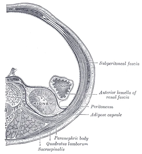
Long description
Transverse section, showing the relations of the capsule of the kidney. (After Gerota.)

Figure 3: Transverse section, showing the relations of the capsule of the kidney (After gerota.).
Figure 4Kidney-numbered
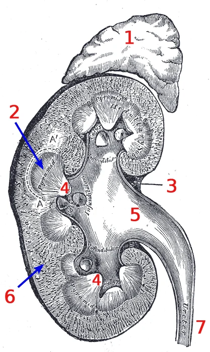
Long description
Vertical section of kidney. A version of File:Gray1127.png with some text removed and numbers added instead, changes done by Daedalus . For non-English projects. English: 1) Adrenal gland 2) Pyramid 3) Renal hilum 4) Papilla 5) Renal pelvis 6) Renal cortex (the medulla is pretty much everything that

Figure 4: Vertical section of kidney.
This position allows them to move slightly with respiration but keeps them relatively fixed against the posterior abdominal wall. The figure labels the position relations, hilum segments, blood supply, ureter course, and constrictions. It also details the hilum and relations on each side, as well as the ureter course, constrictions, and relations.
The kidneys are retroperitoneal structures. The peritoneum covers the anterior surface of the kidneys but does not enclose them completely. Visceral relations differ between the two sides. On the right side, the kidney lies adjacent to the hepatic flexure of the colon and the duodenum. On the left side, it relates to the splenic flexure of the colon, the stomach, the spleen, and the pancreas. Posteriorly, both kidneys rest on the diaphragm and the psoas major muscle. The superior poles are related to the adrenal glands. [3]
Figure 5Gray1120
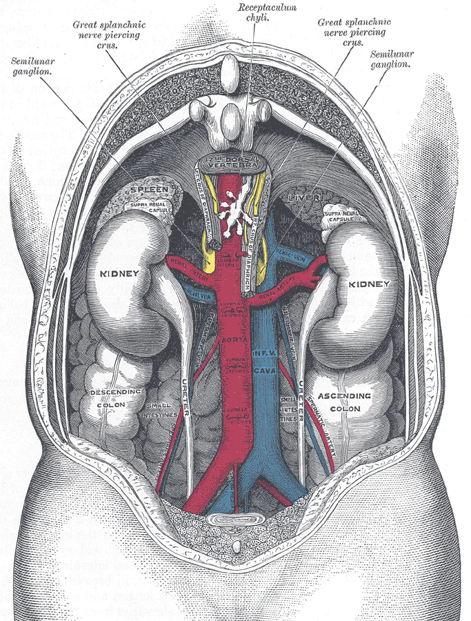
Long description
The relations of the viscera and large vessels of the abdomen. (Seen from behind, the last thoracic vertebra being well raised.)

Figure 5: The relations of the viscera and large vessels of the abdomen.
The right kidney is typically slightly lower than the left due to the presence of the liver. The hilum is the medial indentation where vessels and the ureter enter and exit. The renal artery enters the hilum and divides into anterior and posterior branches. The renal vein exits the hilum and drains into the inferior vena cava. The ureter descends from the renal pelvis. The blood supply is segmental; each segmental artery supplies a specific wedge of renal tissue. This segmental arrangement is important for surgical planning. [1] [3]
The ureter travels from the renal pelvis to the bladder. It has three natural constrictions where stones are likely to lodge. The first is at the ureteropelvic junction. The second is where the ureter crosses the pelvic brim. The third is at the vesicoureteral junction. The ureter descends along the psoas major muscle, crosses the bifurcation of the common iliac artery, and then enters the bladder posteriorly. Children born lacking the oblique course of the ureter through the bladder wall are susceptible to vesicoureteral reflux, which dramatically increases their risk of serious urinary tract infection. [3]
Figure 6Gray1134
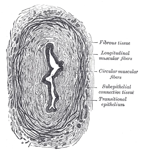
Long description
Transverse section of ureter. Labelled illustration from Wikimedia Commons, used under its open licence.

Figure 6: Transverse section of ureter.
| Constriction | Location | Clinical Significance |
|---|---|---|
| Ureteropelvic junction | Where renal pelvis meets ureter | Common site for stone impaction |
| Pelvic brim | Crossing common iliac artery | Second most common stone site |
| Vesicoureteral junction | Entry into bladder | Third constriction; reflux risk if absent |
Exam trapReflux Risk
The oblique course of the ureter through the bladder wall acts as a valve. Its absence leads to vesicoureteral reflux and increased infection risk.
Figure 7Gray1122
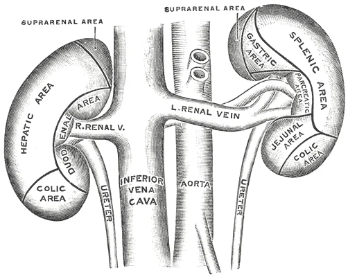
Long description
The anterior surfaces of the kidneys, showing the areas of contact of neighboring viscera.

Figure 7: The anterior surfaces of the kidneys, showing the areas of contact of neighboring viscera.
The kidney's internal structure reveals specialized tissues responsible for filtration and urine formation. The functional unit is the nephron, numbering approximately 1 million per kidney. Each nephron consists of a renal corpuscle and a renal tubule. The renal corpuscle contains the glomerulus, a tuft of capillaries surrounded by Bowman's capsule. Blood enters the glomerulus via the afferent arteriole and exits through the efferent arteriole. [1]
The renal tubule begins with the proximal convoluted tubule, which reabsorbs approximately 65% of the filtered water, sodium, and glucose. It then continues as the loop of Henle, which has a thin descending limb, a thin ascending limb, and a thick ascending limb. The loop of Henle creates the medullary osmotic gradient essential for urine concentration. [1]
The distal convoluted tubule follows, connecting to the collecting duct system. The collecting ducts merge to form papillary ducts, which empty urine into the minor calyces. The kidney's parenchyma is divided into the outer cortex and inner medulla. The cortex contains the renal corpuscles and proximal/distal convoluted tubules, while the medulla contains the loops of Henle and collecting ducts. [1]
The kidney receives about 25% of cardiac output, approximately 1.2-1.3 L/min. This blood flows through the renal artery, which branches into segmental, interlobar, arcuate, and interlobular arteries before forming the afferent arterioles. The renal vein exits the kidney at the hilum and drains into the inferior vena cava. [1]
The cortex appears as the outer darker region, while the medulla consists of the renal pyramids with their bases facing the cortex and apices pointing toward the renal sinus.
The kidneys receive their blood supply from the renal arteries, which arise directly from the abdominal aorta at the level of the L1–L2 intervertebral disc. Each renal artery is a large, high-flow vessel because the kidneys receive about 20–25% of the cardiac output, despite constituting less than 0.5% of body weight. Before entering the renal hilum, each renal artery divides into anterior and posterior divisions, which in turn give rise to segmental arteries. [1] [3]
The segmental arteries are end arteries: they do not anastomose with one another. This means that occlusion of a segmental artery produces a well-demarcated area of infarction. There are five renal segments, each supplied by its own segmental artery: the superior (apical), anterior superior, anterior inferior, inferior, and posterior segments. The posterior segment is supplied by the posterior division, while the anterior division supplies the remaining four. The segmental arteries enter the renal sinus and divide into lobar arteries, one for each renal pyramid. Each lobar artery gives off two or three interlobar arteries that run between the pyramids toward the cortex.
At the corticomedullary junction, the interlobar arteries branch into arcuate arteries, which arch over the bases of the pyramids. From the arcuate arteries arise interlobular (cortical radiate) arteries that pass outward through the cortex. Each interlobular artery gives off afferent arterioles, which supply the glomerular capillary tufts. The efferent arteriole leaves the glomerulus and, for cortical nephrons, forms a peritubular capillary network around the tubules; for juxtamedullary nephrons, it descends as the vasa recta into the medulla. This arrangement—a glomerular capillary bed drained by an arteriole that forms a second capillary bed—constitutes a portal system, the only one in which an arteriole lies between the two capillary beds. [1]
Figure 8Kidney nephron
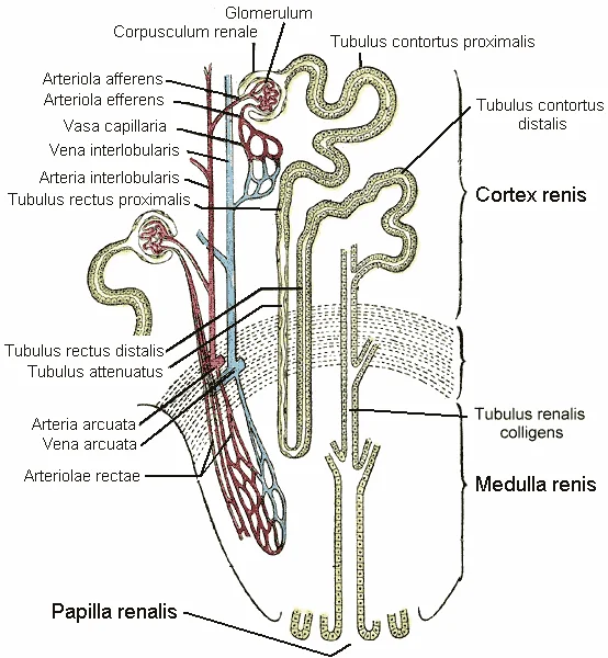
Long description
A nephron of the kidney. from Gray's Anatomy 1918. The illustration includes an important anatomical error in that it fails to depict the distal tubulus ( tubulus rectus distalis ) passing by vascular pole of the glomerulus before becoming the distal convoluted tubule ( tubulus contortus distalis ).

Figure 8: A nephron of the kidney (from gray's anatomy 1918).
Each renal artery divides near the hilum into the anterior and posterior divisions. The anterior division branches into the superior, anterior superior, anterior inferior, and inferior segmental arteries, which fan out over the anterior surface of the kidney. The posterior division continues as the posterior segmental artery, which curves around the lateral border to supply the posterior surface. From the segmental arteries, interlobar arteries ascend between the pyramids, arcuate arteries arch over their bases, and interlobular arteries radiate into the cortex, ending in afferent arterioles that enter the glomeruli.
Figure 9Gray1132
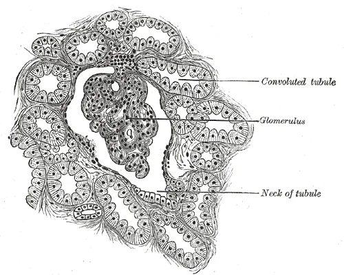
Long description
Section of cortex of human kidney. Labelled illustration from Wikimedia Commons, used under its open licence.

Figure 9: Section of cortex of human kidney.
The venous system of the kidney is distinct because it lacks valves, a feature that permits retrograde flow of infection or tumour thrombus from the renal vein into the inferior vena cava and subsequently to the heart or lungs. This anatomical arrangement is critical for understanding the spread of renal cell carcinoma. [1]
The right renal vein is shorter than its left counterpart. It arises from the posterior aspect of the right kidney, specifically from the renal hilum. It terminates by draining directly into the inferior vena cava. Due to its short length, it is more susceptible to injury during surgical procedures involving the retroperitoneum. The right renal vein receives no significant tributaries before its termination, although it may occasionally receive a small lumbar vein.
The left renal vein is significantly longer than the right. It originates from the left renal hilum and travels horizontally to the right, passing anterior to the aorta and the left crus of the diaphragm. A key anatomical landmark is its relationship with the superior mesenteric artery. The left renal vein passes between the abdominal aorta posteriorly and the superior mesenteric artery anteriorly. This space is known as the aortomesenteric angle. The left renal vein terminates by draining into the anterior aspect of the inferior vena cava. Before its termination, it receives several tributaries, including the left gonadal vein and the left suprarenal vein. The left gonadal vein drains into the left renal vein, whereas the right gonadal vein drains directly into the inferior vena cava. This difference explains why varicoceles are more common on the left side.
Within the kidney, the venous drainage follows a pattern parallel to the arterial supply but in reverse. Blood flows from the interlobular veins into the arcuate veins. The arcuate veins run along the boundary between the renal cortex and the renal medulla, arching over the bases of the renal pyramids. From the arcuate veins, blood drains into the interlobar veins. The interlobar veins run between the renal pyramids, accompanying the interlobar arteries. They converge at the renal hilum to form the single renal vein.
Understanding this drainage is vital for interpreting imaging. On computed tomography, the left renal vein is often visualised passing between the aorta and the superior mesenteric artery. Compression of the left renal vein in this space can lead to nutcracker syndrome, characterised by haematuria and left-sided varicocele. The absence of valves means that increased pressure in the inferior vena cava, such as in right heart failure, can lead to renal congestion.
Figure 10Gray1123
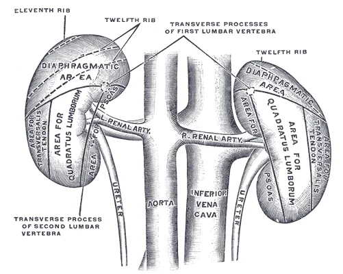
Long description
The posterior surfaces of the kidneys, showing areas of relation to the parietes.

Figure 10: The posterior surfaces of the kidneys, showing areas of relation to the parietes.
The lymphatic drainage of the kidneys follows a predictable pathway that mirrors the vascular supply, ensuring efficient removal of interstitial fluid and immune surveillance. Understanding this route is essential for appreciating how renal pathology, such as carcinoma or infection, may spread to regional nodal groups.
The lymphatic vessels originate from a dense network within the renal sinus and hilum. These initial vessels accompany the renal arteries and veins as they exit the kidney. The lymphatics are thin-walled and valvulated, preventing backflow towards the organ. They collect fluid from the peritubular capillaries and the interstitial spaces of the renal cortex and medulla. [1]
The efferent lymphatic vessels from the kidney drain primarily into the lateral aortic lymph nodes, also known as the lumbar lymph nodes. These nodes are situated on either side of the abdominal aorta and inferior vena cava. Specifically, the right renal lymphatics drain into the right lateral aortic nodes, while the left renal lymphatics drain into the left lateral aortic nodes. This arrangement is ipsilateral, meaning the drainage does not cross the midline at this stage. The lumbar nodes receive lymph from the kidneys, adrenal glands, and gonads. [1]
From the lateral aortic nodes, lymph flows into the cisterna chyli, a dilated sac located anterior to the second lumbar vertebra. The cisterna chyli is formed by the union of the left and right lumbar trunks and the intestinal trunk. From the cisterna chyli, lymph ascends through the aortic opening of the diaphragm to become the thoracic duct. The thoracic duct then drains into the junction of the left internal jugular and left subclavian veins. In rare anatomical variations, right-sided drainage may enter the right lymphatic duct, which empties into the right venous angle. [2]
Figure 11Gray1124
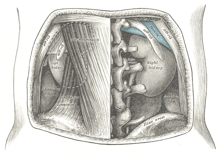
Long description
The relations of the kidneys from behind.

Figure 11: The relations of the kidneys from behind.
Clinical pearlClinical Relevance
Renal cell carcinoma often metastasises to the lateral aortic lymph nodes before spreading distantly.
| Structure | Destination |
|---|---|
| Renal sinus | Lateral aortic nodes |
| Lateral aortic nodes | Cisterna chyli |
| Cisterna chyli | Thoracic duct |
The renal innervation is derived from the autonomic nervous system, specifically the sympathetic division, which plays a critical role in regulating renal blood flow and glomerular filtration rate. The sympathetic fibres originate from the T10 to L1 spinal segments. These preganglionic neurons synapse in the coeliac and superior mesenteric ganglia. Postganglionic fibres then travel along the renal arteries to reach the kidney parenchyma. This pathway allows for vasoconstriction of the afferent arterioles during stress or hypovolaemia, thereby reducing renal perfusion to preserve systemic blood pressure. The density of sympathetic innervation is highest in the juxtaglomerular apparatus, where it stimulates renin release. This mechanism is fundamental to the regulation of blood pressure via the renin-angiotensin-aldosterone system.
DefinitionSympathetic Control
Sympathetic innervation to the kidney arises from T10-L1, synapsing in the coeliac and superior mesenteric ganglia, and travels with the renal arteries to modulate blood flow and renin secretion.
In addition to autonomic input, the kidneys receive somatic sensory innervation. This is crucial for the perception of pain. The afferent pain fibres travel alongside the sympathetic efferent fibres, returning to the T10 to L1 spinal segments. Because these spinal segments also receive somatic sensory input from the skin of the upper lumbar region and the flank, pain originating from the kidney is often perceived in these distant areas. This phenomenon is known as referred pain. For instance, a renal calculus obstructing the ureter may cause severe flank pain that radiates towards the groin. Understanding this pathway is essential for clinical diagnosis, as it explains why patients with renal colic often present with pain in the loin rather than the abdomen.
Figure 12Gray1125
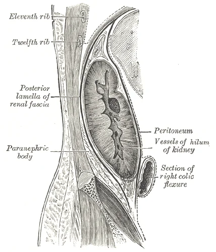
Long description
Sagittal section through posterior abdominal wall, showing the relations of the capsule of the kidney. (After Gerota).

Figure 12: Sagittal section through posterior abdominal wall, showing the relations of the capsule of the kidney (After gerota).
Dotted lines extend from these spinal levels to the skin of the flank and upper lumbar region, indicating the dermatomes associated with referred pain.
NoteClinical Relevance
Referred pain from the kidney is felt in the T10-L1 dermatomes, typically the flank and upper lumbar region, due to shared spinal cord segments with somatic sensory nerves.
The somatic component also includes the phrenic nerve, which provides sensory innervation to the diaphragmatic surface of the kidney capsule. Irritation of this area, such as from a subcapsular haematoma, can cause shoulder tip pain via the phrenic nerve referral to the C3-C5 dermatomes. This distinct pattern helps differentiate renal pathology from other abdominal conditions. The integration of these autonomic and somatic pathways ensures that the kidney is both functionally regulated and sensitively monitored for injury or obstruction.
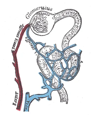
The kidney is composed of approximately one million functional units called nephrons. Each nephron represents the fundamental structural and functional component responsible for blood filtration and urine formation. The nephrons are distributed throughout the renal cortex and medulla, with approximately 80-85% located in the cortex and the remaining 15-20% extending into the medulla. [1]
Figure 13Gray1129
Long description
Distribution of bloodvessels in cortex of kidney.

Figure 13: Distribution of bloodvessels in cortex of kidney.
Each nephron consists of a renal corpuscle and a renal tubule. The renal corpuscle, located in the renal cortex, contains a tuft of capillaries called the glomerulus, which is surrounded by Bowman's capsule. The glomerular filtration barrier consists of three layers: fenestrated endothelium, basement membrane, and podocytes with foot processes. This specialized structure allows for the filtration of water, electrolytes, and small molecules while preventing the passage of larger proteins and blood cells. [1]
The renal tubule begins with the proximal convoluted tubule, which has a simple cuboidal epithelium with prominent brush border microvilli. These microvilli significantly increase the surface area for reabsorption. This segment is responsible for the reabsorption of approximately 65% of filtered sodium, water, and glucose. Additionally, it reabsorbs almost all of the filtered amino acids, vitamins, and proteins through endocytosis. [1] [2]
The loop of Henle has a thin descending limb, a thin ascending limb, and a thick ascending limb. The thin segments consist of simple squamous epithelium, while the thick ascending limb contains mitochondria-rich cells that actively transport sodium and chloride. This segment is impermeable to water but actively transports solutes, contributing to the concentration gradient in the medulla. [1]
The distal convoluted tubule has a simple cuboidal epithelium without brush borders. It contains fewer mitochondria compared to the proximal tubule and is involved in fine-tuning sodium and potassium balance under hormonal control. The collecting duct system begins with connecting tubules and leads to cortical and medullary collecting ducts. These ducts are responsive to antidiuretic hormone (ADH) and aldosterone, regulating final urine concentration and electrolyte balance. [1] [2]
The kidney also contains specialized cells such as juxtaglomerular cells that produce renin in response to low blood pressure. These cells are located at the vascular pole of the glomerulus and play a crucial role in the renin-angiotensin-aldosterone system. Interstitial cells between tubules produce erythropoietin, which stimulates red blood cell production. The renal medulla has a unique arrangement of loops of Henle and vasa recta to create the osmotic gradient for urine concentration, ranging from approximately 300 mOsm/kg in the cortex to 1200-1400 mOsm/kg in the inner medulla. [3] [2]
The kidneys originate from the intermediate mesoderm, with the process beginning in the fourth week of embryonic development. This development occurs in three successive, overlapping stages: the pronephros, the mesonephros, and the metanephros. The pronephros is the most primitive stage; it appears first but is non-functional and degenerates by the end of the fourth week. [1]
The mesonephros follows the pronephros and functions temporarily as an interim kidney during early development. While it largely degenerates by the sixth week, it is not entirely lost; some of its structures persist in the male reproductive system, contributing to the ductal system. [1]
The permanent kidneys, or metanephros, develop from two distinct sources: the metanephric blastema and the metanephric duct, also known as the ureteric bud. The ureteric bud grows out from the mesonephric duct and penetrates the metanephric blastema. This interaction is critical, as the ureteric bud induces the metanephric blastema to differentiate and form the nephrons. Specifically, the metanephric duct gives rise to the collecting system, including the ureter, renal pelvis, major and minor calyces, and collecting ducts, while the metanephric blastema forms the renal parenchyma, including the glomeruli and tubules. [1]
Between the fifth and eighth weeks, the kidneys undergo a process of ascent. They move from their initial pelvic position to their final location in the retroperitoneum. During this migration, the kidneys rotate medially. This rotation ensures that the renal pelvis faces anteriorly and the hilum faces medially. [1]
The vascular supply evolves during this ascent. The kidneys receive blood from successive branches of the aorta as they move upward. Normally, the lower vessels degenerate, leaving a single renal artery. However, if these lower vessels persist, it results in aberrant renal artery patterns, such as accessory renal arteries. Development is complete by the 34th week of gestation, although nephrogenesis continues until birth. [1] [2]
NoteEmbryological Link
For a detailed study of these processes, refer to the embryology page.
Renal Development
- Intermediate MesodermOrigin of all three renal systems
- PronephrosNon-functional; degenerates by end of week 4
- MesonephrosTemporary function; degenerates by week 6
- MetanephrosPermanent kidney; formed by blastema and ureteric bud
- Ascent and RotationPelvis to retroperitoneum; medial rotation
The kidneys are paired retroperitoneal organs located in the upper abdomen, typically between the T12 and L3 vertebral levels. On cross-sectional imaging, the right kidney is usually slightly lower than the left due to the liver's position. Each kidney is approximately 10-12 cm in length, 5-7 cm in width, and 3 cm in thickness. [3]
The renal parenchyma is divided into an outer cortex and an inner medulla. The medulla contains the renal pyramids, which are triangular structures that drain into the minor calyces. These minor calyces merge to form major calyces, which then converge to form the renal pelvis. The renal pelvis funnels urine into the ureter, which courses inferiorly to the urinary bladder. [3]
On a CT scan, the kidneys appear as bilateral, bean-shaped structures with homogeneous attenuation values of 30-50 Hounsfield units (HU) in non-contrast images. The renal cortex appears slightly more dense than the medulla. [3]
In an intravenous urogram, the kidneys are visualized as the contrast agent is filtered by the glomeruli and excreted. The normal nephrogram phase shows uniform opacification of the renal parenchyma, followed by visualization of the calyces, renal pelvis, and ureters. The ureters course inferiorly along the psoas muscle, crossing over the iliac vessels to enter the bladder at the ureteric orifices. [3]
On CT, the bladder appears as a fluid-filled structure with walls measuring approximately 3-5 mm in thickness when empty. When distended, its dome extends superiorly, and its capacity can reach 400-500 ml in adults. [3]
Figure 14Blausen 0592 KidneyAnatomy 01
Long description
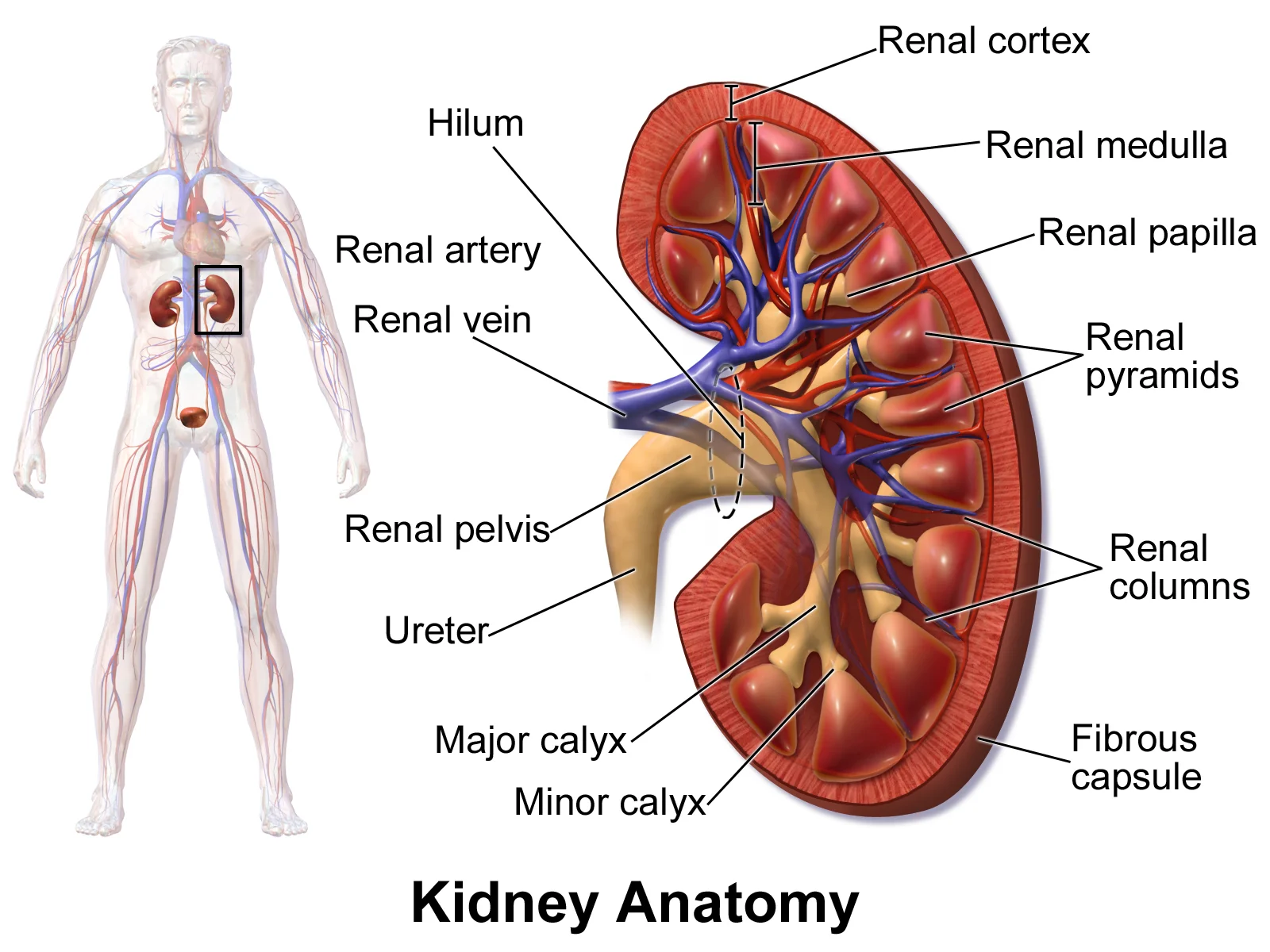
Kidney Anatomy. See a related animation of this medical topic.

Figure 14: Kidney Anatomy.
The kidneys lie retroperitoneally on the posterior abdominal wall, one on each side of the vertebral column, extending from the level of the T12 vertebra down to L3. The right kidney sits slightly lower than the left because of the liver above it. Each kidney measures roughly 11 cm in length, 6 cm in width and 3 cm in anteroposterior thickness. The hilum, where the renal vessels and pelvis enter, faces anteromedially. [1]
The renal artery arises from the abdominal aorta at L1–L2 level, just below the superior mesenteric artery. The right renal artery is longer and passes posterior to the inferior vena cava. Each artery divides into anterior and posterior divisions, which further split into segmental arteries. Five renal segments exist: apical, upper anterior, middle anterior, lower and posterior. These segments are clinically crucial because they have no collateral anastomosis; occlusion of a segmental artery causes infarction of that segment alone. [1]
The renal vein lies anterior to the artery at the hilum. The left renal vein is longer, crosses anterior to the aorta, and receives the left gonadal vein and left suprarenal vein. The right renal vein is short and receives no tributaries. This asymmetry matters: thrombosis of the left renal vein may present with haematuria and a varicocele on the left side. [1]
The ureter passes downwards retroperitoneally, crossing the pelvic brim at the bifurcation of the common iliac artery. Three constrictions occur: at the pelviureteric junction, where it crosses the iliac vessels, and at the vesicoureteric junction. Renal calculi commonly lodge at these sites, causing colic. [1]
The renal cortex forms the outer rim; the medulla contains 8–12 renal pyramids, whose apices (papillae) project into minor calyces. Two or three minor calyces join to form a major calyx; two or three major calyces unite to form the renal pelvis, which narrows into the ureter. The renal sinus contains fat, vessels and the calyces. [1]
The kidney is surrounded by three layers: the fibrous renal capsule, the perirenal fat, and the renal fascia (Gerota's fascia). The fascia is open inferiorly, allowing the kidney to move with respiration. A renal injury may cause blood to track inferiorly along the psoas muscle. [1]
The suprarenal glands sit on the upper poles. The right gland is pyramidal, the left crescentic. Each has a cortex (corticosteroids) and medulla (catecholamines). The arterial supply comes from the inferior phrenic, aorta and renal artery; venous drainage is to the inferior vena cava on the right and renal vein on the left.
Clinical pearlClinical pearl
A stab wound in the posterior axillary line at the 12th rib level may injure the lower pole of the kidney, the ureter or the renal pelvis, because these structures lie deep to the costovertebral angle.
- The kidney lies retroperitoneally at the T12–L3 level, with the right kidney slightly lower than the left because of the liver; the upper pole of the left kidney reaches the 11th rib, the right the 12th rib.
- The renal hilum receives, from anterior to posterior: renal vein, renal artery, and renal pelvis (mnemonic: VAP). The ureter passes posterior to the renal vessels.
- The renal artery divides into five segmental branches that are end arteries — occlusion of any segmental artery causes a wedge-shaped infarct; there is no collateral circulation between segments.
- The renal cortex contains glomeruli and convoluted tubules; the medulla contains 8–18 renal pyramids, each ending in a papilla that projects into a minor calyx.
- Two or three minor calyces join to form a major calyx; 2–3 major calyces form the renal pelvis, which narrows at the ureteropelvic junction — a common site of stone impaction.
- The renal capsule is a tough fibrous layer; perinephric fat surrounds it, and Gerota's fascia (renal fascia) encloses the kidney and adrenal gland, with the anterior and posterior layers fusing superiorly and laterally but remaining open inferiorly — allowing fluid to track downward.
- The ureter has three physiological narrowings: at the pelviureteric junction, where it crosses the pelvic brim over the iliac vessels, and at the vesicoureteric junction — the most common sites for stone lodgement.
- The suprarenal (adrenal) gland sits on the upper pole of each kidney; the right is pyramidal and related anteriorly to the inferior vena cava, the left is semilunar and related anteriorly to the pancreas and splenic vessels.
- The renal plexus (sympathetic from T10–L1 via the lesser and least splanchnic nerves) and parasympathetic vagal fibres supply the kidney; pain from the kidney refers to the loin and radiates to the groin and scrotum/labium majus along the genitofemoral and ilioinguinal nerves.
- The lymphatic drainage of the kidney goes to the lateral aortic (para-aortic) nodes at L1–L2; the ureter drains to the common, external and internal iliac nodes.
- A common trap: the right renal artery passes posterior to the inferior vena cava, and the left renal vein crosses anterior to the aorta, posterior to the superior mesenteric artery — compression here causes the nutcracker phenomenon.
- Another trap: the ureter runs anterior to the psoas major and is crossed by the gonadal vessels anteriorly; during pelvic surgery, the ureter is at risk where it passes under the uterine artery ("water under the bridge").
A common mistake is confusing the structures involved in urine formation. The glomerulus is not a standalone structure but a network of capillaries surrounded by Bowman's capsule. Students often overlook that filtration is regulated by fenestrations in capillary endothelial cells, podocytes with filtration slits, membrane charge, and the basement membrane. These components work together to create filtrate without cells or large proteins. [2]
Another error is misunderstanding the juxtaglomerular apparatus (JGA). The JGA includes the macula densa, which is a cluster of cuboidal epithelial cells in the distal convoluted tubule that monitors fluid composition. Students may not realize that the macula densa releases paracrine signals (ATP and adenosine) in response to Na+ concentration and fluid flow rate. [2]
Exam trapMisconception about filtration
The glomerulus does not act as a simple sieve; its filtration is highly regulated by multiple cellular and molecular mechanisms.
Students often confuse the proximal convoluted tubule (PCT) with other nephron segments. The PCT has simple cuboidal cells with prominent microvilli forming a brush border, which maximizes absorption and secretion of solutes. Its high metabolic activity is indicated by the abundance of mitochondria for ATP production. [2]
A frequent error is misunderstanding aquaporins. These are water channels that allow only water to pass through cell membranes. Six types of aquaporins are found in the kidney, each facilitating water movement across lipid-rich membranes. Students may incorrectly believe aquaporins transport other substances. [2]
Urine Formation Process
- Glomerular FiltrationBlood filtered through glomerulus and Bowman's capsule
- Tubular Reabsorption & SecretionOccurs mainly in proximal convoluted tubule
- ConcentrationMediated by aquaporins in collecting ducts
- StorageUrine stored in bladder until elimination
- EliminationUrine expelled through urethra
In clinical practice, knowledge of kidney anatomy is essential for understanding urinary function. The juxtaglomerular apparatus's role in monitoring Na+ concentration and fluid flow rate is crucial for managing patients with electrolyte imbalances or hypertension. Clinicians use this knowledge when prescribing medications that affect renal blood flow or tubular function.
In the operating theatre, understanding the male urethra's anatomy is vital during urological procedures. The male urethra averages 20 cm in length and is divided into four regions: preprostatic, prostatic, membranous, and spongy urethra. Surgeons must be aware that the prostatic urethra receives sperm via ejaculatory ducts and secretions from seminal vesicles, which is important during prostate surgery. [3]
On imaging, knowledge of kidney structures helps interpret radiological findings. The detrusor muscle's function in bladder emptying is assessed in urodynamic studies. Clinicians recognize that bladder strength diminishes with age, but voluntary contractions of abdominal muscles can increase intra-abdominal pressure to promote emptying, which is relevant in evaluating urinary retention. [3]
In examinations, students must demonstrate understanding of renal anatomy through various question formats. Common questions involve identifying structures on diagrams, describing the path of urine formation, or explaining the functional significance of different nephron segments. Students are often asked to differentiate between male and female urethral anatomy, as this is a key anatomical difference in the urinary system.
NoteClinical relevance
Knowledge of aquaporins in the kidney helps understand how certain medications (like vasopressin analogs) affect water reabsorption in conditions like diabetes insipidus.
- The glomerulus and Bowman's capsule work together to filter blood, creating filtrate without cells or large proteins
- The juxtaglomerular apparatus monitors Na+ concentration and fluid flow rate, releasing ATP and adenosine as signals
- The proximal convoluted tubule has microvilli forming a brush border to maximize absorption and secretion
- Six types of aquaporins in the kidney facilitate water movement across cell membranes
- The male urethra averages 20 cm and has four distinct regions [3]
- Bladder emptying involves detrusor muscle contraction, which can be assisted by voluntary abdominal muscle contraction
- The urethra is the only urinary structure with significant anatomical differences between males and females [3]
Answer each one in your head first, then open the answer.
What structures regulate glomerular filtration?
Show answerHide answer
Fenestrations in capillary endothelial cells, podocytes with filtration slits, membrane charge, and the basement membraneWhat signals does the macula densa release?
Show answerHide answer
ATP and adenosineWhat is the function of the microvilli in the proximal convoluted tubule?
Show answerHide answer
They create a large surface area to maximize absorption and secretion of solutesHow many types of aquaporins are found in the kidney?
Show answerHide answer
SixWhat are the four regions of the male urethra?
Show answerHide answer
Preprostatic, prostatic, membranous, and spongy (penile) urethra
- Betts JG, Young KA, Wise JA, et al. Gross Anatomy of the Kidney. OpenStax, Anatomy and Physiology 2e (CC BY-NC-SA 4.0). Accessed .
- Betts JG, Young KA, Wise JA, et al. Microscopic Anatomy of the Kidney. OpenStax, Anatomy and Physiology 2e (CC BY-NC-SA 4.0). Accessed .
- Betts JG, Young KA, Wise JA, et al. Gross Anatomy of Urine Transport. OpenStax, Anatomy and Physiology 2e (CC BY-NC-SA 4.0). Accessed .
Figure credits
- Figure 1: OpenStax College, via Wikimedia Commons, CC BY 3.0, CC BY 3.0. resized.
- Figure 2: Henry Vandyke Carter, via Wikimedia Commons, Public domain, Public domain.
- Figure 3: Henry Vandyke Carter, via Wikimedia Commons, Public domain, Public domain.
- Figure 4: Henry Vandyke Carter, via Wikimedia Commons, Public domain, Public domain.
- Figure 5: Henry Vandyke Carter, via Wikimedia Commons, Public domain, Public domain.
- Figure 6: Henry Vandyke Carter, via Wikimedia Commons, Public domain, Public domain.
- Figure 7: Henry Vandyke Carter, via Wikimedia Commons, Public domain, Public domain.
- Figure 8: Henry Gray, via Wikimedia Commons, Public domain, Public domain.
- Figure 9: Henry Vandyke Carter, via Wikimedia Commons, Public domain, Public domain.
- Figure 10: Henry Vandyke Carter, via Wikimedia Commons, Public domain, Public domain.
- Figure 11: Henry Vandyke Carter, via Wikimedia Commons, Public domain, Public domain.
- Figure 12: Henry Vandyke Carter, via Wikimedia Commons, Public domain, Public domain.
- Figure 13: Henry Vandyke Carter, via Wikimedia Commons, Public domain, Public domain.
- Figure 14: BruceBlaus. When using this image in external sources it can be cited as: Blausen.com staff (2014). "Medical gallery of Blausen Medical 2014". WikiJournal of Medicine 1 (2). DOI:10.15347/wjm/2014.010. ISSN 2002-4436., via Wikimedia Commons, CC BY 3.0, CC BY 3.0.
Educational content for medical students and doctors. It is not a substitute for clinical judgement or local guidelines.