Spleen
The spleen is a vascular secondary lymphoid organ located in the left upper quadrant, responsible for blood filtration and immune response.
The spleen is a major secondary lymphoid organ, often described as the “filter of the blood” due to its extensive vascularization and the presence of macrophages and dendritic cells that remove microbes and dying red blood cells. It is approximately 12 cm (5 in) long and is attached to the lateral border of the stomach via the gastrosplenic ligament. The organ is fragile, lacking a strong capsule, and appears dark red due to its rich blood supply. It serves as a primary site for immune responses to blood-borne pathogens. [1]
DefinitionSplenic Architecture
The spleen is divided by trabeculae of connective tissue into red pulp, consisting mostly of red blood cells, and white pulp, which resembles lymphoid follicles. [1]
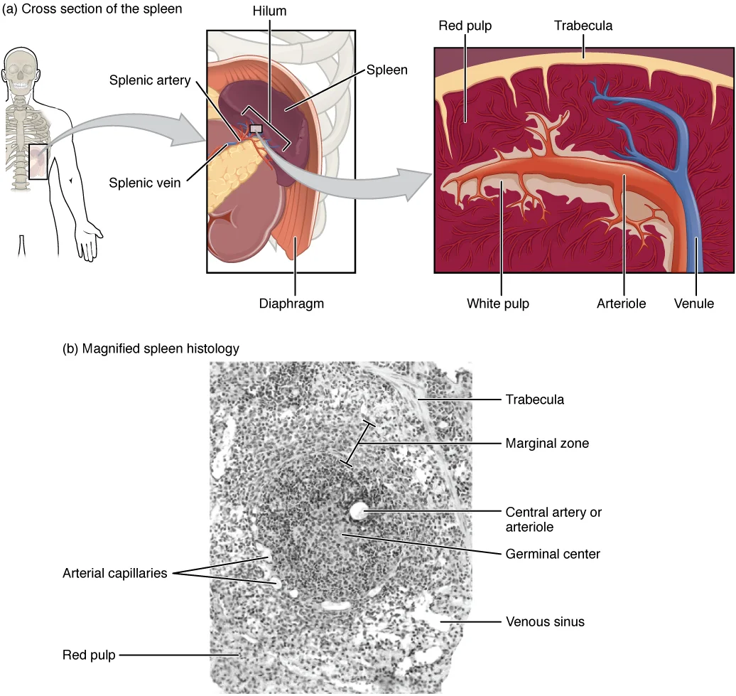
The splenic artery splits into several arterioles surrounded by white pulp, eventually entering sinusoids. Blood from capillaries collects in venous sinuses and leaves via the splenic vein. The red pulp contains reticular fibers with fixed macrophages, free macrophages, and typical blood cells including lymphocytes. The white pulp surrounds a central arteriole and consists of germinal centers of dividing B cells surrounded by T cells and accessory cells. [1]
NoteAccessory Spleens
Accessory spleens are usually surrounded by a fibrotic capsule separating them from adjacent pancreatic parenchyma. They contain red and white pulp similar to the normal spleen. [2]
On ultrasound, an accessory spleen appears as a round or oval mass with mildly echogenic, homogeneous texture and posterior enhancement. A high-amplitude interface, corresponding to a fibrotic capsule, is typically observed. Color or power Doppler ultrasound shows a vascular hilum entering the lesion, a sensitive diagnostic feature with 90% sensitivity. [2]
Technetium-99m HDRBC scintigraphy is highly specific for detecting splenic tissue, as up to 90% of injected HDRBCs are trapped by splenic tissue. The diagnostic criterion for accessory spleen is a marked increase in uptake exceeding that of the cardiac blood pool and major vessels at the suspected site. [2]
Exam trapDifferentiation
Intrapancreatic accessory spleen can be differentiated from nonfunctioning islet cell tumors by lesion size and heterogeneity due to necrosis, but differentiation from functioning tumors is difficult on imaging alone.
Epidermoid cysts of the spleen are rare, accounting for 10 to 20% of non-parasitic splenic cysts. They are lined with stratified squamous epithelium but lack skin appendages. Cysts of accessory spleens are extremely rare, with only about 10 reported cases, all located in the pancreatic tail. [2]
| Feature | Finding |
|---|---|
| US Texture | Round/oval, mildly echogenic, homogeneous |
| Doppler | Vascular hilum (90% sensitivity) |
| Scintigraphy | Uptake exceeds cardiac blood pool |
| CT Enhancement | Similar to spleen in arterial/portal phases |
When epidermoid cysts of an accessory spleen are small and asymptomatic, surgery might not be necessary, but resection is often recommended to exclude malignancy. En-bloc resection including normal pancreatic margins is advised. IPAS shows inhomogeneous enhancement on early phases, a characteristic pattern aiding diagnosis. SPIO-enhanced MRI shows significant signal drop similar to the spleen, while contrast-enhanced US shows prolonged enhancement in the delayed hepatosplenic phase. [2]
Figure 1Gray1188
Long description
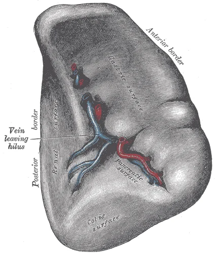
The visceral surface of the spleen. Labelled illustration from Wikimedia Commons, used under its open licence.

Figure 1: The visceral surface of the spleen.
The organ is enclosed by a fibrous capsule. Beneath this capsule lies the parenchyma, which is divided into two distinct functional zones. The outer zone is the white pulp, appearing as lymphoid aggregates. The inner, larger zone is the red pulp, consisting of venous sinuses and splenic cords.
The spleen is an intraperitoneal organ situated in the left hypochondrium. It is covered by peritoneum, except at the hilum where vessels enter and exit. This visceral location allows it to relate closely to several abdominal structures.
The diaphragmatic surface is convex and smooth, facing the diaphragm. It relates to the left lung and pleura superiorly. The visceral surface is irregular and concave. It presents impressions for adjacent organs. The gastric impression faces medially and relates to the stomach. The renal impression lies posteriorly and relates to the left kidney. The colic impression is inferior and relates to the splenic flexure of the colon. The pancreatic impression is medial and relates to the tail of the pancreas.
The spleen is held in position by ligaments. It contains the short gastric vessels and the left gastro-omental vessels. The splenorenal ligament connects the hilum to the left kidney. It contains the tail of the pancreas, the splenic artery, and the splenic vein. These ligaments define the splenic bed.
Accessory spleens may be present. They are small nodules of splenic tissue. They are usually found near the hilum or within the gastrosplenic ligament. They are clinically significant because they may remain after splenectomy.
The blood supply is critical. The splenic artery is a branch of the coeliac trunk. It is tortuous and follows the superior border of the pancreas. It enters the splenorenal ligament. The splenic vein drains the spleen. It runs posterior to the pancreas. [2]
Lymphatic drainage follows the arterial supply. Lymph vessels accompany the splenic artery. They drain into the coeliac lymph nodes. This pathway is important for the spread of malignancy.
The spleen is vulnerable to trauma. Its position makes it susceptible to rupture in blunt abdominal injury. The capsule is thin and fragile. Rupture leads to significant haemorrhage. This is a surgical emergency.
Understanding these relations is essential for surgery. Access to the hilum requires careful dissection. The tail of the pancreas is at risk during splenectomy. Preservation of the pancreas is desirable to prevent diabetes. The short gastric vessels must be ligated. The left gastro-omental vessels must be controlled.
The diaphragmatic surface is smooth. This reduces friction during respiration. The visceral surface is complex. The impressions reflect the pressure from adjacent organs. The gastric impression is large. The renal impression is smaller. The colic impression is variable. The pancreatic impression is distinct.
The ligaments are peritoneal folds. They provide stability. They also contain neurovascular bundles. The gastrosplenic ligament is short. The splenorenal ligament is long. It allows mobility of the spleen. This mobility is important for growth.
The vascular anatomy is variable. The splenic artery may have multiple branches. The splenic vein may have tributaries. These variations affect surgical planning. Imaging is crucial pre-operatively. CT and MRI show the anatomy well.
The lymph nodes are small. They filter lymph from the spleen. They are located along the splenic artery. They are palpable only when enlarged. Enlargement suggests infection or malignancy.
The spleen is a lymphoid organ. It filters blood. It removes old red blood cells. It stores platelets. It produces lymphocytes. These functions depend on its structure. The red pulp filters blood. The white pulp mounts immune responses. [1]
The relations define the surgical approach. The left kidney is posterior. The stomach is anterior. The colon is inferior. The pancreas is medial. The diaphragm is superior. This spatial arrangement guides the surgeon.
The labels identify each structure. The spleen is on the left. The liver is on the right. The stomach is central. The colon is inferior. The kidneys are posterior.
This knowledge is fundamental. It prevents injury during surgery. It aids in diagnosis. It explains clinical signs. It guides treatment. Mastery of these relations is required for safe practice.
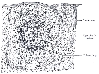
The spleen is a highly vascular organ situated in the left hypochondrium, protected by the rib cage. Its internal architecture is defined by a supportive framework and distinct functional zones. The organ is enclosed by a dense fibrous capsule. This capsule extends inward as trabeculae, which provide structural support and serve as conduits for blood vessels and nerves. The parenchyma is divided into two main components: the red pulp and the white pulp. [1]
The red pulp constitutes the majority of the splenic mass. It consists of splenic cords and venous sinuses. The splenic cords are a loose meshwork of cells and fibres. They contain macrophages, lymphocytes, and blood cells. The venous sinuses are lined by endothelial cells with long, irregular processes. These processes form slits that allow red blood cells to pass through. The red pulp is responsible for filtering blood. It removes old or damaged red blood cells. It also recycles iron from haemoglobin.
The white pulp is scattered throughout the red pulp. It is composed of lymphoid tissue. The central component of the white pulp is the central arteriole. This vessel is surrounded by a sheath of lymphocytes, known as the periarteriolar lymphoid sheath. The periarteriolar lymphoid sheath contains T lymphocytes. Adjacent to the sheath are lymphoid follicles. These follicles contain B lymphocytes. The white pulp is involved in immune responses. It initiates adaptive immunity against blood-borne pat [2]
The vascular supply is unique. The splenic artery branches into trabecular arteries. These enter the white pulp as central arterioles. Blood flows from the central arterioles into the red pulp. It enters the splenic cords directly. This is the open circulation. Some blood flows directly into the venous sinuses. This is the closed circulation. Blood then drains into venous sinuses. These sinuses merge to form trabecular veins. The veins exit the spleen as the splenic vein. [2]
The outer boundary is the fibrous capsule. Beneath the capsule are the trabeculae. The bulk of the tissue is the red pulp. Within the red pulp are the white pulp nodules. The splenic cords surround the venous sinuses. The sinuses are arranged parallel to each other. This arrangement facilitates the filtration of blood. The trabeculae provide a scaffold for these structures.
DefinitionSplenic Architecture
The spleen consists of a fibrous capsule, trabeculae, red pulp (cords and sinuses), and white pulp (periarteriolar sheaths and follicles).
| Component | Location | Primary Function |
|---|---|---|
| Capsule | Outer surface | Structural support |
| Trabeculae | Internal framework | Vascular conduit |
| Red Pulp | Parenchyma | Blood filtration |
| White Pulp | Scattered in red pulp | Immune response |
The spleen is a highly vascular organ, and its blood supply is critical for understanding both its physiology and potential pathologies. The primary source of arterial blood to the spleen is the splenic artery, which is a major branch of the coeliac trunk. This artery typically arises from the coeliac trunk at the level of the first lumbar vertebra. It follows a tortuous course along the superior border of the pancreas, reaching the hilum of the spleen. At the hilum, the splenic artery divides i [2]
Figure 2Gray1190
Long description
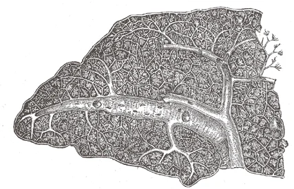
Transverse section of the human spleen, showing the distribution of the splenic artery and its branches.

Figure 2: Transverse section of the human spleen, showing the distribution of the splenic artery and its branches.
DefinitionSplenic Artery
The main arterial supply to the spleen, branching from the coeliac trunk and following a tortuous course along the pancreas.
The splenic artery gives off several branches before reaching the spleen. These include the short gastric arteries, which supply the fundus of the stomach, and the left gastroepiploic artery, which supplies the greater curvature of the stomach. The splenic artery also gives off pancreatic branches, which supply the body and tail of the pancreas. These branches are important to identify during surgical procedures to avoid unnecessary bleeding. [2]
| Branch | Supply |
|---|---|
| Short gastric arteries | Fundus of stomach |
| Left gastroepiploic artery | Greater curvature of stomach |
| Pancreatic branches | Body and tail of pancreas |
| Terminal branches | Spleen |
The spleen is a highly vascular organ, and its blood supply is critical for understanding both its physiology and potential pathologies. The primary source of arterial blood to the spleen is the splenic artery, which is a major branch of the coeliac trunk. This artery typically arises from the coeliac trunk at the level of the first lumbar vertebra. It follows a tortuous course along the superior border of the pancreas, reaching the hilum of the spleen. At the hilum, the splenic artery divides i
NoteClinical Relevance
Understanding the arterial supply of the spleen is essential for diagnosing and managing splenic pathologies, such as splenic infarction or trauma.
In cases of splenic trauma, the integrity of the splenic artery is crucial for maintaining blood flow to the organ. Damage to the splenic artery can lead to significant hemorrhage, requiring immediate intervention. Additionally, the tortuous nature of the splenic artery can make it susceptible to aneurysm formation, which is a rare but serious condition.
Exam trapCommon Misconception
The splenic artery is not a straight vessel; its tortuous course is a key anatomical feature.
The venous drainage of the spleen follows the arterial supply, with the splenic vein joining the superior mesenteric vein to form the hepatic portal vein. This venous system is important for transporting nutrients and immune cells from the spleen to the liver for processing.
Clinical pearlKey Point
The splenic artery's tortuous course is a distinctive feature that aids in its identification during imaging and surgery.
In summary, the arterial supply of the spleen is complex and vital for its function. The splenic artery, with its numerous branches and tortuous course, ensures adequate blood flow to the spleen and surrounding structures. Understanding this anatomy is essential for medical students and healthcare professionals dealing with splenic pathologies.
Figure 3Gray1189
Long description
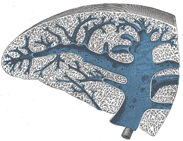
Transverse section of the spleen, showing the trabecular tissue and the splenic vein and its tributaries.

Figure 3: Transverse section of the spleen, showing the trabecular tissue and the splenic vein and its tributaries.
The venous drainage of the spleen is a critical component of the portal system, carrying nutrient-rich but potentially toxin-laden blood from the lymphoid organ towards the liver for filtration. Understanding this pathway requires tracing the vessel from its origin within the splenic hilum to its termination within the hepatic portal vein.
Figure 4Gray1192
Long description
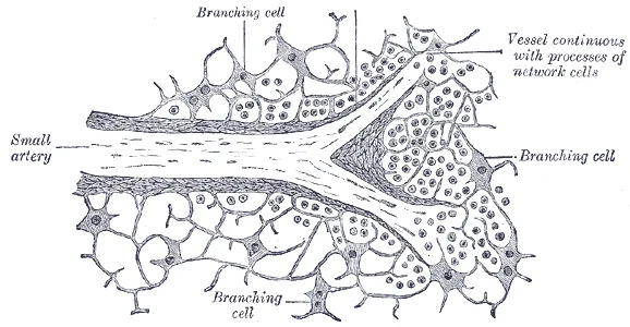
Section of the spleen, showing the termination of the small bloodvessels.

Figure 4: Section of the spleen, showing the termination of the small bloodvessels.
The central structure is the splenic vein, which emerges from the hilum of the spleen. It lies anterior to the pancreas, specifically coursing along the posterior surface of the pancreatic body and tail. To its anterior aspect lies the pancreas, while posteriorly, it is related to the left kidney and the left adrenal gland. The splenic vein travels horizontally to the right, joining the superior mesenteric vein behind the neck of the pancreas to form the hepatic portal vein. This confluence is a key anatomical landmark, situated posterior to the first part of the duodenum.
The splenic vein is formed by the union of several tributaries at the splenic hilum. These include the short gastric veins, which drain the fundus of the stomach, and the left gastroepiploic vein, which drains the greater curvature of the stomach. The splenic vein itself is relatively large and lacks valves, a feature that facilitates retrograde flow in conditions of portal hypertension. As it courses along the pancreas, it receives the posterior superior pancreaticoduodenal vein and sometimes the inferior pancreaticoduodenal vein. The vein terminates by joining the superior mesenteric vein to form the hepatic portal vein.
The hepatic portal vein is formed by the union of the splenic vein and the superior mesenteric vein. It ascends behind the first part of the duodenum and enters the free edge of the lesser omentum. It then divides into right and left branches within the liver. The portal system is unique in that it connects two capillary beds: the gastrointestinal capillaries and the hepatic sinusoids. This arrangement allows the liver to process absorbed nutrients and detoxify substances before they enter the systemic circulation.
| Structure | Origin | Course | Termination |
|---|---|---|---|
| Splenic Vein | Hilum of spleen | Along posterior pancreas | Joins SMV to form portal vein |
| Superior Mesenteric Vein | Intestinal capillaries | Ascends in mesentery | Joins splenic vein to form portal vein |
| Hepatic Portal Vein | Union of splenic and SMV | Behind duodenum, into liver | Divides into right and left branches |
Clinical significance arises when obstruction occurs at the portal vein or its tributaries. Increased pressure in the splenic vein can lead to splenomegaly and the development of collateral circulation, such as esophageal varices. Recognising the anatomical course of these vessels is essential for surgical interventions involving the pancreas or spleen, as inadvertent injury can lead to severe haemorrhage. The close relationship between the splenic vein and the pancreas means that pancreatic pathology, such as pancreatitis or tumours, can compress or invade the vein, further complicating clinical management.
Figure 5Gray1191
Long description
Transverse section of a portion of the spleen.

Figure 5: Transverse section of a portion of the spleen.
The spleen is a secondary lymphoid organ, meaning it is a site where mature lymphocytes congregate to mount immune responses against pathogens circulating in the blood. This drainage is critical for maintaining the homeostasis of the splenic microenvironment and ensuring that immune cells can efficiently survey the blood for threats. [1]
Lymphatic capillaries within the spleen are formed by a one cell-thick layer of endothelial cells, representing the open end of the system. When interstitial pressure is low, the endothelial flaps close to prevent backflow, but as pressure increases, the spaces between the cells open up, allowing fluid to enter. This entry is further enabled by collagen filaments that anchor the capillaries to surrounding structures; as pressure rises, these filaments pull on the endothelial cell flaps, opening them even further to facilitate fluid uptake. [1]
Once inside, the fluid, now termed lymph, travels through larger lymphatic vessels known as lymphatic trunks. These superficial and deep lymphatics eventually merge to form these larger conduits. The lymphatic vessels of the spleen follow the splenic artery and vein, draining towards the hilum of the organ. At the hilum, the lymphatic vessels exit the spleen to join the splenic lymph nodes, which are located along the splenic vessels and the greater curvature of the stomach. [1]
These nodes are sometimes referred to as the “filters of the lymph”. Any bacteria or cellular debris that infect the interstitial fluid of the spleen are taken up by the lymphatic capillaries and transported to these regional lymph nodes. Within these nodes, dendritic cells and macrophages internalize and kill many of the pathogens that pass through, thereby removing them from the body. [1]
The cisterna chyli is a sac-like chamber that receives lymph from the lower abdomen, pelvis, and lower limbs by way of the left and right lumbar trunks and the intestinal trunk. From the cisterna chyli, the lymph enters the thoracic duct. The thoracic duct itself begins just beneath the diaphragm and drains into the left subclavian vein. This final step returns the filtered fluid and immune cells to the bloodstream, completing the lymphatic circuit. [1]
The lymph then flows towards the celiac lymph nodes. The labels indicate the splenic artery, splenic vein, and splenic lymphatic vessels.
The innervation of the spleen is a composite of autonomic and somatic fibres, a duality that explains both its physiological regulation and its characteristic pain referral patterns. Understanding this supply requires tracing the pathways from the central nervous system to the splenic hilum, noting how these nerves travel alongside the vascular structures.
These preganglionic neurons synapse in the coeliac ganglia. Postganglionic sympathetic fibres then travel along the branches of the coeliac trunk, including the splenic artery, to reach the spleen. This sympathetic input is primarily vasoconstrictive, regulating blood flow within the splenic pulp. Parasympathetic fibres, conversely, are derived from the vagus nerve (cranial nerve X). These fibres also pass through the coeliac plexus, accompanying the arterial supply. The parasympathetic role is less defined in terms of direct motor function but is thought to modulate vascular tone and potentially influence immune cell activity. The balance between sympathetic and parasympathetic tone helps regulate the release of stored blood cells and the contraction of the splenic capsule.
This phenomenon is known as Kehr’s sign. It is a critical clinical indicator of splenic rupture or subcapsular haematoma. The lower intercostal nerves (T7–T11) provide somatic sensation to the lateral and posterior aspects of the spleen. Pain from these nerves is typically felt in the left upper quadrant of the abdomen.
The nerves and lymphatic vessels follow a similar course to the splenic artery and its branches. Lymphatic drainage occurs via efferent lymphatic vessels that emerge from the hilum. These vessels accompany the splenic artery and drain into the coeliac lymph nodes. The proximity of nerves to the vascular pedicle means that surgical dissection of the splenic hilum requires careful identification to avoid inadvertent injury to the autonomic fibres, which could theoretically affect splenic function, although the clinical significance of isolated nerve injury is often minimal compared to vascular compromise.
The lymphatic vessels are traced emerging from the hilum, following the arterial path towards the coeliac nodes.
The spleen is a highly vascularised organ composed of two distinct types of parenchyma: red pulp and white pulp. These regions perform separate but complementary functions in blood filtration and immune surveillance. The structural organisation allows for efficient removal of aged erythrocytes while maintaining a reservoir for immune cells ready to respond to systemic antigens. [2]
The red pulp constitutes the majority of the splenic mass and is responsible for filtering blood. It consists of splenic cords (cords of Billroth) and venous sinuses. The cords are reticular connective tissue filled with blood cells, macrophages, and plasma cells. Macrophages within the cords phagocytose old or damaged red blood cells and remove pathogens from the circulation. The venous sinuses are lined by specialized endothelial cells with long, parallel processes that form slit-like openings. Red blood cells must squeeze through these narrow slits to enter the venous circulation. This process allows macrophages to inspect and remove defective cells. If a red blood cell is rigid or damaged, it becomes trapped in the cords and is subsequently phagocytosed. This mechanism ensures that only healthy, flexible erythrocytes re-enter the systemic circulation. The red pulp also serves as a reservoir for platelets and monocytes, which can be released into the bloodstream during times of physiological stress.
The white pulp is the lymphoid component of the spleen and is organised around central arterioles. It is primarily involved in immune responses to blood-borne antigens. The central arterioles are surrounded by periarteriolar lymphoid sheaths (PALS), which are predominantly T-cell zones. Branching off from the PALS are lymphoid follicles, also known as Malpighian corpuscles, which are primarily B-cell zones. These follicles may contain germinal centres where B cells proliferate and differentiate in response to antigenic stimulation. The white pulp is strategically positioned to intercept antigens entering the spleen via the splenic artery. When pathogens are detected, immune cells within the white pulp activate and mount an adaptive immune response. This includes the production of antibodies and the activation of T cells. The close proximity of the white pulp to the red pulp allows for the rapid interaction between immune cells and blood-borne antigens. This integration ensures that the spleen can respond quickly to systemic infections.
Figure 62208 Spleen
Long description
(a) The spleen is attached to the stomach. (b) A micrograph of spleen tissue shows the germinal center. The marginal zone is the region between the red pulp and white pulp, which sequesters particulate antigens from the circulation and presents these antigens to lymphocytes in the white pulp. EM × 6

Figure 6: (a) The spleen is attached to the stomach.
Understanding the histological distinction between red and white pulp is crucial for interpreting splenic pathology. Conditions affecting the red pulp, such as haemolytic anaemias, lead to increased workload for macrophages and potential splenomegaly. Disorders of the white pulp, such as lymphomas, disrupt immune surveillance and can impair the body's ability to fight infections. The unique vascular architecture also makes the spleen susceptible to certain types of infarction, particularly in conditions causing hypercoagulability or embolic events.
The spleen originates from mesenchymal tissue within the dorsal mesogastrium during the fifth week of gestation. This mesenchymal condensation forms the splenic anlage, which subsequently differentiates into the functional parenchyma of the organ.
As the splenic anlage develops, it becomes vascularised by branches of the splenic artery, which arise from the celiac trunk. The mesenchymal cells differentiate into the red pulp and white pulp. The white pulp consists of lymphoid follicles, which are primarily B-cell zones, and periarteriolar lymphoid sheaths, which are T-cell zones. This organisation allows for the efficient interaction between blood-borne antigens and immune cells.
It is composed of dense connective tissue and smooth muscle fibres. The smooth muscle fibres allow the spleen to contract and release stored red blood cells when necessary. The capsule extends into the parenchyma as trabeculae, which provide structural support and carry blood vessels and nerves into the organ.
The hilum of the spleen is located on the visceral surface, where the splenic artery and vein enter and exit. The splenic vein joins the superior mesenteric vein to form the hepatic portal vein. The lymphatic vessels drain into the celiac lymph nodes. [2]
DefinitionPrimary Lymphoid Organs
The bone marrow and thymus gland are the primary lymphoid organs where lymphocytes mature, proliferate, and are selected. [1]
The development of the spleen is complete by the end of the first trimester. However, the functional maturation of the immune components continues throughout life. The spleen plays a crucial role in the immune response, particularly against encapsulated bacteria. It filters blood and removes old or damaged red blood cells. The macrophages in the red pulp phagocytose these cells, while the lymphocytes in the white pulp mount an immune response.
| Structure | Origin | Function |
|---|---|---|
| Capsule | Mesenchyme | Protection and contraction |
| Red Pulp | Sinusoidal system | Blood filtration |
| White Pulp | Lymphoid tissue | Immune response |
| Trabeculae | Mesenchyme | Structural support |
The spleen is a vital organ for immune function and blood filtration. Its development is a complex process involving the differentiation of mesenchymal cells into various functional components. Understanding this development is essential for appreciating the spleen's role in health and disease.
The spleen is a solid, vascular organ situated in the left hypochondrium. It lies deep to the upper left ribs. Its long axis follows the line of one of these ribs. The organ is protected by the rib cage. This position shields it from direct anterior trauma. However, it remains vulnerable to lateral impacts. [2]
The spleen appears as a homogeneous, soft-tissue density structure on the left side of the body. The left kidney is positioned posterior and inferior to the spleen. The stomach is located anteriorly, separated by the gastrosplenic ligament. The diaphragm forms the superior boundary. The colon splenic flexure lies inferiorly. Understanding these relations is vital for interpreting cross-sectional imaging.
DefinitionSplenic Hilum
The medial concave surface where vessels and nerves enter and exit the spleen.
The splenic artery is the largest branch of the coeliac trunk. It courses tortuously along the superior border of the pancreas. It reaches the splenic hilum by dividing into terminal branches. These branches enter the hilum and penetrate the parenchyma. It joins the superior mesenteric vein to form the portal vein. This venous drainage is critical for portal hypertension assessment.
| Structure | Relation to Spleen | Clinical Significance |
|---|---|---|
| Stomach | Anterior | Gastrosplenic ligament attachment |
| Left Kidney | Posterior/Inferior | Splenorenal ligament attachment |
| Pancreas | Anterior/Medial | Tail of pancreas contact |
| Diaphragm | Superior | Pleural reflection boundary |
It has a smooth, convex outer contour. The internal echotexture is homogeneous and fine. Size is measured in the longitudinal plane. Normal dimensions vary, but enlargement suggests pathology such as infection or haematological disorder.
Exam trapSize Thresholds
Do not confuse normal dimensions with pathological enlargement. A length over 12 cm is generally considered splenomegaly. [2]
CT imaging provides superior anatomical detail. The spleen enhances brightly in the arterial phase. This hypervascularity distinguishes it from the liver. In the portal venous phase, enhancement becomes homogeneous. Heterogeneity may indicate infarction or infiltration. The tail of the pancreas is often indistinguishable from the splenic hilum on axial slices. Coronal and sagittal reconstructions clarify this relationship.
NoteImaging Modality
CT is the gold standard for trauma assessment. Ultrasound is preferred for initial screening in stable patients.
MRI offers excellent soft-tissue contrast. The spleen is hypointense on T1-weighted images. It is hyperintense on T2-weighted images. This signal intensity helps differentiate cysts from solid masses. Cysts appear very bright on T2. Solid lesions may show variable enhancement patterns. Functional imaging can assess splenic sequestration. This is useful in sickle cell disease. [2]
DefinitionSplenomegaly
Enlargement of the spleen beyond normal limits, often palpable below the left costal margin.
The radiological anatomy of the spleen is defined by its vascular supply and surrounding structures. Knowledge of these features allows accurate diagnosis. It prevents misinterpretation of normal variants. It also aids in surgical planning. The spleen’s position makes it accessible to ultrasound. Its vascularity makes it visible on CT. These imaging characteristics are fundamental to clinical practice.
The spleen is a visceral organ situated in the left hypochondrium, protected by the rib cage. It lies between the ninth and eleventh ribs, with its long axis parallel to the eleventh rib. The organ is intraperitoneal, suspended by ligaments that connect it to surrounding structures. Understanding its position is vital for interpreting imaging and performing surgical procedures.
The diaphragm lies superior to the spleen, separating it from the left lung base. The left kidney is posterior and inferior to the spleen. The tail of the pancreas extends towards the splenic hilum. The stomach lies anterior to the spleen. The colon, specifically the splenic flexure, is inferior and anterior. The ninth, tenth, and eleventh ribs provide the primary bony protection. The spleen’s hilum faces medially, where the splenic vessels enter and exit. The splenorenal ligament connects the hilum to the left kidney.
The spleen has two surfaces: diaphragmatic and visceral. The diaphragmatic surface is convex and smooth, facing the diaphragm. The visceral surface is irregular and concave, bearing impressions from adjacent organs. The gastric impression is on the anterior part of the visceral surface. The renal impression is on the posterior part. The colic impression is on the inferior part. The pancreatic impression is near the hilum.
The spleen has two poles: anterior and posterior. The spleen has two borders: superior and inferior.
Clinical pearlClinical Pearl
The spleen is highly vascular and fragile. Trauma to the left lower chest can rupture the spleen, causing life-threatening haemorrhage. Always consider splenic injury in left-sided trauma.
The blood supply comes from the splenic artery, a branch of the coeliac trunk. It is tortuous and runs along the superior border. The splenic vein drains the spleen and joins the superior mesenteric vein to form the hepatic portal vein.
| Structure | Relation |
|---|---|
| Diaphragm | Superior and posterior |
| Stomach | Anterior |
| Left Kidney | Posterior and inferior |
| Splenic Flexure of Colon | Inferior |
| Tail of Pancreas | Medial (hilum) |
The lymphatic drainage follows the splenic vessels to the coeliac lymph nodes. The innervation is from the splenic plexus, derived from the coeliac plexus. This provides autonomic control and sensory information.
DefinitionHilum
The hilum is the medial depression where vessels, nerves, and lymphatics enter and exit the spleen. It is the site of attachment for the splenorenal and gastrosplenic ligaments.
The spleen’s size varies. It is approximately 11 cm in length, 7 cm in width, and 4 cm in thickness. It weighs about 150 grams. These dimensions are useful for assessing splenomegaly on physical examination or imaging. Enlargement can indicate various pathologies, including infections, haematological disorders, and portal hypertension. [2]
It is protected by the rib cage, specifically ribs nine to eleven. This anatomical position makes it vulnerable to trauma during blunt abdominal injury. These trabeculae provide structural support and carry blood vessels into the parenchyma.
These follicles are rich in B lymphocytes and surround central arterioles. They are involved in the immune response to blood-borne antigens. The red pulp is responsible for filtering blood and removing old or damaged erythrocytes. It consists of splenic cords and venous sinuses.
This artery is often tortuous and can be compressed against the posterior abdominal wall during emergency splenectomy. The splenic vein drains the organ and joins the superior mesenteric vein to form the hepatic portal vein. This anatomical relationship is critical in understanding portal hypertension.
Lymphatic drainage from the spleen follows the splenic vessels to the coeliac lymph nodes. This ligament is a key landmark during surgical procedures.
Clinical pearlSplenic Rupture
Trauma to the left lower chest or upper abdomen can cause splenic rupture, leading to haemoperitoneum and shock. Immediate recognition is vital for survival.
Exam trapAccessory Spleens
Accessory spleens are common congenital variants. They may be mistaken for lymphadenopathy or masses on imaging. Surgeons must identify them to prevent incomplete splenectomy.
Macrophages in the red pulp phagocytose old red blood cells and debris. The white pulp initiates adaptive immune responses via T and B cells.
DefinitionHypersplenism
Hypersplenism is a condition where the spleen sequesters and destroys blood cells excessively, leading to cytopenias. It can occur in various liver diseases.
NoteSplenomegaly
Enlargement of the spleen, or splenomegaly, can result from infections, haematological disorders, or liver disease. It may cause early satiety due to gastric compression.
| Structure | Relation |
|---|---|
| Stomach | Anterior |
| Left Kidney | Posterior |
| Colon | Inferior |
| Diaphragm | Superior |
Splenic Blood Flow
- Splenic ArteryBranch of coeliac trunk
- Question: Enters hilum
Yes
- Branches into trabecular arteries
No
- Error in anatomy
Students frequently confuse the functional anatomy of the spleen with that of other lymphoid organs, leading to significant errors in clinical reasoning and examination answers. A primary mistake is attributing adaptive immune responses to the red pulp. The red pulp is not the site of specific lymphocyte activation; rather, it primarily functions as a filtration system of the blood, using cells of the relatively nonspecific immune response. In contrast, white pulp is where adaptive T and B cell responses are mounted. Confusing these two zones leads to incorrect localisation of pathology and misunderstanding of splenic physiology. [1]
Another common error involves the morphological identification of plasma cells. Students often overlook the distinct cytoplasmic features that define a plasma cell. A plasma cell is a B cell that has differentiated in response to antigen binding, and has thereby gained the ability to secrete soluble antibodies. These cells differ in morphology from standard B and T cells in that they contain a large amount of cytoplasm packed with the protein-synthesizing machinery known as rough endoplasmic reticulum. Failure to recognise this structural adaptation results in an inability to identify active humoral immune sites on histology slides. [1]
Misunderstanding the role of T cells is also prevalent. A frequent pitfall is assuming T cells secrete antibodies to neutralise pathogens. The T cell, on the other hand, does not secrete antibody but performs a variety of functions in the adaptive immune response. Different T cell types have the ability to either secrete soluble factors that communicate with other cells of the adaptive immune response or destroy cells infected with intracellular pathogens. Recognising this distinction is vital for understanding cell-mediated immunity versus humoral immunity. [1]
Students also struggle with the anatomical boundaries of the thymus, often misplacing it within the mediastinum without precision. The thymus gland is a bilobed organ found in the space between the sternum and the aorta of the heart. Connective tissue holds the lobes closely together but also separates them and forms a capsule. This precise anatomical description is necessary for interpreting anterior mediastinal masses and understanding the site of T-cell maturation. [1]
Finally, confusion often arises between splenic pathology and gastrointestinal lymphoid structures, particularly regarding MALT. Mucosa-associated lymphoid tissue (MALT) consists of an aggregate of lymphoid follicles directly associated with the mucous membrane epithelia. MALT makes up dome-shaped structures found underlying the mucosa of the gastrointestinal tract, breast tissue, lungs, and eyes. Peyer’s patches, a type of MALT in the small intestine, are especially important for immune responses against ingested substances. Peyer’s patches contain specialized endothelial cells called M (or microfold) cells that sample material from the intestinal lumen and transport it to nearby follicles so that adaptive immune responses to potential pathogens can be mounted. A similar process occurs involving MALT in the mucosa and submucosa of the appendix. A blockage of the lumen triggers these cells to elicit an inflammatory response that can lead to appendicitis. Students must not attribute these specific sampling mechanisms to the spleen, which operates via blood filtration rather than luminal sampling. [1]
Exam trapRed vs White Pulp
The red pulp filters blood via nonspecific mechanisms, while the white pulp mounts adaptive T and B cell responses.
DefinitionPlasma Cell Morphology
A plasma cell is a B cell that has differentiated in response to antigen binding, and has thereby gained the ability to secrete soluble antibodies.
Knowledge of splenic and associated lymphoid anatomy is critical at the bedside, in the operating theatre, on imaging, and in examinations. At the bedside, understanding the immune phases helps explain clinical presentations. The immune system is a collection of barriers, cells, and soluble proteins that interact and communicate with each other in extraordinarily complex ways. The modern model of immune function is organized into three phases based on the timing of their effects. The three temporal phases consist of the following. Recognising these phases allows clinicians to anticipate the progression of infections and the efficacy of immunotherapies.
In the operating theatre, precise anatomical knowledge prevents injury to surrounding structures. The thymus gland is a bilobed organ found in the space between the sternum and the aorta of the heart. Connective tissue holds the lobes closely together but also separates them and forms a capsule. Surgeons must respect these boundaries during mediastinal procedures to avoid damaging the thymic lobes or the great vessels. Similarly, splenectomy requires careful dissection of the splenic hilum, respecting the vascular supply that feeds the red and white pulp.
On imaging, distinguishing splenic lesions from other pathologies is essential. Such persistent enhancement of IPAS on the hepatosplenic parenchymal phase is thought to occur due to prolonged entrapment of Levovist by the RES cells in the splenic tissue. The mechanism of Levovist-enhanced US on the hepatosplenic phase is theoretically similar to those of Tc-99m HDRBC scintigraphy and superparamagnetic iron oxide (SPIO)-enhanced MRI. These modalities are used as a confirmatory tool to diagnose IPAS. On all dynamic US phases, the echoenhancement of IPAS is usually greater than that of the pancreas. The lesion conspicuity was less dramatic on the 60-sec portal phase images and this was diminished even further on the 120-sec delayed phase images. Radiologists use these temporal dynamics to differentiate splenic lesions from pancreatic or hepatic abnormalities. [2]
In examinations, students must accurately describe lymphoid tissue functions. Mucosa-associated lymphoid tissue (MALT) consists of an aggregate of lymphoid follicles directly associated with the mucous membrane epithelia. MALT makes up dome-shaped structures found underlying the mucosa of the gastrointestinal tract, breast tissue, lungs, and eyes. Peyer’s patches, a type of MALT in the small intestine, are especially important for immune responses against ingested substances. Peyer’s patches contain specialized endothelial cells called M (or microfold) cells that sample material from the intestinal lumen and transport it to nearby follicles so that adaptive immune responses to potential pathogens can be mounted. A similar process occurs involving MALT in the mucosa and submucosa of the appendix. A blockage of the lumen triggers these cells to elicit an inflammatory response that can lead to appendicitis. Examiners test the ability to distinguish these mucosal sampling mechanisms from the blood filtration role of the spleen.
NoteImaging Dynamics
On all dynamic US phases, the echoenhancement of IPAS is usually greater than that of the pancreas.
Clinical pearlT Cell Function
The T cell, on the other hand, does not secrete antibody but performs a variety of functions in the adaptive immune response.
The red pulp primarily functions as a filtration system of the blood, using cells of the relatively nonspecific immune response. White pulp is where adaptive T and B cell responses are mounted. A plasma cell is a B cell that has differentiated in response to antigen binding, and has thereby gained the ability to secrete soluble antibodies. These cells differ in morphology from standard B and T cells in that they contain a large amount of cytoplasm packed with the protein-synthesizing machinery known as rough endoplasmic reticulum. The T cell, on the other hand, does not secrete antibody but performs a variety of functions in the adaptive immune response. The thymus gland is a bilobed organ found in the space between the sternum and the aorta of the heart. Connective tissue holds the lobes closely together but also separates them and forms a capsule. Mucosa-associated lymphoid tissue (MALT) consists of an aggregate of lymphoid follicles directly associated with the mucous membrane epithelia.
Answer each one in your head first, then open the answer.
What is the primary function of the red pulp?
Show answerHide answer
It primarily functions as a filtration system of the blood, using cells of the relatively nonspecific immune response.Where are adaptive T and B cell responses mounted?
Show answerHide answer
White pulp is where adaptive T and B cell responses are mounted.What defines a plasma cell morphologically?
Show answerHide answer
They contain a large amount of cytoplasm packed with the protein-synthesizing machinery known as rough endoplasmic reticulum.What is the anatomical location of the thymus?
Show answerHide answer
It is a bilobed organ found in the space between the sternum and the aorta of the heart.What triggers an inflammatory response in the appendix MALT?
Show answerHide answer
A blockage of the lumen triggers these cells to elicit an inflammatory response that can lead to appendicitis.
- Betts JG, Young KA, Wise JA, et al. Anatomy of the Lymphatic and Immune Systems. OpenStax, Anatomy and Physiology 2e (CC BY-NC-SA 4.0). Accessed .
- Kim SH, Lee JM, Han JK, et al. Intrapancreatic accessory spleen: findings on MR Imaging, CT, US and scintigraphy, and the pathologic analysis. Korean journal of radiology 2008;9(2):162-174. doi:10.3348/kjr.2008.9.2.162. Accessed .
Figure credits
- Figure 1: Henry Vandyke Carter, via Wikimedia Commons, Public domain, Public domain.
- Figure 2: Henry Vandyke Carter, via Wikimedia Commons, Public domain, Public domain.
- Figure 3: Henry Vandyke Carter, via Wikimedia Commons, Public domain, Public domain.
- Figure 4: Henry Vandyke Carter, via Wikimedia Commons, Public domain, Public domain.
- Figure 5: Henry Vandyke Carter, via Wikimedia Commons, Public domain, Public domain.
- Figure 6: OpenStax College, via Wikimedia Commons, CC BY 3.0, CC BY 3.0.
Educational content for medical students and doctors. It is not a substitute for clinical judgement or local guidelines.