Pancreas
The pancreas is a retroperitoneal gland with exocrine and endocrine functions, located behind the stomach.
Figure 1Gray1097
Long description
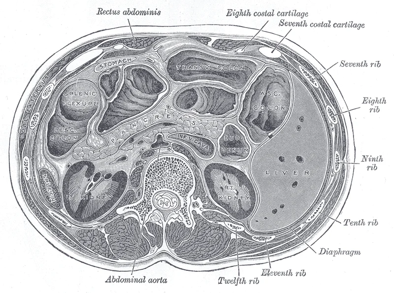
Transverse section through the middle of the first lumbar vertebra, showing the relations of the pancreas. (Braune.)

Figure 1: Transverse section through the middle of the first lumbar vertebra, showing the relations of the pancreas (Braune.).
The pancreas is a soft, oblong, glandular organ situated transversely in the retroperitoneum, lying directly behind the stomach. It functions as a curious mix of exocrine tissue, which secretes digestive enzymes, and endocrine tissue, which releases hormones into the bloodstream. The organ extends approximately 15.2 cm (6 in) from right to left, ending as a tapering tail in the hilum of the spleen. Its head is nestled into the “c-shaped” curvature of the duodenum. [1]
The pancreas originates from the endodermal lining of the foregut. This epithelial tissue proliferates to form two distinct outpouchings, known as pancreatic buds. As the stomach and duodenum rotate during embryonic development, the ventral bud moves dorsally to lie adjacent to the dorsal bud. These two buds eventually fuse to form the single functional organ. The d [2]
Figure 2Gray1103
Long description
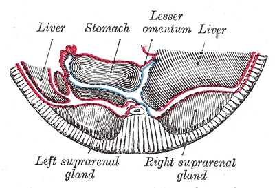
Schematic and enlarged cross-section through the body of a human embryo in the region of the mesogastrium. Beginning of third month. (Toldt.)

Figure 2: Schematic and enlarged cross-section through the body of a human embryo in the region of the mesogastrium (beginning of third month).
Figure 3Gray1102
Long description
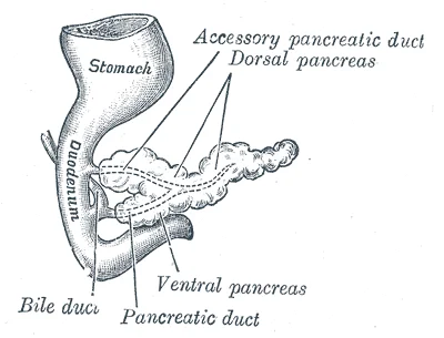
Pancreas of a human embryo at end of sixth week. (Kollmann.)

Figure 3: Pancreas of a human embryo at end of sixth week (Kollmann.).
Figure 4Gray1101
Long description
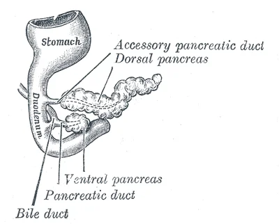
Pancreas of a human embryo of five weeks. (Kollmann.)

Figure 4: Pancreas of a human embryo of five weeks (Kollmann.).
An annular pancreas is a congenital anomaly where a ring of pancreatic tissue surrounds the duodenum, potentially causing obstruction. Pancreas divisum is a developmental variant where the dorsal and ventral pancreatic ducts fail to fuse, resulting in the majority of pancreatic drainage occurring through the smaller accessory duct rather than the main duct.
The anatomical relationship between the pancreas and the duodenum is critical for understanding biliary and pancreatic pathology. The proximity of the head of the pancreas to the common bile duct means that tumours in this region can obstruct bile flow, leading to jaundice. The dual ductal system allows for collateral drainage, which may influence the presentation of ductal obstructions.
DefinitionPancreatic Juice
A clear fluid composed mostly of water, salts, sodium bicarbonate, and digestive enzymes, produced by the pancreas to aid in digestion.
The pancreas produces over a liter of pancreatic juice each day. Unlike bile, it is clear and composed mostly of water along with some salts, sodium bicarbonate, and several digestive enzymes. Sodium bicarbonate is responsible for the slight alkalinity of pancreatic juice (pH 7.1 to 8.2), which serves to buffer the acidic gastric juice in chyme, inactivate pepsin from the stomach, and create an optimal environment for the activity of pH-sensitive digestive enzymes in the small intestine. Pancreatic enzymes are active in the digestion of sugars, proteins, and fats. [1]
| Enzyme (Inactive) | Active Form | Activating Enzyme |
|---|---|---|
| Trypsinogen | Trypsin | Enteropeptidase |
| Procarboxypeptidase | Carboxypeptidase | Trypsin |
| Chymotrypsinogen | Chymotrypsin | Trypsin |
The pancreas produces protein-digesting enzymes in their inactive forms. These enzymes are activated in the duodenum. If produced in an active form, they would digest the pancreas (which is exactly what occurs in the disease, pancreatitis). [1]
Exam trapEnzyme Activation
Remember that trypsinogen is activated by enteropeptidase, not by trypsin itself. Trypsin then activates other zymogens.
Regulation of pancreatic secretion is the job of hormones and the parasympathetic nervous system. The entry of acidic chyme into the duodenum stimulates the release of secretin, which in turn causes the duct cells to release bicarbonate-rich pancreatic juice. The presence of proteins and fats in the duodenum stimulates the secretion of CCK, which then stimulates the acini to secrete enzyme-rich pancreatic juice and enhances the activity of secretin. Parasympathetic regulation occurs mainly during the cephalic and gastric phases of gastric secretion, when vagal stimulation prompts the secretion of pancreatic juice. [1]
Pancreatic Secretion Regulation
- StartFood entry
- Question: Acidic chyme in duodenum?
Yes
- Secretin release
- Bicarbonate-rich juice
No
- No secretin effect
- Question: Proteins/fats in duodenum?
Yes
- CCK release
- Enzyme-rich juice
No
- No CCK effect
The pancreas lies transversely across the posterior abdominal wall, extending from the C-shaped curve of the duodenum on the right to the spleen on the left. The head of the pancreas is the broadest part and lies within the concavity of the duodenum. It is related anteriorly to the transverse colon and posteriorly to the inferior vena cava and the right crus of the diaphragm. The uncinate process extends medially from the head, lying posterior to the superior mesenteric vessels. The neck of the pancreas is the constricted part connecting the head to the body, lying anterior to the superior mesenteric vein and artery. The body of the pancreas crosses the midline, lying anterior to the aorta, the left renal vein, and the left kidney.
The duodenum is the first and shortest part of the small intestine, receiving chyme from the stomach and secretions from the liver and pancreas. It is divided into four parts: superior, descending, horizontal, and ascending. The superior part begins at the pylorus and passes superiorly and posteriorly to the head of the pancreas. The ascending part passes upwards to the left of the aorta, reaching the second lumbar vertebra, where it becomes the jejunum at the duodenojejunal flexure.
The pancreas produces pancreatic juice, which contains digestive enzymes and bicarbonate ions, and delivers it to the duodenum. Usually, the pancreas secretes just enough bicarbonate to counterbalance the amount of HCl produced in the stomach. Chemical digestion in the small intestine cannot occur without the help of the liver and pancreas. [1]
DefinitionPancreatic Juice
A fluid containing digestive enzymes and bicarbonate ions produced by the pancreas and delivered to the duodenum.
| Part | Anterior Relations | Posterior Relations |
|---|---|---|
| Head | Transverse colon | Inferior vena cava, right crus |
| Neck | Transverse mesocolon | Superior mesenteric vessels |
| Body | Stomach, peritoneum | Aorta, left renal vein, left kidney |
| Tail | Spleen, stomach | Splenic vessels |
Figure 52424 Exocrine and Endocrine Pancreas
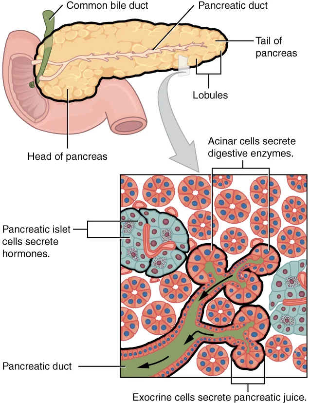
Long description
The pancreas has a head, a body, and a tail. It delivers pancreatic juice to the duodenum through the pancreatic duct.

Figure 5: The pancreas has a head, a body, and a tail.
The pancreas is a retroperitoneal organ, lying transversely across the posterior abdominal wall at the level of the first and second lumbar vertebrae. It is covered by peritoneum only anteriorly, forming part of the posterior wall of the lesser sac. The gland is divided into four distinct regions: the head, neck, body, and tail.
The head is the widest part of the pancreas, nestled within the C-shaped curve of the duodenum. Posterior to the head lies the inferior vena cava and the right crus of the diaphragm. The common bile duct and the main pancreatic duct (of Wirsung) typically unite to form the hepatopancreatic ampulla, which empties into the second part of the duodenum at the major duodenal papilla.
The neck is the narrowest portion, connecting the head to the body. It lies anterior to the superior mesenteric vessels. Specifically, the superior mesenteric vein and the splenic vein join behind the neck to form the portal vein. This relationship is crucial for surgical dissection and CT interpretation.
The body extends to the left, crossing the midline anterior to the aorta and the left crus of the diaphragm. Posteriorly, the splenic artery runs along the superior border of the body. Anteriorly, it is related to the stomach and the lesser sac. The left renal vessels and the hilum of the left kidney lie posterior to the body.
The tail is the narrow, tapered end that reaches the splenic hilum. It lies within the splenorenal ligament, alongside the splenic artery and vein. The tail is the most mobile part of the pancreas and is intraperitoneal, unlike the rest of the gland.
The main pancreatic duct runs the length of the gland, draining into the duodenum. Accessory ducts may drain the superior aspect of the head. The arterial supply is derived from the celiac trunk and the superior mesenteric artery. The head receives blood from the superior and inferior pancreaticoduodenal arteries, while the body and tail are supplied by the splenic artery and its branches. Venous drainage follows the arterial supply into the portal system. [2]
On contrast-enhanced CT, the pancreas is identified by its lobulated texture and specific vascular relationships. As noted in clinical guidelines, contrast-enhanced CT (MDCT is desirable) is recommended for the staging of pancreatic cancer and for assessment of its resectability. In standard clinical imaging modalities, pancreatic ductal adenocarcinomas are identified as regions that take up less injected contrast material than the normal surrounding tissues. This hypoperfusion i [2]
The pancreas is a composite gland situated in the retroperitoneal space, lying deep to the stomach and anterior to the vertebral column. It consists of three main parts: the head, the body, and the tail. The head is the widest portion, nestled within the C-loop of the duodenum. The body extends horizontally to the left, crossing the midline, while the tail tapers to a point near the splenic hilum. This organ is not a simple tube but a lobulated mass of tissue that houses both exocrine and endocrine functions.
The majority of the pancreatic parenchyma is dedicated to exocrine function. This tissue is organized into acini, which are clusters of secretory cells. These acini drain into a network of ducts that eventually merge to form the main pancreatic duct. The duct runs through the length of the gland and typically joins the common bile duct to form the hepatopancreatic ampulla, also known as the ampulla of Vater. This junction opens into the second part of the duodenum at the major duodenal papilla. The exocrine cells produce digestive enzymes, including amylase, lipase, and proteases, which are essential for nutrient breakdown in the small intestine. [1]
Scattered throughout the exocrine tissue are clusters of endocrine cells known as the islets of Langerhans. These islands are not connected to the ductal system and release their products directly into the bloodstream. The islets contain several distinct cell types, each with a specific hormonal role. Alpha cells produce glucagon, which raises blood glucose levels. Beta cells produce insulin, which lowers blood glucose. Delta cells produce somatostatin, which inhibits the release of both insulin and glucagon. These cells work in concert to maintain metabolic homeostasis.
Figure 6Gray1056
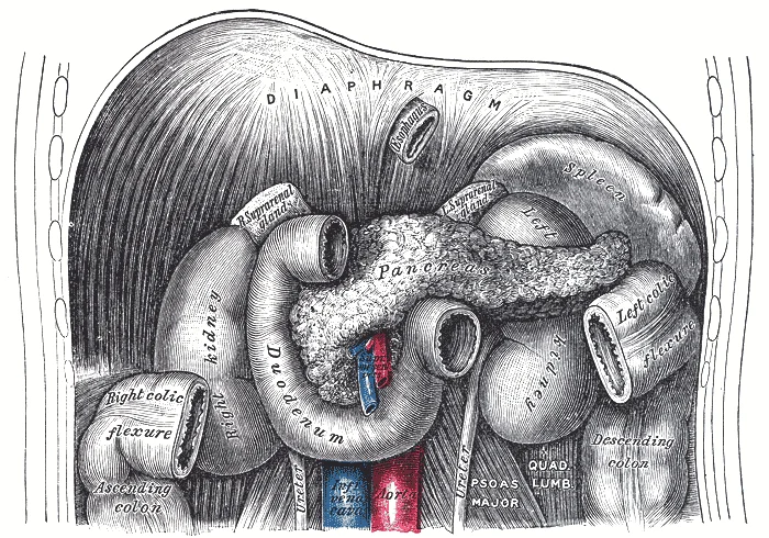
Long description
The duodenum and pancreas. Labelled illustration from Wikimedia Commons, used under its open licence.

Figure 6: The duodenum and pancreas.
The body extends to the left, and the tail reaches toward the spleen. The islets of Langerhans are represented as small, darker clusters within the lighter exocrine tissue.
Understanding the internal structure is crucial for interpreting imaging studies. The head of the pancreas is closely related to the superior mesenteric vessels, which can be compressed by tumors in this region. The body and tail are more mobile and can shift with respiration. The exocrine tissue is susceptible to inflammation, leading to pancreatitis, while the endocrine tissue is the target of type 1 diabetes. The ductal system is prone to obstruction by stones or tumors, which can cause jaundice and pain. [2]
The pancreas is a complex organ with distinct exocrine and endocrine components. The exocrine tissue forms the bulk of the gland and is organized into acini and ducts. The endocrine tissue consists of the islets of Langerhans, which regulate blood glucose. The main pancreatic duct joins the common bile duct to drain into the duodenum. This structural arrangement allows the pancreas to perform its vital roles in digestion and metabolism.
The arterial supply of the pancreas is derived primarily from the coeliac trunk and the superior mesenteric artery. This dual supply ensures robust perfusion but creates complex anastomotic networks that are critical for surgical planning. The head and uncinate process receive blood from branches of both systems, while the body and tail are predominantly supplied by the splenic artery. Understanding these pathways is essential for managing haemorrhage during pancreatic surgery or trauma.
DefinitionPancreaticoduodenal Arcades
The anterior and posterior pancreaticoduodenal arteries form anastomotic loops between the coeliac trunk and the superior mesenteric artery, providing collateral circulation to the head of the pancreas and duodenum.
It follows a winding course along the superior margin of the pancreas, giving off the dorsal pancreatic artery, the great pancreatic artery, and the caudal pancreatic arteries. These branches penetrate the gland to supply the parenchyma.
| Region | Primary Source | Key Branches |
|---|---|---|
| Head (Anterior) | Gastroduodenal Artery | Superior pancreaticoduodenal (anterior) |
| Head (Posterior) | Gastroduodenal Artery | Superior pancreaticoduodenal (posterior) |
| Head (Inferior) | Superior Mesenteric Artery | Inferior pancreaticoduodenal |
| Body and Tail | Splenic Artery | Dorsal, great, caudal pancreatic arteries |
It divides into the right gastro-omental artery and the superior pancreaticoduodenal artery. This anastomosis is crucial for maintaining blood flow if one source is compromised. The uncinate [2]
During distal pancreatectomy, ligation of the splenic artery must be performed carefully to avoid avulsion or injury to the pancreatic parenchyma, which can lead to significant haemorrhage or pancreatic fistula.
Clinical relevance is highest in pancreatitis, where inflammation can erode these vessels, leading to pseudoaneurysm formation. The splenic artery is the most common site for such aneurysms due to its tortuous course and proximity to inflammatory exudate. Recognition of these anatomical relationships on imaging is critical for interventional radiology procedures.
Figure 7Gray1098
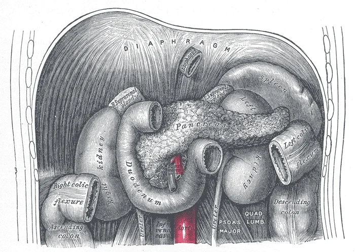
Long description
The duodenum and pancreas. Labelled illustration from Wikimedia Commons, used under its open licence.

Figure 7: The duodenum and pancreas.
Venous drainage follows the arterial supply. Lymph from the head drains to the pylor
Venous drainage follows the arterial supply. Lymph from the head drains to the pylor
This anastomosis is clinically significant in cases of portal hypertension. [2]
The veins draining the body and tail of the pancreas are more variable. It runs horizontally behind the pancreas and joins the superior mesenteric vein to form the portal vein. [2]
DefinitionPortal System
A system of veins that carry blood from one capillary bed to another, such as from the gastrointestinal tract to the liver.
Knowledge of these anatomical relationships aids in surgical planning and interpretation of imaging studies. The precise identification of these veins during pancreaticoduodenectomy is critical to prevent catastrophic haemorrhage.
| Structure | Drains Into |
|---|---|
| Superior Pancreaticoduodenal Veins | Portal Vein or Superior Mesenteric Vein |
| Inferior Pancreaticoduodenal Veins | Superior Mesenteric Vein |
| Splenic Vein Tributaries | Splenic Vein |
| Short Gastric Veins | Splenic Vein |
| Left Gastroepiploic Vein | Splenic Vein |
The lymphatic drainage of the pancreas follows a predictable pattern that mirrors its arterial supply, ensuring that lymph from specific regions drains into corresponding nodal groups. Understanding this pathway is critical for appreciating the spread of pancreatic malignancies and the staging of disease. These nodes are numerous and lie closely associated with the pancreatic tissue.
Figure 8Gray1099
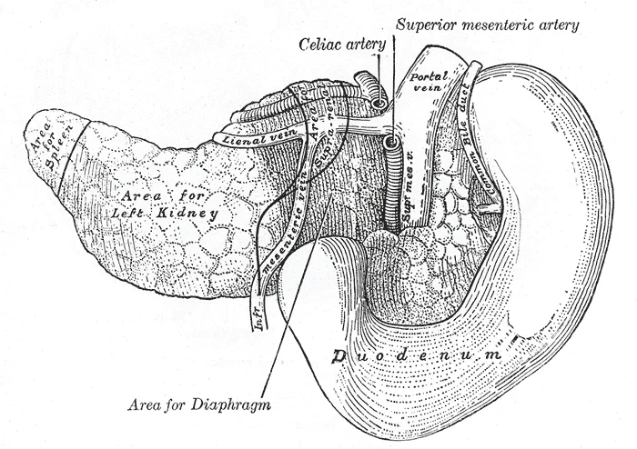
Long description
The pancreas and duodenum from behind. (From model by His.)

Figure 8: The pancreas and duodenum from behind (From model by his.).
The body extends towards the left, crossing the midline. The tail reaches the hilum of the spleen.
The clinical significance of this drainage pattern cannot be overstated. Metastatic spread from pancreatic cancer often follows these lymphatic channels. Tumours in the body or tail may involve the splenic nodes. Involvement of the celiac nodes indicates more advanced disease. Surgeons must carefully dissect these nodal groups during pancreatectomy to achieve clear margins. Preoperative imaging aims to detect enlarged nodes in these specific locations. The presence of metastatic disease in the lymph nodes significantly impacts prognosis and treatment planning.
They are embedded in the splenorenal ligament. This anatomical relationship means that splenectomy may be required if the tail of the pancreas is involved. They lie anterior to the aorta and the crura of the diaphragm. Enlargement of these nodes can compress adjacent structures, including the coeliac axis. This compression can lead to vascular complications. Awareness of these relationships aids in surgical planning and radiological interpretation.
| Pancreatic Region | Primary Nodal Group | Secondary Nodal Group |
|---|---|---|
| Head | Pyloric nodes | Superior mesenteric nodes |
| Uncinate process | Inferior mesenteric nodes | Superior mesenteric nodes |
| Body | Splenic nodes | Celiac nodes |
| Tail | Splenic nodes | Celiac nodes |
This systematic drainage ensures that lymph from the pancreas is filtered through specific nodal stations before entering the thoracic duct. This route explains why distant metastases can occur in unusual sites, such as the supraclavicular nodes. Virchow's node, located in the left supraclavicular fossa, may indicate abdominal malignancy. Recognising this connection is vital for comprehensive physical examination.
The autonomic nervous system comprises sympathetic and parasympathetic fibres that regulate exocrine and endocrine secretion, as well as vascular tone. Sympathetic fibres originate from the thoracic spinal segments and travel via the splanchnic nerves to the coeliac plexus. From this plexus, nerves f [1][2]
Pain from the pancreas is often perceived in the epigastric region but can radiate to the back. This referral occurs because the visceral afferent fibres accompanying the sympathetic nerves enter the spinal cord at similar levels to the somatic afferents from the back. [1]
The nerve supply is intimately associated with the blood supply, lymphatic drainage, and surrounding structures. It receives input from the greater, lesser, and least splanchnic nerves. The nerves to the pancreas branch from this plexus and accompany the arterial supply.
Understanding the nerve supply is essential for surgical procedures and pain management. Coeliac plexus block is a therapeutic intervention used to relieve severe pain in pancreatic cancer. This procedure involves injecting a neurolytic agent, such as alcohol, into the coeliac plexus to interrupt pain transmission. The effectiveness of this block depends on the precise anatomical location of the plexus and the distribution of the nerves to the pancreas. [1]
Figure 9Gray1100
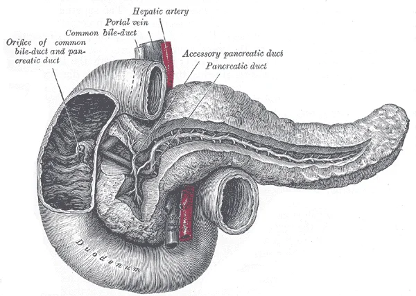
Long description
The pancreatic duct. Labelled illustration from Wikimedia Commons, used under its open licence.

Figure 9: The pancreatic duct.
Branches from the plexus follow the hepatic artery to the pancreatic head and the splenic artery to the pancreatic tail.
Clinical pearlReferred Pain
Pain from the pancreas often radiates to the back due to shared spinal segments between visceral afferents from the pancreas and somatic afferents from the posterior abdominal wall.
The pancreas is a dual-function organ, serving both exocrine and endocrine roles within the abdominal cavity. Its structural integrity relies on a precise arrangement of glandular tissue and vascular networks, which are best understood by examining its microscopic architecture. The exocrine component constitutes the majority of the organ’s mass and is responsible for the production and secretion of digestive enzymes. These enzymes are synthesized within the acinar cells, which are arranged in clusters known as acini. Each acinus drains into a small intercalated duct, which subsequently merges with larger intralobular and interlobular ducts. These ducts eventually coalesce to form the main pancreatic duct, also known as the duct of Wirsung, which runs the length of the gland. In the majority of individuals, this duct joins the common bile duct to form the hepatopancreatic ampulla, which empties into the duodenum at the major duodenal papilla. A secondary accessory duct, the duct of Santorini, may also be present, draining the head of the pancreas independently. [1]
DefinitionAcinar Cells
Specialized epithelial cells responsible for synthesizing and secreting digestive proenzymes such as trypsinogen and chymotrypsinogen into the pancreatic ductal system.
The endocrine component is distributed throughout the exocrine tissue in discrete clusters known as the islets of Langerhans. These islets are richly vascularized and contain several distinct cell types, each secreting specific hormones directly into the bloodstream. The beta cells are the most numerous and produce insulin, which lowers blood glucose levels. Alpha cells secrete glucagon, which raises blood glucose levels. Delta cells produce somatostatin, which regulates the secretion of both insulin and glucagon. PP cells, or pancreatic polypeptide cells, are less common and secrete pancreatic polypeptide, which inhibits exocrine secretion. This close proximity ensures rapid hormonal response to changes in blood nutrient levels.
Figure 10Gray1104
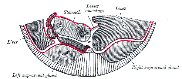
Long description
Section through same region as in Fig. 1103, at end of third month. (Toldt.)

Figure 10: Section through same region as in Fig.
The stroma of the pancreas consists of connective tissue septa that divide the gland into lobules. These septa contain blood vessels, nerves, and larger ducts. The blood supply to the pancreas is derived from branches of the celiac trunk and the superior mesenteric artery. The arterial supply includes the splenic artery, which gives off the dorsal pancreatic artery, the great pancreatic artery, and the caudal pancreatic arteries. Venous drainage follows the arterial pattern, with blood flowing into the splenic vein and the superior mesenteric vein, which unite to form the portal vein. This portal drainage ensures that hormones produced by the islets are delivered directly to the liver for first-pass metabolism. [2]
| Cell Type | Primary Hormone | Main Function |
|---|---|---|
| Beta | Insulin | Lowers blood glucose |
| Alpha | Glucagon | Raises blood glucose |
| Delta | Somatostatin | Inhibits insulin and glucagon |
| PP | Pancreatic Polypeptide | Inhibits exocrine secretion |
The pancreas originates from the endodermal lining of the foregut. This epithelial tissue proliferates to form two distinct outpouchings, known as pancreatic buds. As the stomach and duodenum rotate during embryonic development, the ventral bud moves dorsally to lie adjacent to the dorsal bud. These two buds eventually fuse to form the single functional organ. The d
This specific anatomical arrangement explains the relationship between the superior mesenteric vessels and the head of the pancreas. Consequently, the head of the pancreas lies within the C-shaped curve of the duodenum. [2]
The vascular supply develops in conjunction with the gut tube. The splenic artery supplies the body and tail. This vessel arises from the celiac trunk. These vessels form anastomotic arcs along the medial aspect of the head. [2]
Venous drainage follows the arterial supply. Lymph from the head drains to the pylor
These fibres promote secretion and motility. [2] [1]
Disorders of development are rare but significant. Pancreas divisum results from failure of the dorsal and ventral ducts to fuse. [2]
DefinitionPancreatic Buds
Two endodermal outpouchings from the foregut that fuse to form the pancreas.
| Structure | Arterial Supply | Venous Drainage |
|---|---|---|
| Body and Tail | Splenic artery | Splenic vein |
| Head and Uncinate | Superior and inferior pancreaticoduodenal arteries | Posterior superior pancreaticoduodenal vein |
The pancreas is divided into the head, uncinate process, neck, body, and tail. In the radiological context, the head of the pancreas lies within the C-loop of the duodenum. It is intimately associated with the common bile duct, which passes posterior to the head before joining the pancreatic duct. The uncinate process extends posteriorly and medially, hooking behind the superior mesenteric vessels. This anatomical position is crucial because tumours in this region may encase the superior mesenteric vein or artery, a key factor in assessing resectability.
It lies anterior to the superior mesenteric vein and the portal vein confluence. Radiological assessment of the neck is vital for identifying involvement of these major vessels.
It is the most mobile part of the gland. In cases of pancreatic cancer, particularly those involving the body or tail, the proximity to the splenic vessels is a significant consideration for surgical planning.
However, its invasive nature requires careful determination of indications. Understanding these anatomical relationships allows clinicians to predict the extent of disease and plan appropriate interventions, such as combined portal vein resection if R0 resection is expected. [2]
The pancreas is a retroperitoneal organ situated across the posterior abdominal wall, spanning from the duodenal C-loop to the splenic hilum. Its anatomical position places it in close proximity to major vascular structures, including the superior mesenteric vessels and the inferior vena cava. Understanding these relations is critical for interpreting imaging studies and planning surgical interventions.
The body of the pancreas extends transversely, lying anterior to the superior mesenteric artery and vein. The tail of the pancreas reaches towards the splenic hilum, resting against the anterior surface of the left kidney. Recognising these spatial relationships aids in the localisation of pathology on CT or MRI scans.
DefinitionPancreatic Relations
The pancreas is retroperitoneal, lying anterior to the aorta, inferior vena cava, and kidneys, with the splenic vein tracing its posterior border.
It is enclosed by the C-shaped curve of the duodenum. The uncinate process projects posteriorly and inferiorly from the head, hooking behind the superior mesenteric vessels. This anatomical configuration is significant because tumours in the uncinate process can encase these vessels, complicating resection. It lies anterior to the superior mesenteric vessels and the portal vein confluence. The body extends to the left, crossing the midline. It has an anterior, posterior, and inferior surface. The anterior surface is covered by peritoneum and forms part of the posterior wall of the omental bursa.
It terminates at the splenic hilum, lying within the splenorenal ligament. This ligament contains the splenic vessels and the tail of the pancreas. The proximity of the tail to the spleen is clinically relevant, as distal pancreatectomy often requires splenectomy to ensure complete removal of the organ and its vascular supply.
| Structure | Relation | Clinical Significance |
|---|---|---|
| Head | Enclosed by duodenum | Tumours may cause obstructive jaundice |
| Neck | Anterior to SMV/PV | Key landmark for vascular control |
| Body | Posterior to SMA | Encasement indicates unresectability |
| Tail | In splenorenal ligament | Proximity to spleen affects surgical approach |
The pancreas is a retroperitoneal organ situated transversely across the posterior abdominal wall, typically spanning from the duodenum on the right to the spleen on the left. It is divided into head, neck, body, and tail. The head lies within the C-shaped curve of the duodenum and is the widest part of the gland. The neck is the constricted portion connecting the head to the body, lying anterior to the superior mesenteric vessels.
DefinitionPancreatic Relations
The pancreas is retroperitoneal, except for its tail which is intraperitoneal within the splenorenal ligament.
Exam trapDuctal Anatomy Trap
Do not confuse the major and minor duodenal papillae. The major papilla is in the second part of the duodenum and carries the main duct. The minor papilla is superior and anterior to the major papilla.
The blood supply is derived from branches of the celiac trunk and the superior mesenteric artery.
Clinical pearlVenous Drainage
The splenic vein runs posterior to the body of the pancreas and joins the superior mesenteric vein to form the portal vein.
NoteClinical Correlation
Pancreatitis pain is typically epigastric and radiates to the back. This is due to inflammation of the retroperitoneal pancreas irritating the posterior parietal peritoneum.
| Region | Key Anterior Relation | Key Posterior Relation |
|---|---|---|
| Head | Duodenum | Inferior Vena Cava |
| Neck | Transverse Colon Mesocolon | Superior Mesenteric Vessel |
| Body | Stomach (Lesser Sac) | Aorta and Left Kidney |
| Tail | Stomach (Splenorenal Ligament) | Left Kidney and Spleen |
Common exam questions focus on the anatomical relationships that explain clinical symptoms. For instance, obstruction of the common bile duct by a pancreatic head tumour causes jaundice. Compression of the duodenum can cause vomiting. Involvement of the coeliac axis may cause intractable pain.
DefinitionRetroperitoneal Significance
Being retroperitoneal means the pancreas is fixed and less mobile, which influences the spread of disease and surgical access.
Remember the vascular anatomy carefully. This relationship is vital for understanding portal hypertension and thrombosis.
The lymphatic drainage is extensive. This pathway is relevant for staging pancreatic cancer.
Clinical pearlLymphatic Drainage
Pancreatic lymph drains to the coeliac nodes. This is why coeliac node involvement is a sign of advanced disease.
Finally, note the embryological origin. The pancreas develops from two buds, dorsal and ventral, which fuse. This fusion explains the position of the ducts and the uncinate process.
NoteEmbryology Note
The uncinate process arises from the ventral bud. It hooks behind the superior mesenteric vessels. This is a key anatomical landmark.
Understanding these anatomical details is essential for interpreting imaging and managing pancreatic pathology. The fixed retroperitoneal position, complex vascular supply, and dual ductal system are high-yield topics for medical examinations.
Students frequently confuse the functional anatomy of the biliary tree with the endocrine pancreas, leading to errors in localising pathology. A primary error is assuming the gallbladder possesses a submucosa. The gallbladder wall lacks a submucosa; its structure consists of mucosa, a muscular coat of smooth muscle, and an outer coat formed by visceral peritoneum reflected from the liver. This anatomical distinction is critical when interpreting histological slides or surgical findings, as the absence of a submucosal layer affects how inflammation or tumour invasion progresses through the wall. [1]
Another frequent mistake involves the mechanism of lipid digestion. Students often overlook the physical necessity of emulsification before enzymatic action can occur. Lipids are hydrophobic and do not dissolve in water, so large lipid globules must be broken down into smaller globules, a process called emulsification, before digestion in the watery small intestine can proceed. Bile, secreted by the liver, is the agent responsible for this emulsification. Failing to link bile directly to emulsification rather than enzymatic hydrolysis is a common conceptual gap.
Students also misinterpret the concentration capacity of the gallbladder. It is not merely a storage sac but an active concentrator. The gallbladder's mucosa absorbs water and ions from bile, concentrating it by up to 10-fold. Underestimating this concentration factor leads to incorrect calculations of bile salt availability during cholecystectomy or in cases of biliary obstruction.
In the context of pancreatic ductal adenocarcinoma (PDA), a significant pitfall is assuming that anti-angiogenic therapies are universally effective due to the tumour's vascular nature. Agents that inhibit or normalise angiogenesis are not effective in treating PDA in either humans or mice, despite their proven utility in typically hypervascular pancreatic neuroendocrine tumours. This is because the tumour microenvironment is characterised by high interstitial fluid pressure (IFP) and a dense desmoplastic stroma, which impede drug delivery more than vascular density alone.
Furthermore, students often confuse the types of pressure within the tumour bed. Solid stress (SS) is exerted only at points of direct contact, whereas fluid pressure transmits force in all vectorial directions. In PDA, IFP appears to predominate over SS in causing vascular collapse, contrary to the assumption that solid tumour mass directly crushes vessels. This distinction explains why mechanical stenting or pressure-relief strategies might be considered differently than in other solid tumours.
Finally, there is confusion regarding stent selection in palliative care. When the waiting period for surgery is short, use of a plastic stent is recommended.
This knowledge is applied at the bedside to understand symptoms of biliary obstruction. When a patient presents with jaundice, understanding that bile is concentrated by up to 10-fold helps explain the potency of even small amounts of obstructed bile salts in causing pruritus. Clinicians must recognise that the gallbladder contracts via its smooth muscle coat to eject contents through the cystic duct and into the bile duct. This physiological insight guides the interpretation of ultrasound findings, where a distended gallbladder suggests obstruction distal to the cystic duct.
In the operating theatre, the anatomical fact that the gallbladder is held against the liver by visceral peritoneum is crucial for safe dissection. Surgeons must identify the plane between the liver capsule and the gallbladder's outer coat to avoid liver injury. Misidentifying this plane can lead to bleeding or bile leakage. Additionally, knowing the gallbladder lacks a submucosa helps in assessing the depth of invasion in gallbladder cancer, as invasion through the muscular coat directly contacts the liver bed.
On imaging, the distinction between hydrophobic lipids and the aqueous environment of the intestine is relevant for interpreting MRCP (Magnetic Resonance Cholangiopancreatography). The presence of bile, which facilitates emulsification, is visualised as high-signal intensity fluid. Radiologists look for the absence of this fluid in obstructed ducts. Furthermore, understanding that PDA has a dense desmoplastic response containing significant levels of fibrous collagen helps explain the hypovascular appearance of these tumours on CT scans. The dense collagen and microfibrillary matrices impede diffusion, which correlates with the poor uptake of contrast agents in PDA compared to neuroendocrine tumours.
In examinations, students are tested on the specific recommendations for advanced pancreatic cancer. For microsatellite instability-high cases, pembrolizumab monotherapy is recommended. This is a specific immunotherapy indication that must be recalled precisely. For other advanced cases, combined gemcitabine hydrochloride + erlotinib hydrochloride therapy is recommended. These are specific first-line or second-line regimen criteria that do not vary by institution. [2]
The management of biliary obstruction also relies on these principles. If a patient is not a surgical candidate, the decision between stenting and gastrojejunostomy depends on the patient's condition and wishes. The knowledge that plastic stents are recommended for short waiting periods guides pre-operative planning. This prevents unnecessary placement of expensive metal stents in patients who will undergo surgery soon.
DefinitionEmulsification
The breakdown of large lipid globules into smaller ones by bile, enabling digestion in the watery small intestine.
Clinical pearlGallbladder Wall
The gallbladder has no submucosa; its wall consists of mucosa, smooth muscle, and visceral peritoneum.
Biliary Obstruction Management
- Diagnose Biliary ObstructionJaundice, dilated ducts
- Question: Surgical Candidate?
Yes
- Assess Wait TimeShort vs Long
- Question: Wait Time Short?
Yes
- Place Plastic StentRecommended for short waits
No
- Proceed to SurgeryCholecystectomy
No
- Palliative OptionStent vs Gastrojejunostomy
The gallbladder lacks a submucosa, consisting of mucosa, smooth muscle, and visceral peritoneum. Bile emulsifies hydrophobic lipids into smaller globules for digestion in the watery small intestine. [1] The gallbladder concentrates bile by up to 10-fold by absorbing water and ions. [1] Pembrolizumab is recommended for microsatellite instability-high pancreatic cancer. Gemcitabine plus erlotinib is a recommended therapy for advanced pancreatic cancer. Plastic stents are recommended when the waiting period for surgery is short. Either stenting or gastrojejunostomy may be selected for palliation based on patient wishes. Anti-angiogenic agents are not effective in PDA due to high interstitial fluid pressure.
Answer each one in your head first, then open the answer.
What structural layer is absent in the gallbladder wall?
Show answerHide answer
SubmucosaBy what factor does the gallbladder concentrate bile?
Show answerHide answer
Up to 10-foldWhich therapy is recommended for microsatellite instability-high cases?
Show answerHide answer
Pembrolizumab monotherapyWhat type of stent is recommended for short waiting periods for surgery?
Show answerHide answer
Plastic stentWhy are anti-angiogenic agents ineffective in PDA?
Show answerHide answer
High interstitial fluid pressure and dense stroma impede delivery
- Betts JG, Young KA, Wise JA, et al. Accessory Organs in Digestion: The Liver, Pancreas, and Gallbladder. OpenStax, Anatomy and Physiology 2e (CC BY-NC-SA 4.0). Accessed .
- Okusaka T, Nakamura M, Yoshida M, et al. Clinical Practice Guidelines for Pancreatic Cancer 2019 From the Japan Pancreas Society: A Synopsis. Pancreas 2020;49(3):326-335. doi:10.1097/mpa.0000000000001513. Accessed .
Figure credits
- Figure 1: Henry Vandyke Carter, via Wikimedia Commons, Public domain, Public domain.
- Figure 2: Henry Vandyke Carter, via Wikimedia Commons, Public domain, Public domain.
- Figure 3: Henry Vandyke Carter, via Wikimedia Commons, Public domain, Public domain.
- Figure 4: Henry Vandyke Carter, via Wikimedia Commons, Public domain, Public domain.
- Figure 5: OpenStax College, via Wikimedia Commons, CC BY 3.0, CC BY 3.0.
- Figure 6: Henry Vandyke Carter, via Wikimedia Commons, Public domain, Public domain.
- Figure 7: Henry Vandyke Carter, via Wikimedia Commons, Public domain, Public domain.
- Figure 8: Henry Vandyke Carter, via Wikimedia Commons, Public domain, Public domain.
- Figure 9: Henry Vandyke Carter, via Wikimedia Commons, Public domain, Public domain.
- Figure 10: Henry Vandyke Carter, via Wikimedia Commons, Public domain, Public domain.
Educational content for medical students and doctors. It is not a substitute for clinical judgement or local guidelines.