Gallbladder and biliary tree
Anatomical layout of the biliary tree and gallbladder.
The biliary tree is a conduit system responsible for transporting bile from the liver to the small intestine. Bile is produced by hepatocytes, which are the primary functional cells of the liver. Between adjacent hepatocytes, grooves in the cell membranes provide room for each bile canaliculus. These small ducts accumulate the bile produced by hepatocytes. From here, bile flows first into bile ductules and then into bile ducts. The bile ducts unite to form the larger right and left hepatic ducts, which themselves merge and exit the liver as the common hepatic duct. [1][3]
The common bile duct travels posterior to the first part of the duodenum and then passes behind the head of the pancreas. It usually joins with the main pancreatic duct before entering the duodenum. [1] [3]
The gallbladder is a pear-shaped muscular sac located in a fossa on the visceral surface of the liver. It serves as a reservoir for bile. The simple columnar epithelium of the gallbladder mucosa is organized in rugae, similar to those of the stomach. There is no submucosa in the gallbladder wall. The wall’s middle, muscular coat is made of smooth muscle fibers. When these fibers contract, the gallbladder’s contents are ejected through the cystic duct and into the bile duct. Visceral peritoneum reflected from the liver capsule holds the gallbladder against the liver and forms the outer coat of the gallbladder. [1]
This emulsification is a necessary step in lipid digestion and absorption. [1]
DefinitionBiliary Anatomy
The biliary tree transports bile from hepatocytes via canaliculi, ductules, and ducts to the duodenum, with the gallbladder acting as a storage and concentration organ.
The anatomical continuity ensures that bile is available during meals. The cystic duct connects the gallbladder to the common hepatic duct. This connection allows for the regulation of bile flow. When the gallbladder contracts, bile is expelled into the common bile duct. The common bile duct then delivers bile to the duodenum. This system works in concert with pancreatic secretions.
| Structure | Function |
|---|---|
| Hepatocytes | Produce bile |
| Bile canaliculi | Collect bile from hepatocytes |
| Common hepatic duct | Transport bile from liver |
| Gallbladder | Store and concentrate bile |
| Common bile duct | Transport bile to duodenum |
Understanding these structures is essential for clinical practice. Surgeons must identify the cystic duct and artery during cholecystectomy. Radiologists interpret imaging based on these anatomical landmarks. Pathologists examine bile ducts for signs of disease. The integrity of the biliary tree is crucial for digestion. [3]
NoteClinical Note
The gallbladder wall lacks a submucosa, which allows for direct spread of pathology in some conditions.
Students frequently confuse the anatomical relationships of the biliary tree with those of the pancreas, leading to errors in surgical planning and imaging interpretation. A primary error is assuming the pancreas is an intraperitoneal organ. The pancreas is actually retroperitoneal, lying transversely behind the stomach. This distinction is critical because retroperitoneal structures are fixed in position, whereas intraperitoneal organs move with peristalsis and respiration. Another common mista [1]
Figure 12425 Gallbladder
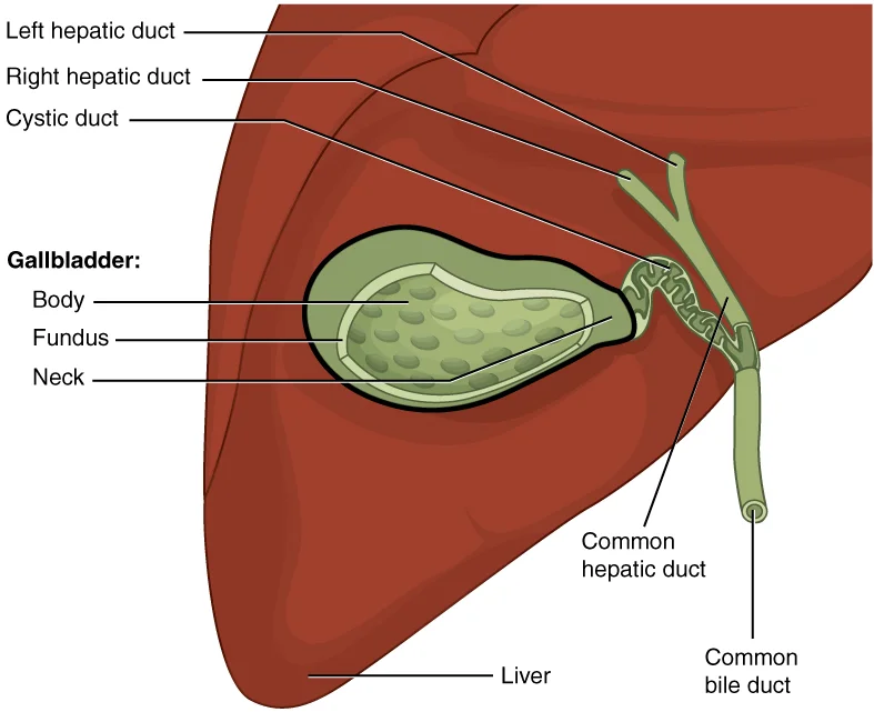
Long description
Illustration from Anatomy & Physiology, Connexions Web site. http://cnx.org/content/col11496/1.6/, Jun 19, 2013.

Figure 1: Illustration from Anatomy & Physiology, Connexions Web site (http://cnx.org/content/col11496/1.6/, jun 19, 2013).
The gallbladder is a pear-shaped muscular sac located on the visceral surface of the liver, specifically within the gallbladder fossa. It serves as a reservoir for bile produced by the hepatocytes. The organ consists of four distinct regions: the fundus, body, neck, and cystic duct. The fundus is the widest portion and tapers medially into the body, which in turn narrows to become the neck. The neck angles slightly superiorly as it approaches the hepatic duct. The cystic duct is 1–2 cm (less than 1 in) long and turns inferiorly as it bridges the neck and hepatic duct. [1]
DefinitionGallbladder Parts
The gallbladder has a fundus, body, neck, and cystic duct. The fundus projects beyond the liver edge. The neck joins the cystic duct. The body lies against the duodenum and colon.
The cystic duct is approximately 4 cm long. It has a spiral valve of Heister. This valve prevents kinking. The hepatopancreatic ampulla is dilated. It is surrounded by the sphincter of Oddi. The duodenal wall surrounds the ampulla. The liver is located superiorly. The stomach is located medially. The transverse colon is located inferiorly. The duodenum is located medially. The portal vein is located posteriorly. The he [3]
| Structure | Relation |
|---|---|
| Fundus | Anterior abdominal wall |
| Body | Transverse colon, duodenum |
| Neck | Cystic duct |
| Cystic duct | Hepatoduodenal ligament |
| Common bile duct | Duodenum, portal vein |
Figure 2Anatomy of Gallbladder and Bile Ducts Diagram
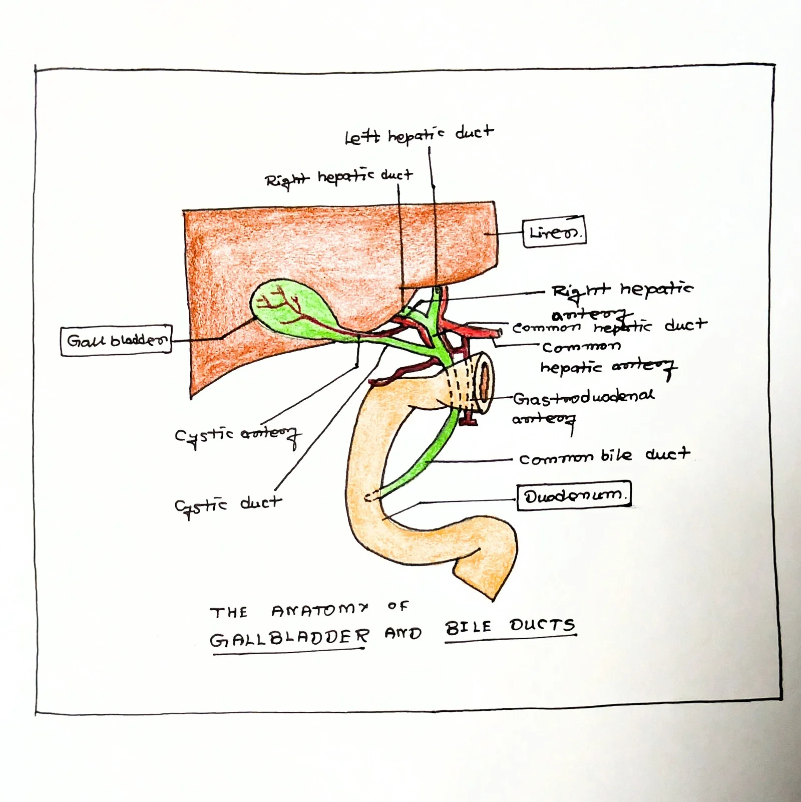
Long description
A detailed, hand-drawn anatomical diagram of the hepatobiliary system, specifically illustrating the gallbladder, left and right hepatic ducts, common hepatic duct, cystic duct, common bile duct, and the first part of th

Figure 2: A detailed, hand-drawn anatomical diagram of the hepatobiliary system, specifically illustrating the gallbladder, left and right hepatic ducts, common hepatic duct, cystic duct, common bile duct, and the first part of th.
The fundus is the widest portion and tapers medially into the body, which
The cystic duct joins the common hepatic duct to form the common bile duct. This union occurs within the triangle of Calot. The cystic artery typically runs within this triangle, supplying the gallbladder. Knowledge of this triangle prevents injury to the bile ducts during cholecystectomy. [3]
The common bile duct descends behind the first part of the duodenum. It lies in the free edge of the lesser omentum. It is accompanied by the hepatic artery and portal vein. The duct then passes posterior to the head of the pancreas. It joins the pancreatic duct at the ampulla of Vater. The pancreatic and common bile ducts normally are confluent at the ampulla of Vater. This structure opens into the duodenum at the major duodenal papilla. The sphincter of Oddi controls the flow of bile and pancr [2][3][4]
The gallbladder is peritoneal on its anterior surface, covered by visceral peritoneum. Its posterior surface is bare and adheres directly to the liver bed. This relation means inflammation can spread directly to the liver. The fundus relates to the transverse colon and the duodenum. The neck relates to the duodenum and the pylorus. These visceral relations are important for understanding referred pain and complications of cholecystitis.
Figure 3Gray1096
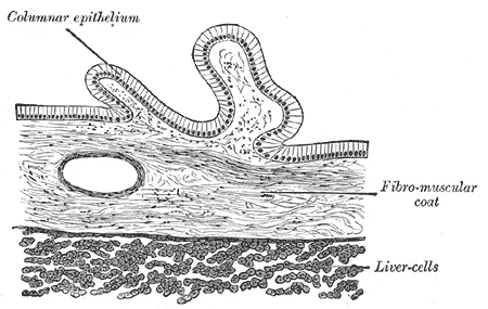
Long description
Transverse section of gall-bladder. Labelled illustration from Wikimedia Commons, used under its open licence.

Figure 3: Transverse section of gall-bladder.
The biliary tree includes the intrahepatic and extrahepatic ducts. The intrahepatic ducts drain bile from hepatocytes. Hepatocytes secrete about one liter of bile each day. This bile flows through canaliculi into larger ducts. The extrahepatic ducts transport bile to the duodenum. The common hepatic duct carries bile from the liver. The cystic duct connects the gallbladder to the common hepatic duct. The common bile duct carries bile to the ampulla. [1]
The triangle of Calot contains the cystic artery and lymph node of Lund. The cystic artery arises from the right hepatic artery. It runs along the cystic duct. The lymph node drains the gallbladder. Metastasis from gallbladder cancer often involves these nodes. Understanding these relations aids in surgical planning and diagnosis.
The next layer is the muscularis propria, or muscular layer. This layer consists of smooth muscle fibres arranged in longitudinal, circular, and oblique directions. The muscle fibres are not as organised as in the intestine but are sufficient to contract the gallbladder and expel bile into the cystic duct. The contraction is stimulated by cholecystokinin, a hormone released by the duodenum in response to fatty meals.
The absence of a serosal layer on the hepatic side has significant clinical implications. It allows for direct invasion of the liver parenchyma by malignant cells, facilitating early metastatic spread. This anatomical feature contributes to the poor prognosis of gallbladder cancer, as the disease can progress silently until it invades adjacent structures. [4]
DefinitionMucosal Folds
The rugae are permanent folds of the mucosa that allow the gallbladder to accommodate varying volumes of bile.
The biliary tree includes the intrahepatic and extrahepatic ducts. The right and left hepatic ducts emerge from the liver and join to form the common hepatic duct. The cystic duct arises from the gallbladder neck and joins the common hepatic duct to form the common bile duct. The common bile duct then travels through the head of the pancreas and joins the main pancreatic duct to form the ampulla of Vater, which empties into the duodenum. The walls of these ducts contain smooth muscle and connective tissue, but lack the complex muscular layers of the gallbladder. The mucosa of the ducts is lined by simple columnar epithelium, similar to the gallbladder, which aids in the transport and modification of bile.
Figure 4Gray1095
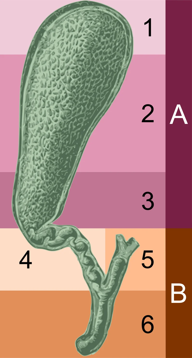
Long description
The gall-bladder and bile ducts laid open. (Spalteholz.)

Figure 4: The gall-bladder and bile ducts laid open (Spalteholz.).
| Layer | Composition | Key Feature |
|---|---|---|
| Mucosa | Simple columnar epithelium, lamina propria | Rugae present; no muscularis mucosae |
| Submucosa | Dense irregular connective tissue | Contains vessels and nerves |
| Muscularis | Smooth muscle (longitudinal, circular, oblique) | Contraction expels bile |
| Serosa/Fibrous | Peritoneum (free surface) or fibrous tissue (liver surface) | No serosa on liver side |
The arterial supply of the gallbladder and biliary tree is derived primarily from the cystic artery, a vessel that exhibits significant anatomical variability. Understanding this supply is critical for safe surgical intervention, particularly during cholecystectomy, to prevent iatrogenic injury to the hepatic artery or bile ducts.
The artery terminates by dividing into anterior and posterior branches that supply the gallbladder body and fundus. Neighbouring structures include the common bile duct, which lies medial to the cystic duct, and the hepatic artery proper, which is located medial to the common hepatic duct.
In some individuals, the cystic artery may arise from the left hepatic artery, the gastroduodenal artery, or the superior mesenteric artery. These variants are clinically significant because they may not follow the expected course through the cystohepatic triangle. The artery runs in the connective tissue of the cystohepatic triangle, lying anterior to the common hepatic duct and posterior to the cystic duct. It divides into two main branches: an anterior branch that supplies the anterior surface of the gallbladder and a posterior branch that supplies the posterior surface. These branches anastomose with each other and with branches from the cystic duct arteries. [4]
The biliary tree itself receives its blood supply from peribiliary vascular plexuses. The common hepatic duct and common bile duct are supplied by branches from the right gastric artery, the gastroduodenal artery, and the superior pancreaticoduodenal artery. These vessels form a rich network around the bile ducts, ensuring adequate perfusion even if individual branches are compromised. The ampulla of Vater, where the common bile duct and pancreatic duct join, is supplied by branches from the superior pancreaticoduodenal artery. This region is highly vascularised, which is relevant in conditions such as pancreatitis or ampullary tumours.
NoteSurgical Pearl
Always identify the cystic artery before clipping or cutting to avoid injury to the right hepatic artery, which may be anomalous.
| Structure | Typical Origin | Variant Origin | Clinical Significance |
|---|---|---|---|
| Cystic Artery | Right Hepatic Artery | Left Hepatic Artery, Gastroduodenal Artery | Risk of injury during cholecystectomy if variant present |
| Common Bile Duct | Peribiliary Plexus | Superior Pancreaticoduodenal Artery | Important in biliary reconstruction |
| Ampulla of Vater | Superior Pancreaticoduodenal Artery | Inferior Pancreaticoduodenal Artery | Relevant in pancreaticoduodenectomy |
The venous drainage of the gallbladder and biliary tree is critical for understanding the spread of disease and surgical anatomy. This direct connection means that malignant cells can spread to the liver without passing through the portal circulation first.
DefinitionDirect Drainage
Veins from the gallbladder wall drain directly into the liver sinusoids, bypassing the portal vein.
This anatomical feature has significant clinical implications. Because the gallbladder drains directly into the liver, local invasion by gallbladder cancer is common. The tumour spreads easily into the adjacent liver parenchyma. This is why staging often considers liver involvement as a key factor. The lack of a serosal layer on the liver-facing side of the gallbladder facilitates this direct invasion.
| Structure | Origin | Course | Termination |
|---|---|---|---|
| Cystic Vein | Gallbladder neck | Variable | Right portal vein branch |
| Direct Hepatic Veins | Gallbladder body/fundus | Through liver bed | Hepatic sinusoids |
| Ductal Plexus | Retroduodenal/R gastric arteries | Along biliary ducts | Portal/Hepatic veins |
Understanding this drainage pattern is essential for safe cholecystectomy. Surgeons must ligate the cystic artery and vein carefully. They must also be aware that bleeding from the gallbladder bed comes from the liver sinusoids. This bleeding is often oozing and difficult to control. Electrocautery is typically required. The direct venous connections also mean that inflammatory mediators from cholecystitis can enter the systemic circulation via the liver. This can contribute to the systemic symptoms seen in severe acute cholecystitis.
The biliary tree's venous return is less distinct. This integration ensures that bile and blood flow are coordinated. Any obstruction in the biliary tree can lead to increased pressure. This pressure can affect the venous drainage. It may lead to congestion and further liver damage.
NoteClinical Pearl
Always consider direct hepatic drainage when assessing gallbladder cancer spread.
In summary, the venous anatomy is complex. The gallbladder drains directly into the liver. The biliary tree has a rich plexus. This anatomy dictates surgical approach and disease spread. Medical students must master these details for both exams and practice. The figures provided help visualise these relationships. This pathway is unique compared to other abdominal organs. It underscores the intimate relationship between the gallbladder and the liver.
From the cystic node, efferent vessels pass posteriorly to the hepatic nodes located along the porta hepatis. These nodes are arranged around the common hepatic artery and the portal vein. Lymph then flows into the celiac lymph nodes, which surround the celiac trunk. From there, drainage continues to the lateral aortic nodes and ultimately into the thoracic duct. This pathway explains why metastatic disease often appears in the liver hilum before distant spread. [3]
Understanding this drainage is critical for surgical planning. During cholecystectomy, identification of the cystic node helps in staging if malignancy is suspected. The proximity of these nodes to the biliary tree means that inflammation or cancer can easily involve them. Clinicians must be aware that lymphatic spread can occur without obvious vascular invasion. This knowledge aids in interpreting imaging studies and planning lymph node dissection if necessary.
The biliary tree itself, including the intrahepatic and extrahepatic ducts, shares similar drainage patterns towards the hepatic and celiac nodes. This consistency allows for a unified approach to managing biliary pathology. However, the gallbladder has a more direct route via the cystic node, making it a key sentinel structure.
DefinitionNode of Lund
A lymph node located in the triangle of Calot that receives primary lymphatic drainage from the gallbladder.
The gallbladder and biliary tree receive a complex innervation that explains both visceral sensation and the phenomenon of referred pain. Understanding this requires looking at the autonomic and somatic components separately, then seeing how they converge.
The autonomic supply is derived from the celiac plexus. The resulting nerve plexus follows the cystic artery. It enters the gallbladder at the neck. This autonomic network primarily mediates motor function and dull visceral pain.
Somatic innervation is crucial for localising pain. The phrenic nerve provides somatic sensory fibres to the diaphragm. The gallbladder lies in close proximity to the diaphragm. Inflammation of the gallbladder irritates the peritoneum covering the diaphragm. The phrenic nerve carries this pain sensation. The phrenic nerve originates from cervical spinal roots C3, C4, and C5. These roots also supply sensation to the shoulder region. Therefore, irritation of the gallbladder can be perceived as pain in the right shoulder. This is referred pain.
Branches extend to the liver and gallbladder.
Benign polyps are frequently (~60%) accompanied by gallstones in those being investigated for upper abdominal pain. [4]
Clinical pearlReferred Pain
Pain from the gallbladder is referred to the right shoulder due to diaphragmatic irritation via the phrenic nerve (C3-C5).
The somatic nerves also supply the abdominal wall muscles. These muscles contract in response to inflammation. This causes guarding and rigidity. This is a local somatic response, distinct from the visceral referred pain.
| Component | Origin | Function |
|---|---|---|
| Parasympathetic | Vagus Nerve | Motor to gallbladder |
| Sympathetic | Greater Splanchnic Nerve | Visceral pain |
| Somatic | Phrenic Nerve (C3-C5) | Diaphragmatic pain referral |
Understanding this anatomy helps in physical examination. It indicates diaphragmatic irritation.
To understand the pathology of the biliary tree, one must first appreciate the normal histological architecture. The gallbladder mucosa is lined by simple columnar epithelium with numerous microvilli, facilitating water and electrolyte absorption. The lamina propria contains loose connective tissue and immune cells. The muscularis propria consists of irregular bundles of smooth muscle, which are thicker near the neck and cystic duct. The outer layer is either adventitia (where the gallbladder attaches to the liver) or serosa. For detailed microscopic features, please refer to the dedicated histology page.
DefinitionGallbladder Tumours
Adenocarcinoma is the most frequent histologic type, accounting for 98% of all gallbladder tumors. [4]
The staging of gallbladder cancer is fundamentally histological and anatomical, determining resectability. In stage I disease, the tumor invades the lamina propria or the muscle layer. T1a lesions are confined to the lamina propria, while T1b lesions extend into the muscle layer. Simple cholecystectomy is reserved for T1a disease and offers a surgical cure. For T1b lesions, the malignancy extends to the muscle layer, requiring a more radical approach. Patients may require cholecystectomy with wedge resections involving segment IVb and V of the liver as well as portal lymphadenectomy. [4]
Exam trapStaging Error
Do not confuse T1a (lamina propria) with T1b (muscle layer); the surgical management differs significantly.
T2 lesions invade perimuscular connective tissue with no extension beyond the serosa or into the liver. This often requires a more formal resection of segments IVb and V. T3 disease involves tumor burden extending to the serosa, liver, and/or adjacent organs/structures. Under these circumstances, resection becomes more radical, including an extended right hepatectomy with possible caudate lobectomy, regional lymphadenectomy, and extirpation of other affected structures. [4]
The other histopathologic variants include the papillary, mucinous, squamous, and adenosquamous subtypes. Two-thirds of adenocarcinomas are moderately or poorly differentiated. If the invasion is limited to the mucosa or subserosa, the 5-year survival rate is over 95%. The disease stage determines the treatment options. The best outcomes are reserved for patients who qualify for cholecystectomy. Both stage I and II are potentially resectable with curative intent. Improved outcomes greatly depend on the recognition of the major risk factors, accurate initial staging, and meticulous surgical excision. [3] [4]
The gallbladder and biliary tree arise from the foregut during the fourth week of embryonic development. This process begins with the formation of the hepatic diverticulum, also known as the liver bud, which protrudes from the ventral wall of the duodenum. The hepatic diverticulum consists of two parts: the cranial pars hepatica and the caudal pars cystica. The pars hepatica gives rise to the liver parenchyma, the intrahepatic bile ducts, and the extrahepatic bile ducts. The pars cystica develops into the gallbladder and the cystic duct.
The liver bud grows into the septum transversum, a mass of mesoderm that separates the thoracic and abdominal cavities. The cells of the pars hepatica proliferate rapidly to form the hepatic cords, which eventually differentiate into hepatocytes and bile duct epithelium. The surrounding mesenchyme of the septum transversum differentiates into the connective tissue, blood vessels, and immune cells of the liver. The hepatic cords anastomose to form a complex network of channels that will become the biliary tree.
The gallbladder develops as an outpouching of the cystic duct. Initially, the lumen of the gallbladder is obliterated by proliferating epithelial cells. Later, recanalization occurs to restore the lumen. This process ensures the formation of a hollow organ capable of storing and concentrating bile. The cystic duct connects the gallbladder to the common hepatic duct, forming the common bile duct. The common bile duct then joins the main pancreatic duct to form the ampulla of Vater, which empties into the duodenum.
The biliary tree is lined by simple columnar epithelium. This epithelium is specialized for the secretion and absorption of water and electrolytes, allowing for the concentration of bile. The gallbladder mucosa is folded into numerous ruffles, which increase the surface area for absorption. The muscularis layer of the gallbladder consists of smooth muscle fibres arranged in longitudinal, circular, and spiral patterns. These fibres contract to expel bile into the cystic duct during digestion.
The blood supply to the gallbladder and biliary tree is derived from the hepatic artery and the portal vein. The cystic artery, a branch of the right hepatic artery, supplies the gallbladder. The cystic vein drains blood from the gallbladder into the portal vein. Lymphatic drainage follows the venous supply, with lymphatics draining into the cystic lymph node and then into the hepatic lymph nodes. Innervation is provided by the celiac plexus, with parasympathetic fibres from the vagus nerve and sympathetic fibres from the splanchnic nerves.
DefinitionHepatic Diverticulum
An outpouching of the ventral foregut that gives rise to the liver, gallbladder, and biliary tree.
| Structure | Embryological Origin |
|---|---|
| Liver Parenchyma | Pars Hepatica |
| Gallbladder | Pars Cystica |
| Cystic Duct | Pars Cystica |
| Common Hepatic Duct | Pars Hepatica |
The development of the biliary tree is a complex process involving precise coordination of cell proliferation, differentiation, and morphogenesis. Any disruption in this process can lead to congenital anomalies such as biliary atresia or choledochal cysts. Understanding the embryological origins of these structures is essential for diagnosing and managing congenital biliary disorders.
The biliary tree and gallbladder are best visualised using cross-sectional imaging and cholangiography. This image illustrates the ERCP and imaging anatomy alongside features relevant to gallstones and cholecystectomy anatomy. The view is a coronal reconstruction, oriented with the patient’s right side on the viewer’s left. The liver parenchyma appears as a homogeneous grey mass superiorly. The gallblad
DefinitionCholecystolithiasis
The presence of gallstones within the gallbladder.
Gallstones are radiolucent or radiopaque depending on their composition. On ultrasound, they appear as mobile, echogenic foci with posterior acoustic shadowing. This is the primary imaging modality for diagnosing cholelithiasis. When stones obstruct the cystic duct, they cause biliary colic or acute cholecystitis. Chronic inflammation, a major risk factor, accompanies cholelithiasis and infection. Diagnosis may come at the time of cholecystectomy for gallstones, although preoperative imaging with transabdominal and endoscopic ultrasound is providing an important advance. Surgery represents the only potential cure. [4]
Clinical pearlAsymptomatic Stones
Routine cholecystectomy is not advised for asymptomatic cholecystolithiasis (recommendation grade C2). [2]
However, in high-risk populations such as American Indians, cholecystectomy in those at risk, such as American Indians, with even asymptomatic cholelithiasis can prevent the tumor from arising, although such an approach carries inherent risks and cost. This is because chronic inflammation increases the risk of gallbladder carcinoma. Thus, early diagnosis is imperative as surgery can be curative. The usual late presentation means an advanced stage with potential nodal involvement and leads to rec [4]
The gallbladder is a pear-shaped muscular sac located on the visceral surface of the liver, specifically within the gallbladder fossa. It serves as a reservoir for bile produced by the hepatocytes. The organ consists of four distinct regions: the fundus, body, neck, and cystic duct. The fundus is the rounded distal end that typically projects beyond the inferior border of the liver. [1][3]
The cystic duct joins the common hepatic duct to form the common bile duct. The common bile duct descends behind the first part of the duodenum and within the free edge of the lesser omentum. It runs anterior to the portal vein and to the right of the hepatic artery proper. The duct then passes posterior to the head of the pancreas. Finally, it joins the main pancreatic duct to form the hepatopancreatic ampulla. This ampulla opens into the second part of the duodenum at the major duodenal papill [3]
Blood supply to the gallbladder arises from the cystic artery, a branch of the right hepatic artery. Venous drainage occurs directly into the liver parenchyma via small veins that bypass the portal system. Lymphatic drainage follows the cystic artery to the cystic lymph node of Lund. This node lies in the triangle of Calot. This anatomical space is c [3]
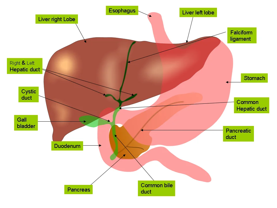
Figure 5Anatomy of liver and gall bladder
Long description
Anatomy of the biliary tree, liver and gall bladder

Figure 5: Anatomy of the biliary tree, liver and gall bladder.
The common hepatic duct descends from the liver hilum. The common bile duct is traced downwards behind the duodenum. The major duodenal papilla is indicated at the duodenal wall. Understanding these relations prevents iatrogenic injury during surgery.
It lies inferior to the diaphragm in the right upper quadrant of the abdominal cavity[1].
The porta hepatis is the gate to the liver where the hepatic artery and hepatic portal vein enter[1]. The hepatic artery delivers oxygenated blood from the heart to the liver[1]. The hepatic portal vein delivers partially deoxygenated blood containing nutrients absorbed from the small intestine[1]. This vein actually supplies more oxygen to the liver than do the much smaller hepatic arteries[1]. With this hepatic portal circulation, all blood from the alimentary canal passes through the liver[1]. This largely explains why the liver is the most common site for the metastasis of cancers that originate in the alimentary canal[1].
Clinical pearlPortal Circulation
The hepatic portal vein supplies more oxygen to the liver than the hepatic artery does, despite carrying nutrient-rich, partially deoxygenated blood from the gut[1].
Exam trapLobe Anatomy
The quadrate and caudate lobes are defined by internal features within the right lobe, not by external surface anatomy alone[1].
DefinitionPorta Hepatis
The porta hepatis is the entry point for the hepatic artery, hepatic portal vein, and common hepatic duct[1].
NoteMetastasis Risk
Because all blood from the alimentary canal passes through the liver, it is the most common site for metastasis from gastrointestinal cancers[1].
Clinical pearlLigament Remnants
The falciform ligament and ligamentum teres hepatis are embryological remnants of the umbilical vein[1].
Exam trapBlood Supply
Do not confuse the oxygenated blood of the hepatic artery with the nutrient-rich blood of the portal vein; the latter provides the majority of hepatic oxygen[1].
DefinitionLesser Omentum
The lesser omentum connects the liver to the lesser curvature of the stomach[1].
NoteAnatomical Relations
The liver receives protection from the surrounding ribs while lying in the right upper quadrant[1].
Clinical pearlVascular Drainage
Blood processed by the liver drains into the central vein, then through the hepatic vein to the inferior vena cava[1].
Exam trapLigament Function
The coronary ligaments and lateral ligaments connect the liver to the diaphragm and abdominal wall, distinct from the falciform ligament[1].
DefinitionHepatic Artery Role
The hepatic artery delivers oxygenated blood from the heart to the liver[1].
Students frequently confuse the anatomical relationships of the biliary tree with those of the pancreas, leading to errors in surgical planning and imaging interpretation. A primary error is assuming the pancreas is an intraperitoneal organ. The soft, oblong, glandular pancreas lies transversely in the retroperitoneum behind the stomach. This distinction is critical because retroperitoneal structures are fixed in position, whereas intraperitoneal organs move with peristalsis and respiration. [1]
A significant conceptual trap involves the origin of bilirubin. Many students attribute bilirubin production to the liver, whereas it is a waste product produced when the spleen removes old or damaged red blood cells from the circulation. The liver merely excretes this pigment. Consequently, students often fail to link haemolysis to jaundice correctly. The breakdown products, including toxic bilirubin, are transported to the liver via the splenic vein of the hepatic portal system. Understanding this pathway clarifies why pre-hepatic causes of jaundice present differently from hepatic or post-hepatic causes. [1]
Students also frequently misinterpret the clinical significance of stool colour. Acholic (white) stool is not merely a sign of liver failure but specifically indicates that bile does not enter the intestine. This is a direct consequence of obstruction, not just reduced production. Furthermore, confusion arises regarding the portal triad. It is not a random collection of vessels but a distinctive arrangement around the perimeter of hepatic lobules. It consists of three basic structures: a bile duct, a hepatic artery branch, and a hepatic portal vein branch. Missing the bile duct in this triad leads to errors in understanding biliary drainage. [1]
Another pitfall is ignoring the role of Kupffer cells in liver pathology. These are star-shaped reticuloendothelial cells that remove dead red and white blood cells, bacteria, and other foreign material that enter the sinusoids. Students often focus only on hepatocytes, neglecting the immune function of the liver. This oversight is dangerous when interpreting signs of sepsis or systemic infection. Additionally, students may confuse the function of pancreatic juice with bile. Pancreatic juice is clear and composed mostly of water along with some salts, sodium bicarbonate, and several digestive enzymes. It is distinct from bile in both appearance and chemical composition. [1]
Finally, students often underestimate the link between chronic inflammation and malignancy in the biliary tree. Chronic inflammation is inexorably linked to malignant transformation, being a major factor in carcinogenesis. Recurrent or chronic inflammatory insults are detrimental, causing deoxyribonucleic acid (DNA) damage, provoking repeated tissue proliferative attempts at restoration, releasing cytokines and growth factors, and thus, predisposing cells to oncogenic transformation. For example, cholelithiasis with its attendant repeated trauma, leading to chronic cholecystitis, might be the mechanism by which cancer develops after many years. Failing to recognise this pathway leads to delayed diagnosis of biliary cancers. [4]
This knowledge is applied at the bedside to interpret jaundice. When a patient presents with yellowing of the skin, the clinician must determine if the cause is pre-hepatic, hepatic, or post-hepatic. If the patient has acholic stool, it suggests a post-hepatic obstruction where bile does not enter the intestine. The clinician recalls that bilirubin is eventually transformed by intestinal bacteria into stercobilin, a brown pigment that gives your stool its characteristic color. Absence of this pigment confirms biliary obstruction.
In the operating theatre, anatomical precision is vital. The surgeon must identify the portal triad, which is a distinctive arrangement around the perimeter of hepatic lobules. It consists of three basic structures: a bile duct, a hepatic artery branch, and a hepatic portal vein branch. Mistaking the bile duct for a vessel can lead to catastrophic bleeding or biliary leak. The surgeon also notes that the pancreas lies transversely in the retroperitoneum behind the stomach. Its head is nestled into the “c-shaped” curvature of the duodenum with the body extending to the left about 15.2 cm (6 in) and ending as a tapering tail in the hilum of the spleen. This anatomy dictates the approach for pancreaticoduodenectomy. [1]
On imaging, radiologists look for specific signs of biliary disease. In primary sclerosing cholangitis, they identify multifocal stricturing with a beaded appearance. This appearance is thought to reflect the fibrous stricture portions and the normal segments of the bile duct. One-fifth of the cases present band-like strictures that are extremely short, and one-fourth present diverticulum-like outpouchings. Recognising this pattern distinguishes it from other causes of ductal dilation. Radiologists also assess for congenital anomalies such as PBM, which is a congenital anomaly consisting of a union of the pancreatic and bile ducts located outside the duodenal wall. This anomaly is a risk factor for biliary cancer. [2]
In examinations, students must distinguish between exocrine and endocrine functions of the pancreas. The pancreas is a curious mix of exocrine (secreting digestive enzymes) and endocrine (releasing hormones into the blood) functions. They must also recall that the pancreas produces over a liter of pancreatic juice each day. This juice has a pH of 7.1 to 8.2, which serves to buffer the acidic gastric juice in chyme, inactivate pepsin from the stomach, and create an optimal environment for the act [1]
DefinitionPortal Triad
A distinctive arrangement around the perimeter of hepatic lobules, consisting of three basic structures: a bile duct, a hepatic artery branch, and a hepatic portal vein branch.
Exam trapBile Function
Students frequently confuse the anatomical relationships of the biliary tree with those of the pancreas, leading to errors in surgical planning and imaging interpretation. A primary error is assuming the pancreas is an intraperitoneal organ. The pancreas is actually retroperitoneal, lying transversely behind the stomach. This distinction is critical because retroperitoneal structures are fixed in position, whereas intraperitoneal organs move with peristalsis and respiration. Bile is a mixture secreted by the liver to accomplish the emulsification of lipids in the small intestine. [1]
Jaundice Assessment
- Patient presents with jaundiceAssess stool and urine colour
- Question: Is stool acholic (white)?
Yes
- Suspect post-hepatic obstructionBile does not enter the intestine
No
- Check for haemolysisBilirubin is a waste product from RBC breakdown
The pancreas is retroperitoneal, lying transversely behind the stomach. Bile emulsifies lipids, which are hydrophobic and do not dissolve in water. Bilirubin is produced when the spleen removes old red blood cells. The portal triad contains a bile duct, hepatic artery branch, and portal vein branch. Kupffer cells are phagocytes that remove foreign material from sinusoids. Pancreatic juice buffers gastric acid and has a pH of 7.1 to 8.2. Chronic inflammation predisposes cells to oncogenic transformation. PBM is a union of pancreatic and bile ducts outside the duodenal wall.
Answer each one in your head first, then open the answer.
What is the primary function of bile in the small intestine?
Show answerHide answer
To accomplish the emulsification of lipidsWhere does bilirubin originate before reaching the liver?
Show answerHide answer
From the removal of old red blood cells by the spleenWhat are the three components of the portal triad?
Show answerHide answer
A bile duct, a hepatic artery branch, and a hepatic portal vein branchWhy is stool white in biliary obstruction?
Show answerHide answer
Bile does not enter the intestineWhat is the pH range of pancreatic juice?
Show answerHide answer
7.1 to 8.2
- Betts JG, Young KA, Wise JA, et al. Accessory Organs in Digestion: The Liver, Pancreas, and Gallbladder. OpenStax, Anatomy and Physiology 2e (CC BY-NC-SA 4.0). Accessed .
- Miyazaki M, Takada T, Miyakawa S, et al. Risk factors for biliary tract and ampullary carcinomas and prophylactic surgery for these factors. Journal of hepato-biliary-pancreatic surgery 2008;15(1):15-24. doi:10.1007/s00534-007-1276-8. Accessed .
- Kamal F, Khan MA, Lee-Smith W, et al. Efficacy and safety of EUS-guided gallbladder drainage for rescue treatment of malignant biliary obstruction: A systematic review and meta-analysis. Endoscopic ultrasound 2023;12(1):8-15. doi:10.4103/eus-d-21-00206. Accessed .
- Hundal R, Shaffer EA. Gallbladder cancer: epidemiology and outcome. Clinical epidemiology 2014;6:99-109. doi:10.2147/clep.s37357. Accessed .
Figure credits
- Figure 1: OpenStax College, via Wikimedia Commons, CC BY 3.0, CC BY 3.0.
- Figure 2: Dr. Swapnil Banerjee, via Wikimedia Commons, CC BY-SA 4.0, CC BY-SA 4.0. resized.
- Figure 3: Henry Vandyke Carter, via Wikimedia Commons, Public domain, Public domain.
- Figure 4: Henry Vandyke Carter, via Wikimedia Commons, Public domain, Public domain.
- Figure 5: Jiju Kurian Punnoose, via Wikimedia Commons, CC BY-SA 4.0, CC BY-SA 4.0.
Educational content for medical students and doctors. It is not a substitute for clinical judgement or local guidelines.