Liver
Anatomy of the liver covering its segments, blood supply, hilum and microscopic structure
The liver is the body's largest gland, lying chiefly in the right upper quadrant beneath the diaphragm; its internal plan matches a rich blood supply with an outward drainage of bile.
Couinaud split the liver into functionally independent segments, each with its own Glissonean pedicle and hepatic venous drainage; the plate system he described is a fibrous thickening of that sheath. At the hilum the hilar plate wraps the outer circumference of the pedicles.
The porta hepatis, the gate to the liver, admits the hepatic artery and the hepatic portal vein and lets the common hepatic duct leave; they run behind the lateral border of the lesser omentum. The portal vein carries partially deoxygenated, nutrient-rich blood and, though the hepatic artery is much smaller, supplies more of the liver's oxygen. At the edge of each lobule the matching trio forms the portal triad.
| Component | Direction | Function |
|---|---|---|
| Hepatic portal vein branch | Entering | Nutrient-rich, partially deoxygenated blood |
| Hepatic artery branch | Entering | Oxygenated blood |
| Bile duct branch | Leaving | Bile towards the common hepatic duct |
Blood from each lobule drains to a central vein, then through a hepatic vein into the inferior vena cava. Canaliculi run bile towards the portal tract while sinusoidal blood flows the other way, so bile and blood move in opposite directions.
Figure 1Gray1089
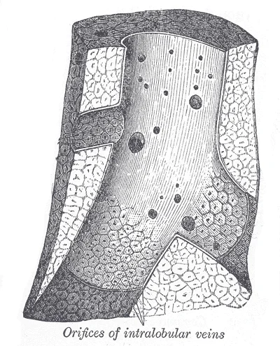
Long description
Longitudinal section of a hepatic vein. (After Kiernan.)

Figure 1: Longitudinal section of a hepatic vein (After Kiernan.).
The liver has three main components: hepatocytes, bile canaliculi and hepatic sinusoids. Hepatocytes dominate, filling around 80 percent of the liver's volume, and form plates called hepatic laminae radiating from the central vein. Sinusoids are open, porous spaces of fenestrated capillaries fed by the afferent vessels, allowing rapid exchange across their leaky wall. Kupffer cells, star-shaped phagocytes in the sinusoids, clear ageing blood cells, bacteria and foreign material. [1]
The acinus, the functional unit around the portal triad, is zoned from the periportal edge towards the draining venule, each zone handling different metabolic tasks.
Glisson described the pedicle sheath in 1642, and Laennec distinguished the true capsule from the serosa in 1802, after which it was neglected for more than 200 years. [2]
Blood and bile in the liver
- Porta hepatisArtery and portal vein enter, bile duct leaves
- Portal triadDuct, artery branch and vein branch
- SinusoidsFenestrated channels with Kupffer cells
- Question: Exchange done
Blood
- Central vein
- Hepatic vein to inferior vena cava
Bile
- CanaliculiRun opposite to blood
- Bile duct
The liver is the largest gland in the body, weighing about three pounds in an adult[1]. It lies inferior to the diaphragm in the right upper quadrant of the abdominal cavity and receives protection from the surrounding ribs[1].
The liver is connected to the abdominal wall and diaphragm by five peritoneal folds referred to as ligaments, including the falciform ligament. [1]
The digestive role of the liver is to produce bile and export it to the duodenum[1]. Bile is a mixture secreted by the liver to accomplish the emulsification of lipids in the small intestine[1]. Bile contains bile salts and phospholipids, which emulsify large lipid globules into tiny lipid droplets, a necessary step in lipid digestion and absorption[1]. Bilirubin, the main bile pigment, is a waste product produced when the spleen removes old or damaged red blood cells from the circulation[1]. These breakdown products, including proteins, iron, and toxic bilirubin, are transported to the liver via the splenic vein of the hepatic portal system[1]. In the liver, proteins and iron are recycled, whereas bilirubin is excreted in the bile[1]. It accounts for the green color of bile[1].
DefinitionBile Function
Bile is a mixture secreted by the liver to accomplish the emulsification of lipids in the small intestine[1].
The liver is also one of the most important organs[1]. In addition to being an accessory digestive organ, it plays a number of roles in metabolism and regulation[1]. Chemical digestion in the small intestine cannot occur without the help of the liver and pancreas[1]. The liver produces bile and delivers it to the common hepatic duct[1]. The gallbladder stores and concentrates bile, releasing it when it is needed by the small intestine[1]. The gal
| Ligament | Description |
|---|---|
| Falciform | Remnant of umbilical vein; separates right and left lobes anteriorly[1]. |
| Ligamentum teres | Remnant of umbilical vein[1]. |
| Coronary | Connects liver to diaphragm[1]. |
| Lateral | Connects liver to diaphragm[1]. |
| Lesser Omentum | Tethers liver to lesser curvature of stomach[1]. |
Figure 2Gray1085
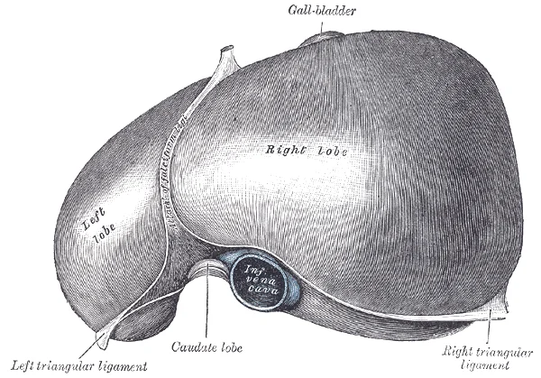
Long description
The superior surface of the liver. (From model by His.)

Figure 2: The superior surface of the liver (From model by his.).
The liver’s internal architecture is defined by its functional units and vascular supply rather than simple lobes. The parenchyma is organised into hexagonal lobules, each centred on a central vein. Blood flows from the portal triads at the periphery toward the centre. The portal triad contains the portal vein, hepatic artery, and bile duct. These structures are embedded in connective tissue within the Glissonean capsule. [1]
The central veins converge to form the hepatic veins. The bile ducts run alongside the portal vessels. This arrangement ensures efficient exchange between blood and hepatocytes.
The vascular supply is dual. The portal vein provides most of the blood volume. The hepatic artery provides oxygen-rich blood. These vessels branch together within the Glissonean pedicles. Couinaud described the three different ways to approach the Glissonean pedicles : the intrafascial, the extrafascial, and the extrafascial and transfissural approach. The intrafascial approach means the individual vascular transection method. The extrafascial approach means the Glissonean pedicle approach. The extrafascial approach is further divided into the intrahepatic and the extrahepatic approach according to the presence or absence of parenchymal destruction. The extrafascial and transfissural approach is the pedicle approach with major liver transection through the main portal or the umbilical fissure. [2]
The biliary system drains bile from the hepatocytes. Bile flows from canaliculi into ducts. These ducts merge to form larger channels. The right and left hepatic ducts join to form the common hepatic duct. This duct joins the cystic duct from the gallbladder. The resulting common bile duct carries bile to the duodenum. The sphincter of Oddi controls the flow. [1]
The liver is divided into segments based on vascular supply. This segmentation allows for precise surgical resection. The ligamentum teres and ligamentum venosum mark the boundaries between functional segments. These ligaments are remnants of fetal circulation. The falciform ligament attaches the liver to the anterior abdominal wall. [1]
The sinusoids are the capillaries of the liver. They are lined by endothelial cells and Kupffer cells. Blood from the portal vein and hepatic artery mixes in the sinusoids. It then flows toward the central vein. This flow direction is critical for metabolic function. Nutrients are processed as blood passes through the liver. [1]
DefinitionGlissonean Pedicle
A bundle containing the portal vein branch, hepatic artery branch, and bile duct branch, enclosed in connective tissue.
| Structure | Function | Location |
|---|---|---|
| Portal Vein | Nutrient-rich blood supply | Periphery of lobule |
| Hepatic Artery | Oxygen supply | Periphery of lobule |
| Central Vein | Drainage to hepatic veins | Centre of lobule |
| Bile Duct | Bile transport | Periphery of lobule |
Figure 3Gray532
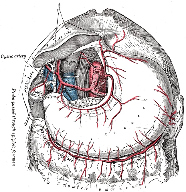
Long description
The celiac artery and its branches; the liver has been raised, and the lesser omentum and anterior layer of the greater omentum removed.

Figure 3: The celiac artery and its branches; the liver has been raised, and the lesser omentum and anterior layer of the greater omentum removed.
The liver receives a dual blood supply, comprising oxygenated arterial blood and nutrient-rich venous blood. This arrangement ensures the organ has sufficient oxygen for its high metabolic rate while processing absorbed nutrients from the gastrointestinal tract. The arterial component originates from the hepatic artery proper, which is a branch of the common hepatic artery arising from the celiac trunk. This vessel travels within the hepatoduodenal ligament, part of the lesser omentum, alongside the portal vein and bile duct.
The left hepatic artery enters the left lobe. The cystic artery, often arising from the right hepatic artery, supplies the gallbladder. The bile ducts accompany the artery. This triad structure is crucial for surgical identification. The remaining seventy-five percent of blood flow comes from the portal vein, which carries blood rich in nutrients and toxins from the spleen, stomach, and intestines. [1]
Lymphatic drainage follows the vascular pathways. Lymph from the liver parenchyma drains into nodes at the porta hepatis. These nodes then drain into the celiac lymph nodes. This pathway is clinically significant in the spread of hepatocellular carcinoma. The lymphatics also drain into the diaphragmatic nodes, which can lead to pleural effusions in advanced disease.
DefinitionHepatic Triad
The portal triad consists of the hepatic artery branch, portal vein branch, and bile duct branch, enclosed in connective tissue within the portal tract.
The anatomical variation of the hepatic artery is common. In some individuals, the right hepatic artery arises from the superior mesenteric artery. This variant is critical to identify during cholecystectomy to avoid iatrogenic injury. The left hepatic artery may arise from the left gastric artery. Surgeons must be aware of these variations to prevent ischaemic complications. The liver's unique blood supply allows it to regenerate rapidly after partial hepatectomy. The portal flow stimulates hepatocyte proliferation, while arterial flow supports metabolic function. Understanding this dual supply is essential for managing liver trauma and planning resections. [1]
| Structure | Origin | Function |
|---|---|---|
| Hepatic Artery Proper | Common Hepatic Artery | Oxygen supply |
| Portal Vein | Superior Mesenteric & Splenic Veins | Nutrient transport |
| Cystic Artery | Right Hepatic Artery | Gallbladder supply |
The liver possesses a dual blood supply, receiving oxygenated blood from the hepatic artery and nutrient-rich, deoxygenated blood from the portal vein. However, the venous drainage is singular, converging into the hepatic veins which empty directly into the inferior vena cava. This arrangement ensures that processed blood leaves the organ efficiently to return to the systemic circulation.
Figure 42423 Microscopic Anatomy of Liver
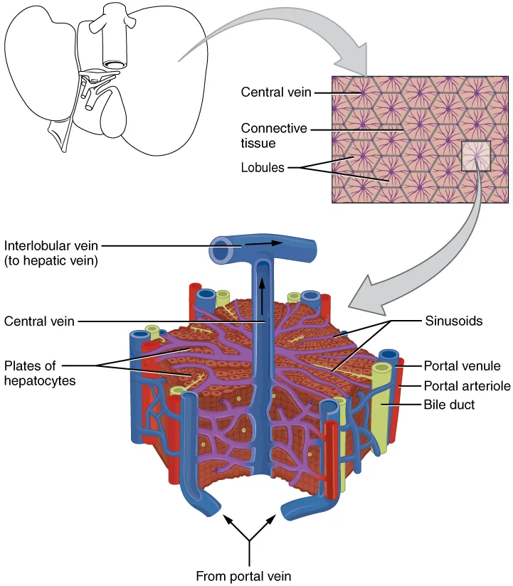
Long description
The liver receives oxygenated blood from the hepatic artery and nutrient-rich deoxygenated blood from the hepatic portal vein.

Figure 4: The liver receives oxygenated blood from the hepatic artery and nutrient-rich deoxygenated blood from the hepatic portal vein.
Figure 5Gray1090
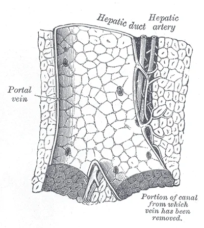
Long description
Longitudinal section of a small portal vein and canal. (After Kiernan.)

Figure 5: Longitudinal section of a small portal vein and canal (After Kiernan.).
DefinitionHepatic Veins
The right, middle, and left hepatic veins drain the liver parenchyma into the inferior vena cava. They are distinct from the portal triad structures and run between the hepatic segments. [1]
The hepatic veins are short, wide, and thin-walled, lacking the robust connective tissue sheath found in the portal triads. This anatomical feature makes them vulnerable to compression or rupture during trauma or surgical manipulation. The right hepatic vein is particularly prone to injury due to its size and direct course into the inferior vena cava.
| Structure | Course | Relations | Termination |
|---|---|---|---|
| Right Hepatic Vein | Runs in right hepatic fissure | Separates right anterior and posterior sections | Inferior vena cava |
| Middle Hepatic Vein | Runs in main portal fissure | Divides left and right functional lobes | Inferior vena cava (often via common trunk) |
| Left Hepatic Vein | Runs in left portal fissure | Drains left lobe | Inferior vena cava (often via common trunk) |
Understanding the spatial relationships of these veins is essential for interpreting cross-sectional imaging. On axial CT or MRI scans, the hepatic veins appear as anechoic or hypointense tubular structures radiating from the inferior vena cava towards the liver capsule. The right hepatic vein is typically the most prominent, followed by the middle and left hepatic veins. Any obstruction or thrombosis in these veins, such as in Budd-Chiari syndrome, leads to characteristic imaging findings inclu
Exam trapBudd-Chiari Syndrome
Obstruction of hepatic venous outflow causes liver congestion. The caudate lobe is often spared because it drains directly into the inferior vena cava, unlike the rest of the liver which drains via the main hepatic veins.
The hepatic veins are not accompanied by nerves or lymphatics in the same way the portal triads are. Their primary function is passive drainage, relying on the pressure gradient between the liver sinusoids and the right atrium. This gradient is maintained by the negative intrathoracic pressure during inspiration and the pumping action of the right heart. Any increase in right atrial pressure, such as in right-sided heart failure, can lead to hepatic congestion and centrilobular necrosis, known as 'nutmeg liver'. This pathological change is a direct consequence of the anatomical arrangement of the venous drainage system.
The lymphatic drainage of the liver is a critical component of its immune surveillance and potential pathway for metastatic spread. Understanding this system requires examining the superficial and deep networks, their origins, courses, and terminations.
The superficial lymphatic vessels originate from the subserous plexus located just beneath the Glisson’s capsule. These vessels are numerous and form a dense network across the entire surface of the liver. They follow the course of the hepatic veins and portal triads towards the porta hepatis.
DefinitionGlisson’s Capsule
The fibrous capsule covering the liver, containing the portal triads.
As these vessels converge, they form larger trunks that accompany the hepatic veins. These trunks pass through the diaphragm via the caval opening or the aortic hiatus, depending on their specific origin within the liver lobes.
The deep lymphatic vessels originate from the perivascular plexuses surrounding the portal triads and hepatic veins within the liver parenchyma. These vessels are less numerous than the superficial ones but are equally important. They follow the portal triads towards the porta hepatis.
NoteClinical Relevance
Metastatic cancer cells often travel via these deep lymphatic channels to reach the regional lymph nodes.
These nodes are located along the common hepatic artery and the portal vein. From here, the lymphatic flow continues towards the celiac lymph nodes. The celiac nodes then drain into the intestinal trunk, which ultimately empties into the cisterna chyli.
| Structure | Origin | Course | Termination |
|---|---|---|---|
| Superficial Lymphatics | Subserous plexus | Along hepatic veins | Mediastinal/Phrenic nodes |
| Deep Lymphatics | Perivascular plexus | Along portal triads | Hepatic nodes |
| Hepatic Nodes | Porta hepatis | Along common hepatic artery | Celiac nodes |
| Celiac Nodes | Celiac trunk | Along abdominal aorta | Cisterna chyli |
The lymphatic drainage of the liver is not uniform. The right lobe has a more extensive superficial drainage network compared to the left lobe. This asymmetry is clinically significant when considering the spread of disease. The lymphatic vessels are closely associated with the blood vessels, which facilitates the rapid transport of fluids and cells.
Clinical pearlKey Point
The liver has a rich lymphatic supply, making it susceptible to metastatic spread via lymphatic channels.
In summary, the lymphatic drainage of the liver involves both superficial and deep networks that converge at the porta hepatis. The superficial network drains into the mediastinal and phrenic nodes, while the deep network drains into the hepatic nodes. From there, the lymph flows into the celiac nodes and eventually into the cisterna chyli. This complex system ensures efficient drainage of interstitial fluid and plays a crucial role in immune response and disease progression.
The innervation of the liver is complex, involving both autonomic and somatic components that serve distinct physiological and sensory functions. Understanding this dual supply is critical for explaining both hepatic regulation and the phenomenon of referred pain.
DefinitionAutonomic Innervation
The liver receives sympathetic and parasympathetic fibres from the autonomic nervous system, which regulate vascular tone and metabolic activity.
Sympathetic fibres originate from the thoracic spinal segments T5 to T9. These preganglionic neurons synapse in the celiac ganglion. Postganglionic sympathetic fibres then travel along the hepatic artery to reach the liver parenchyma. Their primary function is vasoconstriction of the hepatic arterial blood vessels and modulation of metabolic processes within hepatocytes. This sympathetic input is part of the greater splanchnic nerve pathway. [1]
Parasympathetic innervation is derived from the vagus nerve (cranial nerve X). The parasympathetic system generally promotes digestive activities, contrasting with the sympathetic 'fight or flight' response. [1]
Clinical pearlReferred Pain Mechanism
Pain from the liver capsule is referred to the right shoulder due to shared spinal segments with the phrenic nerve.
Somatic sensation is unique to the liver. The liver parenchyma itself lacks pain receptors. Pain arises only when the Glisson’s capsule, the fibrous connective tissue covering the liver, is stretched or inflamed. This somatic pain is transmitted via the phrenic nerve. Because these cervical segments also supply sensation to the shoulder region, irritation of the diaphragmatic peritoneum overlying the liver is perceived as pain i [1]
Hepatic branches are traced along the portal triad.
| Feature | Autonomic | Somatic |
|---|---|---|
| Origin | T5-T9 (Symp), Vagus (Para) | C3-C5 (Phrenic) |
| Target | Parenchyma, Vessels | Glisson’s Capsule |
| Function | Metabolism, Vasoconstriction | Pain Sensation |
| Perception | Visceral (Unlocalised) | Referred (Shoulder) |
This integrated system ensures that while metabolic regulation is automatic, protective pain signals are sharp and localising, albeit referred to distant somatic sites.
The liver is a complex glandular organ composed of functional units known as hepatic lobules. These lobules are hexagonal in shape and consist of plates of hepatocytes radiating from a central vein. Between the plates of hepatocytes lie sinusoids, which are specialized capillaries that allow for the exchange of substances between blood and liver cells. The sinusoids are lined by endothelial cells and contain Kupffer cells, which are macrophages responsible for phagocytosing debris and pathogens. [1]
At the corners of the hexagonal lobules are portal triads, each containing a branch of the hepatic artery, a branch of the portal vein, and a bile duct. The hepatic artery supplies oxygenated blood, while the portal vein brings nutrient-rich blood from the gastrointestinal tract. The bile duct collects bile produced by the hepatocytes. The central vein drains blood from the sinusoids into the hepatic veins, which eventually empty into the inferior vena cava. [1]
The portal triad enters each segment, and the hepatic veins run between segments, draining into the central veins. [1]
DefinitionHepatic Lobule
The functional unit of the liver, consisting of hepatocyte plates radiating from a central vein, surrounded by portal triads at the periphery.
DefinitionPortal Triad
A collection of structures found at the corners of hepatic lobules, comprising a branch of the hepatic artery, a branch of the portal vein, and a bile duct.
| Structure | Description |
|---|---|
| Right Lobe | Largest lobe, located on the right side of the abdomen. |
| Left Lobe | Smaller lobe, located on the left side, connected by the falciform ligament. |
| Caudate Lobe | Posterior segment, drains directly into the inferior vena cava. |
| Quadrate Lobe | Inferior segment, adjacent to the gallbladder. |
| Falciform Ligament | Attaches the liver to the anterior abdominal wall. |
Liver Blood Flow
- Blood EntryPortal vein and hepatic artery
- Question: Where does it go?
Nutrients/Oxygen
- Sinusoids
Exchange
- Hepatocytes
- DrainageCentral vein to hepatic veins
The histological structure of the liver supports its diverse functions, including metabolism, detoxification, and bile production. The arrangement of hepatocytes and sinusoids facilitates efficient exchange of materials. Kupffer cells play a vital role in immune defense within the liver. [1]
The liver originates from the hepatic diverticulum, an outpouching of the foregut endoderm. This structure emerges during the fourth week of gestation. It grows into the septum transversum, the primitive mesoderm that forms the floor of the thoracic cavity. Hepatoblasts migrate from the diverticulum into the surrounding mesenchyme. These cells differentiate into hepatocytes and cholangiocytes. The sinusoids develop from the vitelline veins. These veins anastomose to form the hepatic plexus. The liver parenchyma is thus endodermal in origin. The connective tissue capsule is mesodermal.
Figure 6Gray1088
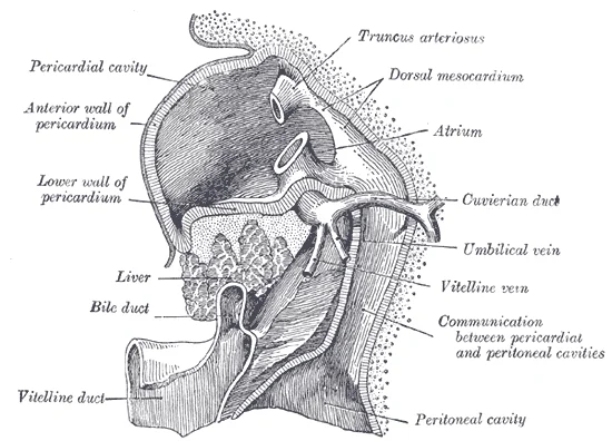
Long description
Liver with the septum transversum. Human embryo 3 mm. long. (After model and figure by His.)

Figure 6: Liver with the septum transversum (human embryo 3 mm).
DefinitionHepatic Diverticulum
An endodermal outgrowth from the foregut that gives rise to the liver parenchyma and biliary tree.
The gallbladder also arises from the hepatic diverticulum. It forms as a bud from the lower part of the diverticulum. The cystic duct connects the gallbladder to the common hepatic duct. The common hepatic duct joins the cystic duct to form the common bile duct. This duct enters the duodenum at the major duodenal papilla. The sphincter of Oddi regulates bile flow. [1]
NoteClinical Pearl
Congenital atresia of the bile ducts leads to neonatal jaundice and requires surgical intervention.
The liver receives dual blood supply. The portal vein provides nutrient-rich blood from the gastrointestinal tract. The hepatic artery provides oxygenated blood from the systemic circulation. These vessels enter the liver at the porta hepatis. The porta hepatis is the transverse fissure on the visceral surface. It contains the portal triad. The triad consists of the portal vein, hepatic artery proper, and common bile duct. Lymphatics and nerves accompany these structures.
| Structure | Origin | Function |
|---|---|---|
| Portal Vein | Gut | Nutrient transport |
| Hepatic Artery | Celiac Trunk | Oxygen supply |
| Common Bile Duct | Liver/Gallbladder | Bile drainage |
The liver lobes are defined by external fissures. The falciform ligament attaches the liver to the anterior abdominal wall. It contains the ligamentum teres hepatis, the remnant of the umbilical vein. The ligamentum venosum is the remnant of the ductus venosus. This shunt bypasses the liver in fetal life. It connects the umbilical vein to the inferior vena cava. After birth, the ductus venosus closes. The liver begins to function independently. [1]
Exam trapCommon Error
Do not confuse the ligamentum teres with the round ligament of the uterus.
These veins drain into the inferior vena cava. The hepatic veins are short and wide. They lack valves. This makes the liver sensitive to right heart failure. [1]
Liver Development Outline
- Foregut EndodermWeek 4
- Question: Hepatic Diverticulum Forms?
Yes
- Growth into Septum Transversum
No
- Wait
The liver is the largest solid organ in the abdomen, occupying the right upper quadrant and extending across the epigastric region. Its radiological anatomy is defined by its lobular architecture and vascular supply, which are critical for surgical planning and diagnostic imaging. The organ sits inferior to the diaphragm and is separated from the right lung by the right pleural cavity. [1]
The large, homogeneous parenchyma of the liver dominates the right upper quadrant.
The true surgical anatomy relies on the Cantlie line, also known as the principal plane. This imaginary plane runs from the gallbladder fossa anteriorly to the inferior vena cava posteriorly. It divides the liver into the right and left functional lobes. The right functional lobe is larger than the left. Within the liver, the hepatic veins drain blood into the inferior vena cava.
The portal triads, consisting of the portal vein, hepatic artery, and bile duct, follow a different distribution. The portal vein bifurcates into right and left branches at the porta hepatis. These branches further divide into segmental branches. Each segment has its own vascular inflow, outflow, and biliary drainage. This segmentation is crucial for hepatic resections, as it allows for the removal of disease [1]
The gallbladder lies in a fossa on the inferior surface of the liver. It is responsible for storing and concentrating bile. The cystic duct joins the common hepatic duct to form the common bile duct. [1]
DefinitionCantlie Line
An imaginary plane running from the gallbladder fossa to the inferior vena cava, dividing the liver into right and left functional lobes.
The liver’s blood supply is unique, receiving dual input from the hepatic artery and the portal vein. The hepatic artery provides oxygenated blood, while the portal vein supplies nutrient-rich blood. This dual supply ensures the liver receives adequate oxygen and nutrients for its metabolic functions. [1]
| Vein | Location | Function |
|---|---|---|
| Right Hepatic Vein | Right portal fissure | Drains right functional lobe |
| Middle Hepatic Vein | Cantlie line | Drains central segments |
| Left Hepatic Vein | Left portal fissure | Drains left functional lobe |
Understanding these radiological landmarks allows clinicians to interpret imaging studies accurately. The relationship between the hepatic veins and portal branches is key to identifying segments. This anatomical arrangement is essential for avoiding complications during surgery. [1]
Figure 7Liver Cirrhosis
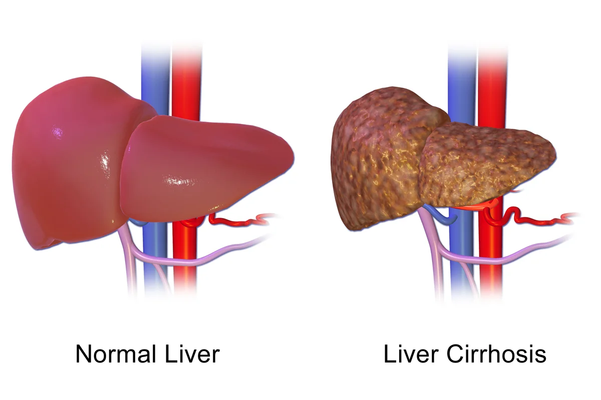
Long description
Liver Cirrhosis. See a full animation of this medical topic.

Figure 7: Liver Cirrhosis.
The liver is the largest internal organ, occupying the right hypochondrium and epigastrium, extending from the fifth intercostal space in the mid-clavicular line to the costal margin. Its superior surface is convex, fitting against the diaphragm, while the inferior surface is concave and rests on abdominal viscera.
The large, homogeneous organ dominating the right upper quadrant is the liver.
The liver's blood supply is unique, receiving dual input from the hepatic artery and the portal vein. The portal vein, formed by the union of the superior mesenteric and splenic veins, carries nutrient-rich, deoxygenated blood from the gastrointestinal tract. These vessels enter the liver at the porta hepatis and branch repeatedly within the liver parenchyma. The hepatic veins, draining the liver, exit posteriorly and empty directly into the inferior vena cava. This arrangement means that the liver is susceptible to both ischemic injury and congestion.
DefinitionPortal Triad
The portal triad consists of the hepatic artery, portal vein, and common bile duct, enclosed within the hepatoduodenal ligament. It is the key structure entering and leaving the liver at the porta hepatis.
The liver is covered by Glisson's capsule, a fibrous connective tissue layer. This capsule is sensitive to stretch, and rapid enlargement of the liver, such as in acute hepatitis or congestion, causes capsular distension and pain. The liver's regenerative capacity is remarkable; it can regenerate to its original size after significant resection, provided the remaining tissue is healthy. This property is fundamental to living donor liver transplantation and surgical management of liver trauma. [1]
| Structure | Location | Key Relations |
|---|---|---|
| Right Lobe | Right hypochondrium | Gallbladder, right kidney, hepatic flexure |
| Left Lobe | Epigastrium | Stomach, diaphragm |
| Caudate Lobe | Posterior | IVC, omental foramen |
| Quadrate Lobe | Inferior | Gallbladder, porta hepatis |
Clinical assessment of the liver often involves palpation and percussion. A normal liver edge is smooth, firm, and non-tender. An enlarged liver may indicate congestion, infiltration, or neoplasia. Tenderness suggests inflammation or capsular stretch. Percussion helps determine the liver span, which is typically 6-12 cm in the mid-clavicular line. Deviations from these norms warrant further investigation with imaging, such as ultrasound or CT, to identify structural abnormalities or lesions.
Understanding this composition is vital for extended resections. [2]
Students frequently confuse the regulatory mechanisms of the liver and pancreas, leading to errors in understanding digestive physiology. A primary error is assuming bile production is constant regardless of dietary intake. In reality, hepatocytes work non-stop, but bile production increases when fatty chyme enters the duodenum and stimulates the secretion of the gut hormone cholecystokinin. Between meals, bile is produced but conserved. The valve-like hepatopancreatic ampulla closes, allowing bile to divert to the gallbladder, where it is concentrated and stored until the next meal. Another common mistake is misidentifying the composition of pancreatic juice. Unlike bile, it is clear and composed mostly of water along with some salts, sodium bicarbonate, and several digestive enzymes. Students often overlook the critical role of sodium bicarbonate, which is responsible for the slight alkalinity of pancreatic juice (pH 7.1 to 8.2), which serves to buffer the acidic gastric juice in chyme, inactivate pepsin from the stomach, and create an optimal environment for the activity of pH-sensitive digestive enzymes in the small intestine. [1]
A significant trap involves the activation of pancreatic enzymes. The pancreas produces protein-digesting enzymes in their inactive forms. These enzymes are activated in the duodenum. If produced in an active form, they would digest the pancreas (which is exactly what occurs in the disease, pancreatitis). Students must remember that the intestinal brush border enzyme enteropeptidase stimulates the activation of trypsin from trypsinogen of the pancreas, which in turn changes the pancreatic enzymes procarboxypeptidase and chymotrypsinogen into their active forms, carboxypeptidase and chymotrypsin. Confusing the triggers for secretin and cholecystokinin (CCK) is another frequent error. Regulation of pancreatic secretion is the job of hormones and the parasympathetic nervous system. The entry of acidic chyme into the duodenum stimulates the release of secretin, which in turn causes the duct cells to release bicarbonate-rich pancreatic juice. The presence of proteins and fats in the duodenum stimulates the secretion of CCK, which then stimulates the acini to secrete enzyme-rich pancreatic juice and enhances the activity of secretin. Parasympathetic regulation occurs mainly during the cephalic and gastric phases of gastric secretion, when vagal stimulation prompts the secretion of pancreatic juice. [1]
Exam trapEnzyme Activation
Pancreatic enzymes are produced inactive to prevent autodigestion. Trypsinogen is activated by enteropeptidase, not by the pancreas itself.
DefinitionBile Conservation
Between meals, bile is produced but conserved. The valve-like hepatopancreatic ampulla closes, allowing bile to divert to the gallbladder, where it is concentrated and stored until the next meal.
This knowledge is applied at the bedside to interpret symptoms of digestive dysfunction. Understanding that pancreatic juice buffers the acidic gastric juice in chyme, inactivates pepsin from the stomach, and enables the optimal functioning of digestive enzymes in the small intestine helps clinicians manage conditions like achlorhydria or pancreatic insufficiency. If a patient presents with steatorrhea, one must consider whether the lack of bicarbonate or enzymes is the cause. The fact that the pancreas produces over a liter of pancreatic juice each day underscores the volume required for effective digestion. [1]
In the operating theatre, anatomical landmarks are crucial for safe dissection. Consequently, a gap exists between the Glissonean pedicle (the plate system) and Laennec's capsule (gray space) as shown in Figure that allows us to isolate the extrahepatic Glissonean pedicle without parenchymal destruction. Because the plate system (yellow line) represents a fibrous thickened part of the Glissonean pedicle including vaginal ductuli , , , it would become a good landmark for the extrahepatic Glissonean pedicle isolation. Similarly, between the parenchyma of the cystic fossa and the subserosal layer of the gallbladder, Laennec's capsule and the cystic plate are interposed and the cystic plate also would become a good landmark. Surgeons use these spaces to avoid damaging the liver parenchyma during cholecystectomy or hepatic resection. [2]
On imaging, recognizing the role of the gallbladder in concentrating and storing bile is essential for diagnosing cholelithiasis. Since bile is concentrated and stored until the next meal, ultrasound findings of a distended gallbladder may indicate obstruction of the cystic duct. The clear nature of pancreatic juice contrasts with the concentrated bile, aiding in the differential diagnosis of fluid collections in the abdomen.
Pancreatic Secretion Regulation
- Start: Chyme enters duodenum...
- Question: Is chyme acidic?
Yes
- Secretin releasedCauses duct cells to release bicarbonate-rich pancreatic juice
No
- Check for proteins/fats...
- Question: Are proteins/fats present?
Yes
- CCK releasedStimulates acini to secrete enzyme-rich pancreatic juice
No
- Minimal secretion...
In examinations, students must distinguish between the roles of secretin and CCK. The entry of acidic chyme into the duodenum stimulates the release of secretin, which in turn causes the duct cells to release bicarbonate-rich pancreatic juice. The presence of proteins and fats in the duodenum stimulates the secretion of CCK, which then stimulates the acini to secrete enzyme-rich pancreatic juice and enhances the activity of secretin. Memorising that the pancreas produces protein-digesting enzymes in their inactive forms is vital for questions on pancreatitis pathophysiology. The intestinal brush border enzyme enteropeptidase stimulates the activation of trypsin from trypsinogen of the pancreas, which in turn changes the pancreatic enzymes procarboxypeptidase and chymotrypsinogen into their active forms, carboxypeptidase and chymotrypsin. This cascade must be understood to answer questions on enzyme activation sequences. [1]
| Enzyme (Inactive) | Activator | Active Enzyme |
|---|---|---|
| Trypsinogen | Enteropeptidase | Trypsin |
| Procarboxypeptidase | Trypsin | Carboxypeptidase |
| Chymotrypsinogen | Trypsin | Chymotrypsin |
Bile production increases when fatty chyme enters the duodenum and stimulates the secretion of the gut hormone cholecystokinin. Between meals, bile is produced but conserved. The valve-like hepatopancreatic ampulla closes, allowing bile to divert to the gallbladder, where it is concentrated and stored until the next meal. The pancreas produces over a liter of pancreatic juice each day. Unlike bile, it is clear and composed mostly of water along with some salts, sodium bicarbonate, and several digestive enzymes. Sodium bicarbonate is responsible for the slight alkalinity of pancreatic juice (pH 7.1 to 8.2), which serves to buffer the acidic gastric juice in chyme, inactivate pepsin from the stomach, and create an optimal environment for the activity of pH-sensitive digestive enzymes in the small intestine. The pancreas produces protein-digesting enzymes in their inactive forms. These enzymes are activated in the duodenum. If produced in an active form, they would digest the pancreas (which is exactly what occurs in the disease, pancreatitis). The intestinal brush border enzyme enteropeptidase stimulates the activation of trypsin from trypsinogen of the pancreas, which in turn changes the pancreatic enzymes procarboxypeptidase and chymotrypsinogen into their active forms, carboxypeptidase and chymotrypsin. Regulation of pancreatic secretion is the job of hormones and the parasympathetic nervous system. The entry of acidic chyme into the duodenum stimulates the release of secretin, which in turn causes the duct cells to release bicarbonate-rich pancreatic juice.
Answer each one in your head first, then open the answer.
What hormone is released when acidic chyme enters the duodenum?
Show answerHide answer
SecretinWhat enzyme activates trypsinogen?
Show answerHide answer
EnteropeptidaseWhere is bile stored between meals?
Show answerHide answer
GallbladderWhat is the pH range of pancreatic juice?
Show answerHide answer
7.1 to 8.2What structure is a good landmark for extrahepatic Glissonean pedicle isolation?
Show answerHide answer
The plate system
- Betts JG, Young KA, Wise JA, et al. Accessory Organs in Digestion: The Liver, Pancreas, and Gallbladder. OpenStax, Anatomy and Physiology 2e (CC BY-NC-SA 4.0). Accessed .
- Sugioka A, Kato Y, Tanahashi Y. Systematic extrahepatic Glissonean pedicle isolation for anatomical liver resection based on Laennec's capsule: proposal of a novel comprehensive surgical anatomy of the liver. Journal of hepato-biliary-pancreatic sciences 2017;24(1):17-23. doi:10.1002/jhbp.410. Accessed .
Figure credits
- Figure 1: Henry Vandyke Carter, via Wikimedia Commons, Public domain, Public domain.
- Figure 2: Henry Vandyke Carter, via Wikimedia Commons, Public domain, Public domain.
- Figure 3: Henry Vandyke Carter, via Wikimedia Commons, Public domain, Public domain.
- Figure 4: OpenStax College, via Wikimedia Commons, Public domain, Public domain.
- Figure 5: Henry Vandyke Carter, via Wikimedia Commons, Public domain, Public domain.
- Figure 6: Henry Vandyke Carter, via Wikimedia Commons, Public domain, Public domain.
- Figure 7: BruceBlaus, via Wikimedia Commons, CC BY-SA 4.0, CC BY-SA 4.0.
Educational content for medical students and doctors. It is not a substitute for clinical judgement or local guidelines.