Large intestine
Anatomy of the large intestine including regions, surface features, and clinical landmarks.
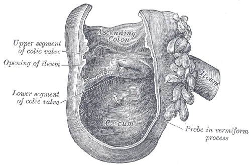
The large intestine frames the small intestine on three sides and runs from the appendix to the anus[1]. It is called large because it is more than twice the diameter of the small intestine, about 3 inches[1]. The ileum joins the cecum, the first portion of the large intestine, at the ileocecal sphincter (or valve)[1]. The ileocecal valve, located at the opening between the ileum and the large intestine, controls the flow of chyme from the small intestine to the large intestine[1].
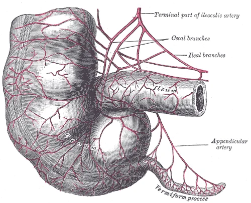
The first part of the large intestine is the cecum, a sac-like structure that is suspended inferior to the ileocecal valve[1]. It is about 6 cm (2.4 in) long, receives the contents of the ileum, and continues the absorption of water and salts[1]. The appendix (or vermiform appendix) is a winding tube that attaches to the cecum[1]. Although the 7.6-cm (3-in) long appendix contains lymphoid tissue, suggesting an immunologic function, this organ is generally considered vestigial[1].
Figure 1Gray1073
Long description
The cecum and vermiform process, with their arteries.

Figure 1: The cecum and vermiform process, with their arteries.
Clinical pearlMcBurney Point
Tenderness at McBurney's point, located one-third of the distance from the anterior superior iliac spine to the umbilicus, suggests appendicitis.
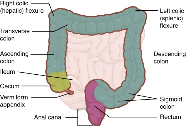
The cecum blends seamlessly with the colon[1]. Upon entering the colon, the food residue first travels up the ascending colon on the right side of the abdomen[1]. At the inferior surface of the liver, the colon bends to form the right colic flexure (hepatic flexure) and becomes the transverse colon[1]. The region defined as hindgut begins with the last third of the transverse colon and continues on[1]. Food residue passing through the transverse colon travels across to the left side of the abdomen, where the colon angles sharply immediately inferior to the spleen, at the left colic flexure (splenic flexure)[1]. From there, food residue passes through the descending colon, which runs down the left side of the posterior abdominal wall[1]. After entering the pelvis inferiorly, it becomes the s-shaped sigmoid colon, which extends medially to the midline[1].
Figure 2Gray1075
Long description
Interior of the cecum and lower end of ascending colon, showing colic valve.

Figure 2: Interior of the cecum and lower end of ascending colon, showing colic valve.
| Region | Location |
|---|---|
| Ascending | Right side of abdomen |
| Transverse | Crosses abdomen |
| Descending | Left side of abdomen |
| Sigmoid | Pelvis, midline |
Three features are unique to the large intestine: teniae coli, haustra, and epiploic appendages[1]. The teniae coli are three bands of smooth muscle that make up the longitudinal muscle layer of the muscularis of the large intestine, except at its terminal end[1]. Tonic contractions of the teniae coli bunch up the colon into a succession of pouches called haustra (singular = haustrum), which are responsible for the wrinkled appearance of the colon[1]. Attached to the teniae coli are small, fat-filled sacs of visceral peritoneum called epiploic appendages[1]. The purpose of these is unknown[1].
DefinitionVolvulus
Twisting of the bowel around its mesentery, causing obstruction and potential ischaemia.
The ascending and descending colon, and the rectum are located in the retroperitoneum[1]. The transverse and sigmoid colon are tethered to the posterior abdominal wall by the mesocolon[1].
Figure 32420 Large Intestine
Long description
The large intestine includes the cecum, colon, and rectum.

Figure 3: The large intestine includes the cecum, colon, and rectum.
The large intestine begins at the ileocecal junction and terminates at the anal canal. It is divided into the caecum, colon, and rectum. The caecum is the first portion of the large intestine, receiving chyme from the ileum via the ileocecal sphincter. The appendix is a narrow, blind tube attached to the caecum. The colon is the longest part, divided into ascending, transverse, descending, and sigmoid sections. The rectum leads to the anal canal, which is 3.8–5 cm long and located in the perineum. [1]
The outer longitudinal muscle layer of the colon is not uniform; it is concentrated into three distinct bands called taeniae coli. These bands are shorter than the colon itself, causing the tube to pucker into pouches known as haustra. This arrangement is unique to the large intestine, which lacks the circular folds and villi found in the small intestine. The haustra are visible on the external surface and are separated by the taeniae.
The blood supply to the large intestine is derived from the superior and inferior mesenteric arteries. The superior mesenteric artery supplies the small intestine and the proximal two-thirds of the transverse colon. The inferior mesenteric artery supplies the distal transverse colon, descending colon, sigmoid colon, and rectum. The marginal artery of Drummond is an anastomotic channel that runs along the inner border of the colon, connecting the superior and inferior mesenteric arterial systems. This marginal artery ensures a continuous blood supply along the length of the colon.
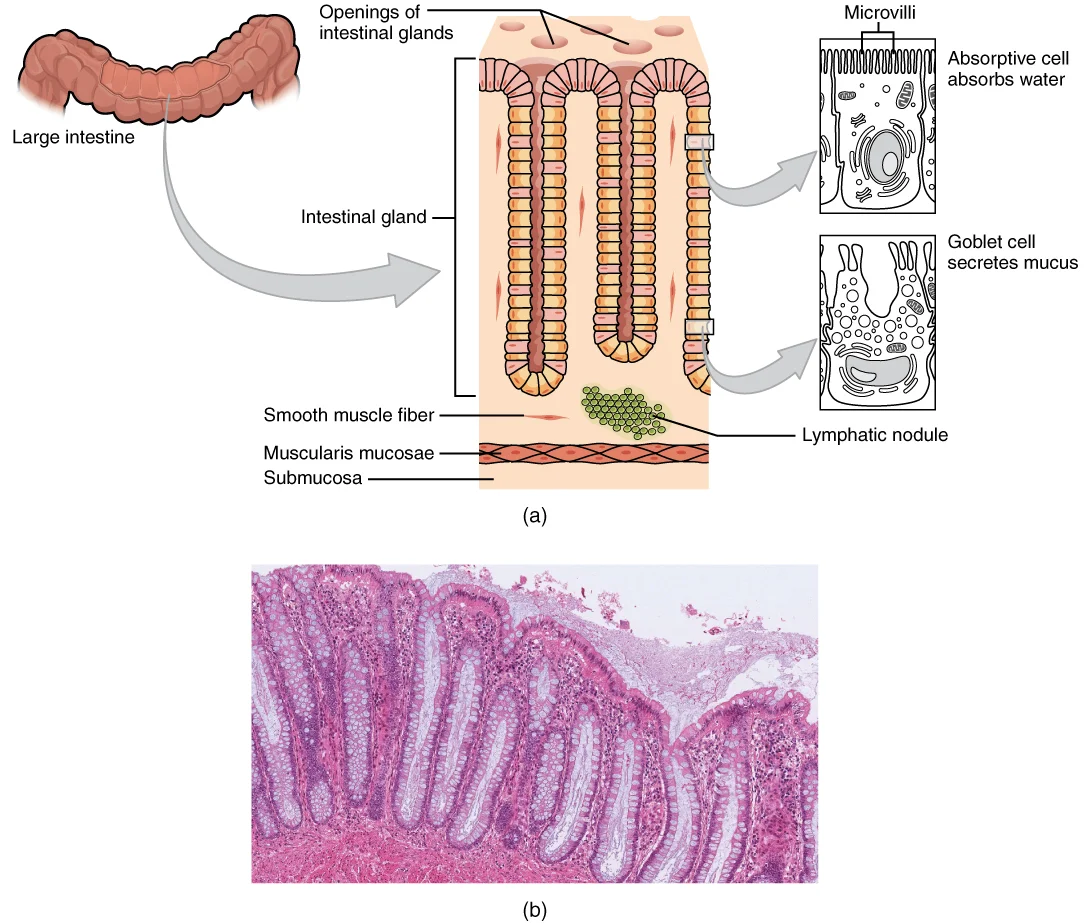
The mucosa of the colon is simple columnar epithelium, rich in goblet cells that secrete mucus to ease fecal movement. Unlike the small intestine, the large intestine has no villi or circular folds. The primary functions are water and salt absorption, and the formation of feces. Bacteria in the lumen perform saccharolytic fermentation, breaking down carbohydrates and producing gases such as hydrogen, carbon dioxide, and methane. Up to 1500 mL of flatus is produced daily in the colon. The enterocytes absorb water, salts, and vitamins synthesized by intestinal bacteria. [1]
Figure 42421 Histology of the Large IntestineN
Long description
(a) The histologies of the large intestine and small intestine (not shown) are adapted for the digestive functions of each organ. (b) This micrograph shows the colon’s simple columnar epithelium and goblet cells. LM x 464. (credit b: Micrograph provided by the Regents of University of Michigan Medic

Figure 4: (a) The histologies of the large intestine and small intestine (not shown) are adapted for the digestive functions of each organ.
The anal canal contains two sphincters: the internal anal sphincter, made of smooth muscle and involuntary, and the external anal sphincter, made of skeletal muscle and voluntary. Both remain closed except during defecation. The entry of feces into the rectum activates the defecation reflex. Mass movements in the colon occur three to four times per day, often triggered by the gastrocolic reflex after eating. [1]
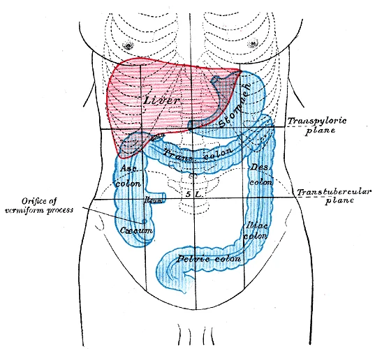
The large intestine forms the terminal part of the alimentary canal, serving as the final segment before elimination. Its primary physiological role is to complete the absorption of water and nutrients, synthesize specific vitamins, and form and expel faeces. The organ is defined by its anatomical relations to surrounding structures, which determine its peritoneal covering and surgical accessibility. [1]
Figure 5Gray1223
Long description
Large Intestine Anatomy. Labelled illustration from Wikimedia Commons, used under its open licence.

Figure 5: Large Intestine Anatomy.
The cecum, located in the right iliac fossa, is typically intraperitoneal, possessing a mesentery known as the mesocecum, although this is often rudimentary or absent. The ascending colon travels superiorly along the right flank. In most individuals, it is retroperitoneal, lying against the posterior abdominal wall without a mesentery, fixed by fusion of its anterior peritoneum to the parietal peritoneum. The transverse colon crosses the abdomen from right to left. It is suspended by the long transverse mesocolon, making it intraperitoneal and mobile. The descending colon runs inferiorly along the left flank. Like the ascending colon, it is generally retroperitoneal, fixed to the posterior abdominal wall. The sigmoid colon descends into the pelvis. It is intraperitoneal, suspended by the sigmoid mesocolon, which allows for significant mobility. The rectum begins at the level of the third sacral vertebra. The upper third is intraperitoneal on its anterior and lateral surfaces. The middle third is retroperitoneal, lying posterior to the peritoneal reflection. The lower third is subperitoneal, lying below the level of the peritoneal reflection in the pelvis.
The peritoneal relations dictate the potential for movement and the spread of pathology. Intraperitoneal segments, such as the transverse and sigmoid colon, are mobile and can undergo torsion or volvulus. Retroperitoneal segments, such as the ascending and descending colon, are fixed and less prone to such mechanical complications but are more difficult to access surgically without entering the peritoneal cavity. The rectum’s subperitoneal position places it within the pelvic cavity, surrounded by pelvic organs and nerves.
DefinitionPeritoneal Relations
The relationship of the large intestine to the peritoneum determines whether it is intraperitoneal (mobile, with a mesentery) or retroperitoneal (fixed, without a mesentery). This distinction is crucial for understanding surgical approaches and the potential for complications like volvulus.
The large intestine’s position is also related to the small intestine. The ileum empties into the cecum at the ileocaecal junction. The caecum is located inferior to the right kidney and lateral to the psoas major muscle. The transverse colon lies anterior to the duodenum, pancreas, and small intestine coils. The sigmoid colon lies anterior to the left ureter, left gonadal vessels, and the left psoas major muscle. The rectum lies anterior to the sacrum and coccyx, and in males, it is related to the bladder, seminal vesicles, and prostate. In females, it is related to the uterus and vagina.
The large intestine’s surface area is vast, facilitating its absorptive functions. Although the small intestine absorbs the majority of nutrients and water, the large intestine absorbs most of the remaining water, converting liquid chyme into semisolid faeces. This process is supported by the extensive bacterial flora residing within the lumen, which synthesizes vitamins such as biotin, pantothenic acid, and vitamin K. The anatomical relations ensure that these processes occur in a protected and organized manner, with the peritoneal coverings providing both structural support and a pathway for vascular and neural supply. [1]
The mucosal surface is notably smooth compared to the small intestine, lacking the prominent circular folds (plicae circulares) and villi that define the absorptive surface of the proximal gut. Instead, the large intestine features straight, tubular intestinal glands (crypts of Lieberkühn) that open directly onto the surface. These glands are lined with abundant goblet cells, which secrete mucus to lubricate the passage of faeces. The absence of villi significantly reduces the surface area, reflecting the organ’s primary role in water absorption and faecal storage rather than nutrient uptake. The transition from the sigmoid colon to the rectum occurs near the third sacral vertebra, where the taeniae coli spread out to form a continuous longitudinal muscle layer. This structural change supports the rectum’s function as a temporary reservoir. The mucosa here is thick and vascular, designed to withstand the pressure of retained material. Understanding these internal features is crucial for interpreting endoscopic images and identifying pathological changes such as polyps or inflammation. The smooth mucosal surface allows for easier visualization of subtle lesions, although the deep crypts can harbor bacteria and debris. The haustral folds create pockets where stool may accumulate, increasing the risk of diverticula formation if intraluminal pressure rises excessively. The rectal valves are particularly important clinically, as their obstruction can lead to difficulties in defecation. The overall design of the large intestine interior prioritizes efficient water reabsorption and safe transit of waste, contrasting sharply with the complex absorptive machinery of the small intestine. [1]
The large intestine receives its blood supply from two major branches of the abdominal aorta, ensuring a robust collateral network that protects the bowel from ischaemia during surgical manipulation or vascular compromise.
The marginal artery connects the branches of the superior and inferior mesenteric arteries, forming a continuous vascular arcade that lies just beneath the serosa. This arrangement allows blood to flow from one system to the other if one main trunk is obstructed. [1]
The superior mesenteric artery supplies the midgut derivatives, including the caecum, ascending colon, and the proximal two-thirds of the transverse colon. It arises from the abdominal aorta at the level of L1, just below the coeliac trunk. Its branches include the ileocolic artery, right colic artery, and middle colic artery. The ileocolic artery supplies the caecum and appendix. The right colic artery runs horizontally to supply the ascending colon. The middle colic artery divides into left and right branches to supply the transverse colon. These arteries form anastomoses with each other and with the marginal artery.
The inferior mesenteric artery supplies the hindgut derivatives, comprising the distal third of the transverse colon, the descending colon, the sigmoid colon, and the upper rectum. It arises from the aorta at the level of L3. Its primary branches are the left colic artery, sigmoid arteries, and the superior rectal artery. The left colic artery ascends to anastomose with the middle colic artery via the marginal artery. The sigmoid arteries branch repeatedly to supply the sigmoid colon. The superior rectal artery continues into the pelvis to supply the upper rectum.
The marginal artery of Drummond is a crucial anastomotic channel that runs along the mesenteric border of the colon. It connects the terminal branches of the superior and inferior mesenteric arteries. This vessel provides collateral circulation, ensuring that the colon remains perfused even if one of the main arterial trunks is compromised. The reliability of this anastomosis varies among individuals, but it is generally well-developed. Surgeons must preserve this artery during colectomy to prevent postoperative ischaemia.
The venous drainage of the large intestine follows a pattern that mirrors its arterial supply, ultimately converging into the portal venous system. This arrangement is critical for transporting absorbed nutrients from the gut to the liver for metabolic processing. The venous network begins with extensive capillary beds within the mucosa and submucosa, which coalesce into larger vessels that run parallel to the corresponding arteries.
The superior mesenteric vein drains the midgut derivatives, including the cecum, appendix, ascending colon, and the proximal two-thirds of the transverse colon. It is formed by the union of the ileocolic, right colic, and middle colic veins. These veins accompany their arterial counterparts, running within the mesocolon. The inferior mesenteric vein drains the hindgut derivatives, comprising the distal one-third of the transverse colon, the descending colon, the sigmoid colon, and the upper part of the rectum. It receives the left colic, sigmoid, and superior rectal veins. Notably, the superior rectal vein, which drains the upper rectum, is a tributary of the inferior mesenteric vein. This connection is clinically significant because it represents a portosystemic anastomosis site.
The anal canal presents a unique venous arrangement. The stratified squamous epithelial mucosa of the anal canal connects to the skin on the outside of the anus. This mucosa varies considerably from that of the rest of the colon to accommodate the high level of abrasion as feces pass through. The anal canal’s mucous membrane is organized into longitudinal folds, each called an anal column, which house a grid of arteries and veins. Two superficial venous plexuses are found in the anal canal: one within the anal columns and one at the anus. The plexus within the anal columns drains into the superior rectal vein, which then joins the inferior mesenteric vein, linking to the portal system. The plexus at the anus drains into the middle and inferior rectal veins, which pass into the internal iliac veins and subsequently the systemic circulation. This dual drainage system explains the potential for portosystemic shunting in conditions such as portal hypertension. [1]
The lymphatic drainage of the large intestine follows a predictable pattern that mirrors its arterial supply, ensuring that immune surveillance and potential metastatic spread are directed to specific nodal stations. Understanding this pathway is critical for surgical oncology and the staging of colorectal malignancies. The drainage begins with the mucosal and submucosal plexuses, which converge into larger collecting vessels that accompany the arterial branches.
The caecum and ascending colon drain primarily into the ileocolic and right colic nodes. These nodes are located along the corresponding arteries. From these initial stations, lymph flows into the superior mesenteric nodes. This pathway is consistent with the embryological origin of the midgut. The vessels run alongside the superior mesenteric artery, ensuring that any pathological process in the right colon is first evaluated in these regional nodes.
The transverse colon presents a dual drainage pattern due to its transitional nature. The proximal two-thirds, derived from the midgut, drain into the middle colic nodes. These nodes lie along the middle colic artery. The distal one-third, derived from the hindgut, drains into the left colic nodes. This distinction is vital because it reflects the different vascular supplies and embryological origins. The lymph from the middle colic nodes also flows into the superior mesenteric nodes, maintaining continuity with the proximal drainage.
The descending colon and sigmoid colon are hindgut derivatives. Their lymphatic drainage follows the inferior mesenteric artery. Lymph from the sigmoid colon first passes through the sigmoid (hilar) nodes, which are located along the sigmoid arteries. From there, it moves to the inferior mesenteric nodes, situated near the origin of the inferior mesenteric artery from the aorta. This sequential drainage ensures that pathology in the left colon is monitored at these specific nodal levels.
The rectum has a complex drainage system that varies by height. The upper third drains into the inferior mesenteric nodes, following the superior rectal artery. The middle third drains into the internal iliac nodes, accompanying the middle rectal vessels. The lower third drains into the inguinal nodes, similar to the perineum. This tripartite drainage explains why rectal cancers can metastasize to distant sites, including the groin.
Figure 6Gray1084
Long description
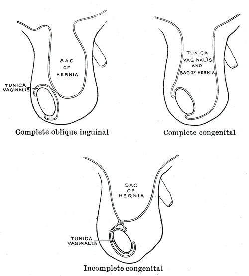
Varieties of oblique inguinal hernia. Labelled illustration from Wikimedia Commons, used under its open licence.

Figure 6: Varieties of oblique inguinal hernia.
DefinitionNodal Stations
Regional lymph nodes that receive drainage from specific segments of the gastrointestinal tract, serving as the first site of potential metastatic spread.
| Structure | Primary Nodes | Secondary Nodes |
|---|---|---|
| Caecum & Ascending Colon | Ileocolic, Right Colic | Superior Mesenteric |
| Transverse Colon (Proximal) | Middle Colic | Superior Mesenteric |
| Transverse Colon (Distal) | Left Colic | Inferior Mesenteric |
| Descending & Sigmoid Colon | Sigmoid, Inferior Mesenteric | Inferior Mesenteric |
| Rectum (Upper) | Superior Rectal | Inferior Mesenteric |
| Rectum (Middle) | Middle Rectal | Internal Iliac |
| Rectum (Lower) | Inferior Rectal | Inguinal |
The innervation of the large intestine is a dual system involving both autonomic and somatic components, which dictates sensory perception and motor control. The autonomic nervous system provides the primary visceral supply. Parasympathetic fibres originate from the vagus nerve for the proximal two-thirds of the colon and from the pelvic splanchnic nerves (S2–S4) for the distal third. These fibres facilitate peristalsis and glandular secretion. Sympathetic innervation arises from the thoracic and lumbar spinal cord, travelling via the superior and inferior mesenteric plexuses. These fibres generally inhibit motility and cause vasoconstriction.
The somatic nervous system becomes critical at the anal canal. The external anal sphincter is skeletal muscle, innervated by the inferior rectal nerve, a branch of the pudendal nerve (S2–S4). This allows for voluntary control of defecation. The internal anal sphincter is smooth muscle, controlled by the autonomic nervous system.
Referred pain patterns are essential for clinical diagnosis. Visceral afferent fibres travel with sympathetic nerves to the spinal cord. This occurs because visceral pain fibres enter the spinal cord at similar levels to somatic fibres from the abdominal wall.
The mucosa above this line is fairly insensitive, whereas the area below is very sensitive. The resulting difference in pain threshold is due to the fact that the upper region is innervated by visceral sensory fibers, and the lower region is innervated by somatic sensory fibers. Lesions above the line are typically painless, while those below are exquisitely painful. [1]
The lymphatic drainage follows the arterial supply, converging on the mesenteric nodes.
Clinical pearlClinical Pearl
Pain above the pectinate line is dull and poorly localised. Pain below it is sharp and well-localised.
The large intestine is lined by a simple columnar epithelium that is distinct from the small intestine due to the absence of villi. This surface is dominated by numerous crypts of Lieberkühn, which house the primary cellular machinery for absorption and secretion. The epithelial cells are specialized for water and electrolyte reabsorption, a function critical for the consolidation of faecal matter before excretion.
DefinitionEpithelial Composition
The mucosa contains abundant goblet cells for mucus secretion and enterocytes for absorption, with a high density of mitochondria in the basal region to support active ion transport.
Under normal physiological conditions, the lamina propria contains a moderate population of immune cells, including lymphocytes and plasma cells, reflecting the gut's role as a major immunological interface. The muscularis mucosae separates the mucosa from the submucosa, allowing for local movement of the epithelial surface. The submucosa is a connective tissue layer containing larger blood vessels, lymphatics, and the submucosal (Meissner’s) plexus, which regulates secretory activity and blood flow.
The muscularis externa consists of an inner circular layer and an outer longitudinal layer. In the large intestine, the outer longitudinal muscle is condensed into three distinct bands known as the taeniae coli. These bands are shorter than the colon itself, causing the bowel to pucker into sacculations called haustra. Between the taeniae coli, the outer longitudinal muscle is thin or absent, which influences the mechanical properties of the colon wall. The myenteric (Auerbach’s) plexus lies between these two muscle layers and controls motility.
| Layer | Description |
|---|---|
| Inner Circular | Thick layer; responsible for segmental contractions and mixing. |
| Outer Longitudinal | Condensed into three taeniae coli; responsible for shortening the bowel. |
The serosa (or adventitia in retroperitoneal segments) covers the outer surface. The histological architecture supports the primary functions of water absorption and storage. Abnormalities in this structure can lead to significant pathology. For instance, neoplastic changes in the colorectal mucosa are often associated with specific molecular alterations.
IGFBP7 functions downstream of p53 to mediate its tumor suppressor function . In serrated polyps that do not mutate p53, methylation of IGFBP7 may be an alternate mechanism for inactivating the p53 pathway. [2]
These molecular pathways highlight the importance of histological assessment in diagnosing precancerous lesions. The distinction between tubular serrated adenomas and sessile serrated adenomas relies on recognizing these subtle architectural and molecular differences. Understanding the normal histology provides the baseline for identifying such deviations, ensuring accurate diagnosis and appropriate clinical management.
The large intestine develops from the hindgut, a caudal portion of the primitive gut tube. This developmental phase is critical for establishing the structural integrity and functional capacity of the adult organ. The hindgut extends from the distal third of the transverse colon to the upper part of the anal canal. During embryogenesis, the gut tube undergoes rapid growth and rotation. This rotation positions the ascending and descending colon against the posterior abdominal wall. The large intestine lumen recanalizes after a period of solid epithelial proliferation. This process ensures the formation of a patent lumen for fecal transport.
Figure 7Gray1074
Long description
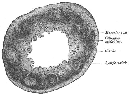
Transverse section of human vermiform process. X 20.

Figure 7: Transverse section of human vermiform process (x 20).
DefinitionHindgut Derivatives
The hindgut gives rise to the distal one-third of the transverse colon, the descending colon, the sigmoid colon, and the upper anal canal.
The mesentery of the hindgut fuses with the posterior parietal peritoneum. This fusion creates the retroperitoneal position of the ascending and descending colon. The sigmoid colon retains a mesentery, the sigmoid mesocolon, allowing for mobility. The rectum develops within the pelvic cavity. It is initially separated from the urogenital sinus by the urorectal septum. This septum divides the cloaca into the urogenital sinus and the anorectal canal. Failure of this separation results in congenital anomalies such as rectourethral fistulas.
The muscular layers of the large intestine differentiate from the splanchnic mesoderm. The outer longitudinal muscle layer is not continuous. It condenses into three distinct bands known as the taeniae coli. These bands are shorter than the colon itself, causing the wall to pucker into haustra. The inner circular muscle layer forms the internal anal sphincter. This sphincter is under involuntary control. It is under voluntary control.
| Structure | Origin | Insertion | Action | Nerve Supply |
|---|---|---|---|---|
| Taeniae Coli | Continuous with longitudinal muscle layer | Appendices epiploicae and haustral folds | Shorten colon, form haustra | Autonomic nervous system |
| Internal Anal Sphincter | Circular muscle layer of rectum | Anoderm | Constricts anal canal | Autonomic nervous system |
| External Anal Sphincter | Coccyx and anococcygeal ligament | Perineal body | Constricts anal canal | Inferior rectal nerve |
The large intestine begins at the caecum, located in the right iliac fossa. On the radiological image, the caecum appears as a dilated pouch-like structure in the right lower quadrant. It is continuous with the ascending colon, which travels upwards along the right lateral abdominal wall. The ascending colon lies posterior to the small intestine loops and anterior to the right kidney and quadratus lumborum muscle. Its radiological appearance is characterized by haustral folds, which are incomplete transverse ridges caused by the taeniae coli. These folds give the colon a segmented appearance, distinct from the smooth wall of the small intestine.
Moving superiorly, the ascending colon reaches the right hypochondrium, where it turns sharply to form the hepatic flexure. The hepatic flexure is situated deep to the liver and the right costal margin. In the axial plane, this flexure appears as a curved transition between the vertical ascending colon and the horizontal transverse colon. The transverse colon then courses across the upper abdomen from right to left. It is suspended by the transverse mesocolon, which attaches it to the posterior abdominal wall. Radiologically, the transverse colon often contains air and fluid levels, making it easily identifiable. It lies anterior to the pancreas, duodenum, and small intestine.
At the left hypochondrium, the transverse colon reaches the splenic flexure. It lies anterior to the left kidney and the tail of the pancreas. From the splenic flexure, the descending colon travels downwards along the left lateral abdominal wall, mirroring the path of the ascending colon. It lies anterior to the left kidney and quadratus lumborum muscle.
The descending colon continues into the left iliac fossa, where it becomes the sigmoid colon. The sigmoid colon is S-shaped and suspended by the sigmoid mesocolon. It lies within the pelvis, anterior to the left psoas major muscle and the left ureter. The sigmoid colon terminates at the rectum, which begins at the level of the third sacral vertebra. The rectum descends through the pelvis, curving around the sacrum and coccyx. It is surrounded by the pelvic fascia and lies anterior to the sacrum and posterior to the bladder in males or the uterus and vagina in females.
DefinitionHaustra
Haustra are the sacculations of the large intestine caused by the taeniae coli, visible as transverse folds on imaging.
NoteKey Landmark
The hepatic and splenic flexures are critical landmarks for identifying the transverse colon on axial imaging.
| Structure | Location | Key Relations |
|---|---|---|
| Caecum | Right iliac fossa | Terminal ileum, appendix |
| Ascending colon | Right lateral wall | Right kidney, liver |
| Transverse colon | Upper abdomen | Pancreas, stomach |
| Descending colon | Left lateral wall | Left kidney, spleen |
| Sigmoid colon | Left iliac fossa | Ureter, psoas major |
The large intestine forms the terminal portion of the gastrointestinal tract, extending from the caecum to the anal canal. Its primary functions include water absorption, electrolyte balance, and the storage and expulsion of faeces. Understanding its anatomical layout is critical for interpreting imaging and performing endoscopic procedures, as lesion detection is operator-dependent.
The appendix, a blind-ended tube, arises from the posteromedial aspect of the caecum. Its base is consistently located at the convergence of the three taeniae coli. Variations in appendix position include retrocaecal, pelvic, and subcaecal orientations. Inflammation of the appendix (appendicitis) typically presents with pain migrating from the periumbilical region to the right lower quadrant.
The ascending colon travels superiorly along the right lateral abdominal wall, from the caecum to the hepatic flexure. It is a retroperitoneal structure, fixed to the posterior abdominal wall. It lies anterior to the quadratus lumborum and the lower pole of the right kidney.
The transverse colon extends from the hepatic flexure to the splenic flexure. Unlike the ascending and descending colon, it is intraperitoneal and suspended by the transverse mesocolon. This mobility allows it to vary in position, often hanging into the pelvis. It lies anterior to the duodenum, pancreas, and small intestine coils.
The sigmoid colon is an S-shaped intraperitoneal segment connecting the descending colon to the rectum. It is suspended by the sigmoid mesocolon. The rectum begins at the level of the third sacral vertebra and descends through the pelvis. It lacks taeniae coli, which spread out to form the longitudinal muscle layer. The anal canal marks the termination of the gastrointestinal tract.
Detection of serrated lesions is clearly operator-dependent, indicating that specific knowledge and skills are required for their detection and recognition. [2]
The anal canal is at the distal end. The hepatic and splenic flexures are marked.
1. Fecal Consistency and Transit Time The consistency of stool is directly determined by the duration of colonic transit. If defecation is delayed for an extended time, additional water is absorbed, making the feces firmer and potentially leading to constipation. Conversely, if waste matter moves too quickly through the intestines, not enough water is absorbed, and diarrhea can result. [1]
2. Bowel Movement Frequency Norms There is no single "normal" frequency for bowel movements. The number of bowel movements varies greatly between individuals, ranging from two or three per day to three or four per week. Diet, health, and stress determine this frequency. [1]
3. Serrated Polyp Prevalence Ratio Sessile serrated adenomas (SSAs) are significantly more common than traditional serrated adenomas (TSAs). This high prevalence makes their detection critical for colorectal cancer prevention. [2]
4. Histological Appearance of Serrated Polyps Serrated polyps are defined by a specific microscopic architecture.
**5. The terms sessile serrated adenoma and sessile serrated polyp are both synonyms and acceptable in diagnostic use. [2]
Students frequently confuse the anatomical attachments of the appendix, leading to errors in surgical planning. The mesoappendix is the mesentery of the appendix, which tethers it to the mesentery of the ileum. It is crucial to remember that the appendix is not free-floating but anchored by this vascularised fold. [1]
Exam trapAppendix Attachment
The mesoappendix, the mesentery of the appendix, tethers it to the mesentery of the ileum.
A second major pitfall involves the classification of serrated polyps, particularly distinguishing hyperplastic polyps (HPs) from sessile serrated adenomas (SSAs). HPs represent the most innocuous subtype of serrated polyps but there are still unresolved questions on their possible evolution to more advanced polyps. Students must recognise that while HPs are generally benign, the distinction between microvesicular HP and goblet cell HP is mostly of academic interest at the present time and is usually not reported by pathologists. [2]
DefinitionHP Subtypes
HPs are further subdivided into microvesicular HP and goblet cell HP. However, this distinction is mostly of academic interest at the present time and is usually not reported by pathologists. [2]
Diagnostic confusion also arises when a polyp displays an overall growth pattern of a TSA with ectopic crypt formations, but with a predominance of goblet cells (as opposed to tall eosinophilic cells with pencillate nuclei), the polyp should be classified as TSA. Students often misinterpret cytological features; for instance, confusion also occurs when a polyp with an overall growth pattern of SSA displays cytologic features of TSA. In such ambiguous cases, a descriptive report is recommended until a better understanding of these lesions is known. [2]
NoteDiagnostic Ambiguity
Confusion also occurs when a polyp with an overall growth pattern of SSA displays cytologic features of TSA. A descriptive report is recommended until a better understanding of these lesions is known.
Another common difficult situation arises when the polyp is obliquely sectioned, not showing the crypt bases to be able to distinguish HP from SSA. If deeper sections do not help, a diagnosis of ‘non-dysplastic serrated polyp, unclassified’ is recommended. Students must not force a diagnosis when histological architecture is obscured. [2]
Exam trapOblique Sectioning
Another common difficult situation arises when the polyp is obliquely sectioned, not showing the crypt bases to be able to distinguish HP from SSA. If deeper sections do not help, a diagnosis of ‘non-dysplastic serrated polyp, unclassified’ is recommended.
Finally, students often underestimate the risk of interval cancers in the proximal colon. It is hypothesized that this in part relates to failures to detect, recognize, and completely resect SSA. Consistent with this hypothesis are data showing that cancers occurring after colonoscopy are more likely to be MSI-high and CIMP-high, suggesting that they originated in unrecognized SSAs. [2]
Knowledge of the mesoappendix is vital in the operating theatre during appendicectomy. The surgeon must ligate the appendicular artery within the mesoappendix to prevent haemorrhage. Understanding that the mesoappendix, the mesentery of the appendix, tethers it to the mesentery of the ileum helps in identifying the correct plane of dissection.
DefinitionSurgical Anatomy
The mesoappendix, the mesentery of the appendix, tethers it to the mesentery of the ileum.
In clinical practice, management of SSAs and TSAs depends on understanding their natural history particularly the transition to malignancy. There is abundant evidence that these lesions are associated with CRC. Perhaps more informative are studies of lesions “caught in the act” of transition to malignancy. As discussed above, histological studies indicate an abrupt transition from SSA to SSA with high-grade cytological dysplasia and invasive malignancy and there is a case report of this transition occurring in an 8-month time period. Three published case series show that the mean size of such lesions is not much greater than the mean size of typical SSAs without cytological dysplasia. [2]
NoteMalignant Transition
As discussed above, histological studies indicate an abrupt transition from SSA to SSA with high-grade cytological dysplasia and invasive malignancy and there is a case report of this transition occurring in an 8-month time period.
Surveillance colonoscopy intervals are determined by polyp characteristics. It is recommended that patients with SSAs smaller than 10 mm and without dysplasia be followed up at 5 years and patients with TSAs or SSAs of at least 10 mm or with dysplasia be followed up at 3 years. This distinction is critical for patient safety and resource allocation. [2]
DefinitionSurveillance Intervals
It is recommended that patients with SSAs smaller than 10 mm and without dysplasia be followed up at 5 years and patients with TSAs or SSAs of at least 10 mm or with dysplasia be followed up at 3 years.
Number of polyps: Almost certainly the risk of subsequent polyps and cancer increases with the number of polyps and the most extreme example of this is serrated polyposis where it is agreed that the surveillance interval should be 1 year. Concomitant conventional adenomas: There is no direct evidence but it is likely that patients with a higher polyp burden due to the presence of both adenomas and serrated polyps are at greater risk. [2]
NotePolyposis Risk
Number of polyps: Almost certainly the risk of subsequent polyps and cancer increases with the number of polyps and the most extreme example of this is serrated polyposis where it is agreed that the surveillance interval should be 1 year.
Surgery is indicated when CRC is diagnosed or when a high polyp burden cannot be controlled by colonoscopy. Until a genetic hallmark of SPS is identified, the criteria for the diagnosis and the surveillance of this syndrome remain rather arbitrary. [2]
DefinitionSurgical Indication
Surgery is indicated when CRC is diagnosed or when a high polyp burden cannot be controlled by colonoscopy.
On imaging and in examinations, recognising the molecular basis of these lesions is key. The Wnt signaling pathway plays an important role in the initiation of conventional adenomas, usually through mutation and deletion of the APC tumor suppressor gene. MCC is another Wnt pathway molecule that directly interacts with β-catenin to dampen the Wnt signal and this is also methylated in HP and SSA but uncommonly in conventional adenomas. [2]
NoteMolecular Pathology
MCC is another Wnt pathway molecule that directly interacts with β-catenin to dampen the Wnt signal and this is also methylated in HP and SSA but uncommonly in conventional adenomas.
Because interobserver variability in histological diagnosis still exists, many experts consider that all serrated polyps in the proximal colon larger than 10 mm in size are likely to be SSAs, even if pathologists interpret them as HPs. This heuristic is frequently tested to ensure clinicians prioritise caution in the proximal colon. [2]
Exam trapProximal Polyp Heuristic
Because interobserver variability in histological diagnosis still exists, many experts consider that all serrated polyps in the proximal colon larger than 10 mm in size are likely to be SSAs, even if pathologists interpret them as HPs.
The mesoappendix tethers the appendix to the mesentery of the ileum. HPs are the most innocuous serrated polyps, with subtypes mostly of academic interest. TSA classification prevails if ectopic crypts are present, regardless of goblet cell predominance. Oblique sectioning may require a diagnosis of ‘non-dysplastic serrated polyp, unclassified’. Interval CRCs are often MSI-high and originate from missed proximal SSAs. SSA to malignancy transition can be abrupt and occur within 8 months. Surveillance is 5 years for small SSA without dysplasia, and 3 years for larger/dysplastic lesions. Serrated polyposis requires 1-year surveillance intervals due to high cancer risk.
Answer each one in your head first, then open the answer.
What structure tethers the appendix to the mesentery of the ileum?
Show answerHide answer
The mesoappendixWhat is the recommended surveillance interval for serrated polyposis?
Show answerHide answer
1 yearWhat diagnosis is recommended if crypt bases cannot be seen due to oblique sectioning?
Show answerHide answer
Non-dysplastic serrated polyp, unclassifiedWhich molecular marker is methylated in HP and SSA but uncommonly in conventional adenomas?
Show answerHide answer
MCCWhat is the surveillance interval for SSAs smaller than 10 mm without dysplasia?
Show answerHide answer
5 years
- Betts JG, Young KA, Wise JA, et al. The Small and Large Intestines. OpenStax, Anatomy and Physiology 2e (CC BY-NC-SA 4.0). Accessed .
- Rosty C, Hewett DG, Brown IS, et al. Serrated polyps of the large intestine: current understanding of diagnosis, pathogenesis, and clinical management. Journal of gastroenterology 2013;48(3):287-302. doi:10.1007/s00535-012-0720-y. Accessed .
Figure credits
- Figure 1: Henry Vandyke Carter, via Wikimedia Commons, Public domain, Public domain.
- Figure 2: Henry Vandyke Carter, via Wikimedia Commons, Public domain, Public domain.
- Figure 3: OpenStax College, via Wikimedia Commons, CC BY 3.0, CC BY 3.0.
- Figure 4: OpenStax College, via Wikimedia Commons, CC BY 3.0, CC BY 3.0.
- Figure 5: Henry Vandyke Carter, via Wikimedia Commons, Public domain, Public domain.
- Figure 6: Henry Vandyke Carter, via Wikimedia Commons, Public domain, Public domain.
- Figure 7: Henry Vandyke Carter, via Wikimedia Commons, Public domain, Public domain.
Educational content for medical students and doctors. It is not a substitute for clinical judgement or local guidelines.