Small intestine
Anatomy of the small intestine including jejunum, ileum, mesentery and common pathologies.
The large intestine frames the small intestine on three sides. [1]
No clear demarcation exists between the jejunum and the ileum. [1]
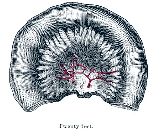
Both segments are tethered to the posterior abdominal wall by the mesentery. The mesentery contains vascular arcades and vasa recta. These structures supply the bowel and allow for mobility. The root of the mesentery attaches obliquely from the left upper quadrant to the right lower quadrant. [1]
Meckel diverticulum is the most prevalent congenital abnormality of the gastrointestinal tract in children. It is located in the distal ileum within 100 cm of the ileocecal valve. The most common presentation is lower gastrointestinal bleeding. It can also serve as a lead point for intussusception. [1]
Small bowel atresia involves obstruction due to failure of recanalisation. Volvulus occurs when the bowel twists around its mesenteric axis. This causes ischaemia if not corrected promptly. Ischaemia results from compromised blood flow through the vasa recta.
DefinitionMeckel Diverticulum
A true diverticulum containing all layers of the bowel wall, arising from the vitelline duct remnant.
The surface marking of the small intestine covers the central abdomen. The duodenum is retroperitoneal. The jejunum and ileum are intraperitoneal. The ileocecal valve controls flow into the large intestine. [1]
| Feature | Jejunum | Ileum |
|---|---|---|
| Length | 0.9 meters | 1.8 meters |
| Wall Thickness | Thinner | Thicker |
| Vascularity | Less vascular | More vascular |
| Mucosal Folds | Prominent | Less developed |
| Location | Upper left quadrant | Lower right quadrant |
Exam trapIschaemia
Twisting of the mesentery compromises blood supply, leading to tissue death.
Small Bowel Pathology
- Acute AbdomenAssess stability
- Question: Unstable?
Yes
- Resuscitate
No
- Imaging
Figure 12417 Small IntestineN
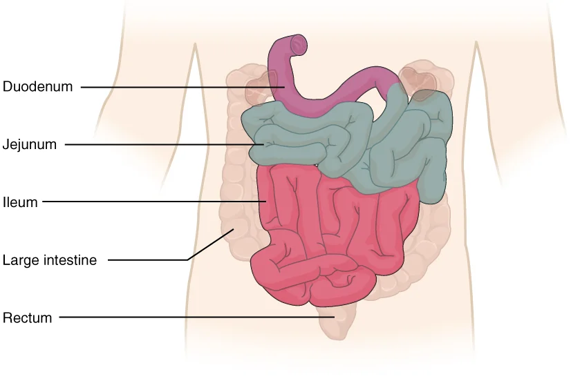
Long description
The three regions of the small intestine are the duodenum, jejunum, and ileum.

Figure 1: The three regions of the small intestine are the duodenum, jejunum, and ileum.
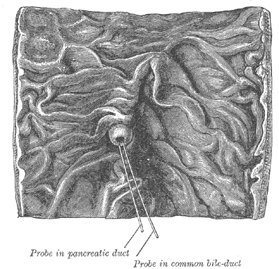
The duodenum is the initial short segment, fixed retroperitoneally. It consists of four parts: superior, descending, horizontal, and ascending. The descending part contains the major duodenal papilla, where the common bile duct and main pancreatic duct usually join to form the ampulla of Vater before entering the lumen. This structure is critical for the delivery of bile and pancreatic enzymes.
Figure 2Gray1057
Long description
Interior of the descending portion of the duodenum, showing bile papilla.

Figure 2: Interior of the descending portion of the duodenum, showing bile papilla.
The jejunum begins at the duodenojejunal flexure, located to the left of the aorta at the level of L2. It occupies the upper left quadrant of the abdomen. The jejunum has a larger diameter and thicker wall than the ileum. Its mucosa features prominent circular folds (plicae circulares) and villi, which increase the absorptive surface area more than 600-fold. These adaptations are most abundant in the proximal two-thirds of the small intestine. The mesentery attaches the jejunum to the posterior abdominal wall, creating a fan-shaped fold of peritoneum that contains vessels and nerves. [1]
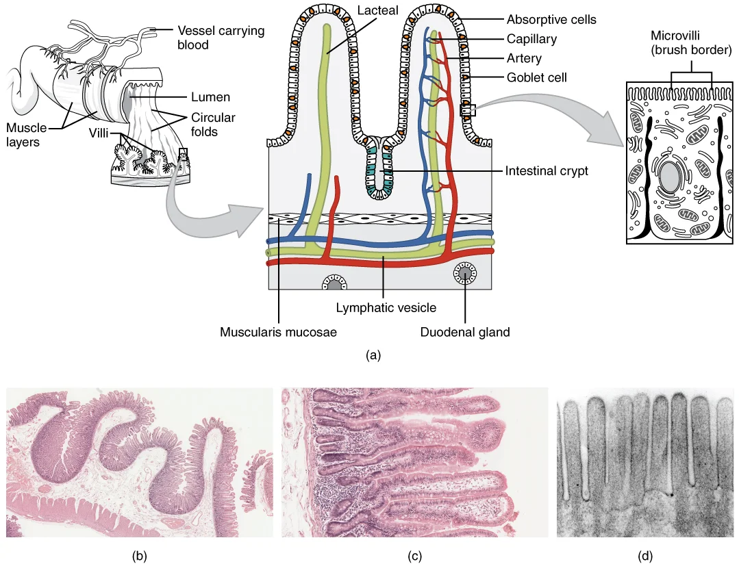
Figure 32418 Histology Small IntestinesN
Long description
(a) The absorptive surface of the small intestine is vastly enlarged by the presence of circular folds, villi, and microvilli. (b) Micrograph of the circular folds. (c) Micrograph of the villi. (d) Electron micrograph of the microvilli. From left to right, LM x 56, LM x 508, EM x 196,000. (credit b-

Figure 3: (a) The absorptive surface of the small intestine is vastly enlarged by the presence of circular folds, villi, and microvilli.
The terminal ileum passes through the ileocaecal valve into the large intestine. Transport movements replace segmentation when chyme absorption is largely complete.
The mesentery is a double layer of peritoneum that suspends the jejunum and ileum from the posterior abdominal wall. It provides a pathway for blood vessels, lymphatics, and nerves. The root of the mesentery extends obliquely from the duodenojejunal flexure to the right sacroiliac joint.
Meckel diverticulum is a congenital remnant of the omphalomesenteric duct. It is located on the antimesenteric border of the ileum, typically within 100 cm of the ileocaecal valve. It is more common in males, with a ratio ranging from 2:1 to 4:1. The incidence in the general population is approximately 2%. It can contain ectopic gastric or pancreatic mucosa, leading to complications such as bleeding or obstruction.
DefinitionMeckel Diverticulum
A true diverticulum resulting from incomplete obliteration of the vitelline duct, located on the antimesenteric border of the ileum.
| Structure | Key Feature | Clinical Relevance |
|---|---|---|
| Duodenum | Retroperitoneal, 4 parts | Ampulla of Vater in descending part |
| Jejunum | Proximal 2/5, thick wall | Major site of absorption |
| Ileum | Distal 3/5, thinner wall | Terminal ileum at ileocaecal valve |
| Meckel Diverticulum | Antimesenteric border | Remnant of omphalomesenteric duct |
Despite its considerable length, it is termed 'small' because of its relatively narrow diameter of about 2.54 cm (1 in), which contrasts sharply with the 7.62 cm (3 in) diameter of the large intestine[1]. The organ serves as the primary site for both di
The jejunum occupies the upper left quadrant, suspended by the mesentery, while the ileum descends into the lower right quadrant and pelvis. The transverse colon lies anterior to the loops of the small intestine, separated by the transverse mesocolon. The liver and gallbladder are positioned superiorly and to the right, influencing the position of the hepatic flexure of the colon which borders the small intestine. The mesentery fans out from the posterior abdominal wall, anchoring the jejunum and ileum and containing the superior mesenteric vessels.
The small intestine is intraperitoneal, except for the first part of the duodenum which is retroperitoneal. This arrangement allows for significant mobility, facilitating the mixing and propulsion of chyme. The mucosal lining features folds and projections that maximize the surface area for nutrient absorption[1]. Segmentation movements occur most rapidly in the duodenum at a rate of about 12 times per minute, while in the ileum, these contractions occur at a slower rate of about eight times per minute[1]. These movements do not propel chyme forward but rather mix it with digestive juices and push food particles against the mucosa for absorption[1].
DefinitionSmall Intestine
The primary digestive organ where most digestion and practically all absorption occur, characterized by a long length and small diameter. [1]
| Feature | Value |
|---|---|
| Living Length | 3.05 meters (10 feet) |
| Cadaver Length | ~6.1 meters (twice living length) |
| Diameter | 2.54 cm (1 in) |
| Surface Area | ~200 m2 |
| Duodenal Segmentation | ~12 times per minute |
| Ileal Segmentation | ~8 times per minute |
The residue of chyme entering the large intestine contains few nutrients except water, which is reabsorbed over 12 to 24 hours[1]. In cases of severe inflammatory bowel disease, the large intestine may be removed via colectomy, and the distal ileum can be brought through the abdominal wall to create an ileostomy[1].
The small intestine is the longest segment of the alimentary canal, occupying the majority of the abdominal cavity. Its primary physiological role is the completion of digestion and the absorption of nutrients. While the stomach initiates the breakdown of proteins and carbohydrates, the small intestine completes this process using intestinal and pancreatic juices. Lipids, which arrive largely undigested, are processed here with the aid of bile and pancreatic lipase. The intestinal juice mixes with pancreatic juice to create a liquid medium that facilitates absorption, and the intestine is the site where most water is absorbed via osmosis. [1]
A key distinction from the stomach is that enzymatic digestion in the small intestine occurs not only in the lumen but also on the luminal surfaces of the mucosal cells. The absorptive cells synthesize digestive enzymes and place them in the plasma membranes of the microvilli. This structural adaptation significantly increases the surface area available for absorption. The absorptive surface is determined by the small intestinal length, the height of the villi, and the length of the microvilli. Enterocyte stem cells located within the crypts divide and migrate onto the villi to maintain this surface. [1]
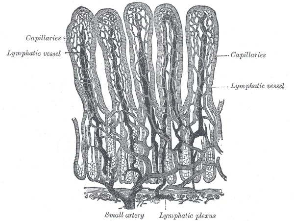
Figure 4Gray1061
Long description
Villi of small intestine, showing bloodvessels and lymphatic vessels. (Cadiat.)

Figure 4: Villi of small intestine, showing bloodvessels and lymphatic vessels (Cadiat.).
The lumen is lined by the mucosa, which projects into finger-like villi. Each villus contains a central lacteal and capillaries. The epithelial cells covering the villi have a brush border of microvilli. Beneath the mucosa lies the submucosa, which contains larger blood vessels and nerves. The muscularis externa consists of an inner circular layer and an outer longitudinal layer. The serosa forms the outermost covering. The villi increase the surface area for absorption, while the crypts of Lieberkühn are located between the villi at the base of the mucosa.
The intestine adapts to energetic demands for monosaccharide absorption through nonspecific and specific mechanisms. Nonspecific adaptations change the overall absorptive capacity, including changes in absorptive surface, enterocyte number, size, and differentiation. Specific adaptations affect the uptake of individual monosaccharides. For instance, the abundance of glucose transporters can change in response to dietary factors or disease states such as diabetes. These adaptations ensure that the small intestine meets the body's metabolic needs.
DefinitionAbsorptive Surface
The total area available for nutrient uptake, determined by intestinal length, villus height, and microvillus length.
| Feature | Location | Function |
|---|---|---|
| Villi | Mucosa | Increase surface area for absorption |
| Microvilli | Apical membrane | Site of brush border enzymes |
| Crypts | Base of mucosa | Site of stem cell division |
| Lacteals | Central core of villi | Absorb dietary lipids |
The vascular architecture of the small intestine is designed to support extensive nutrient absorption while maintaining robust perfusion. The primary arterial supply is derived from the superior mesenteric artery, which arises from the abdominal aorta. This vessel is the main source of oxygenated blood for the jejunum and ileum. It courses anteriorly and inferiorly, passing behind the neck of the pancreas and crossing the third part of the duodenum. From its concave left border, it gives rise to numerous branches that form an intricate network. These branches include the jejunal and ileal arteries, which run between the layers of the mesentery. As they approach the intestinal wall, they form arcades. The first-order arcades are few and simple, while higher-order arcades become more complex and numerous towards the ileum. This arrangement ensures that blood reaches the intestinal villi efficiently. [1]
Figure 5Gray1070
Long description
—Diagrams showing the arrangement and variations of the loops of the mesenteric vessels for various segments of the small intestine of average length. Nearest the duodenum the mesenteric loops are primary, the vasa recta are long and regular in distribution, and the translucent spaces (lunettes) are

Figure 5: —Diagrams showing the arrangement and variations of the loops of the mesenteric vessels for various segments of the small intestine of average length.
Figure 6Gray1056
Long description
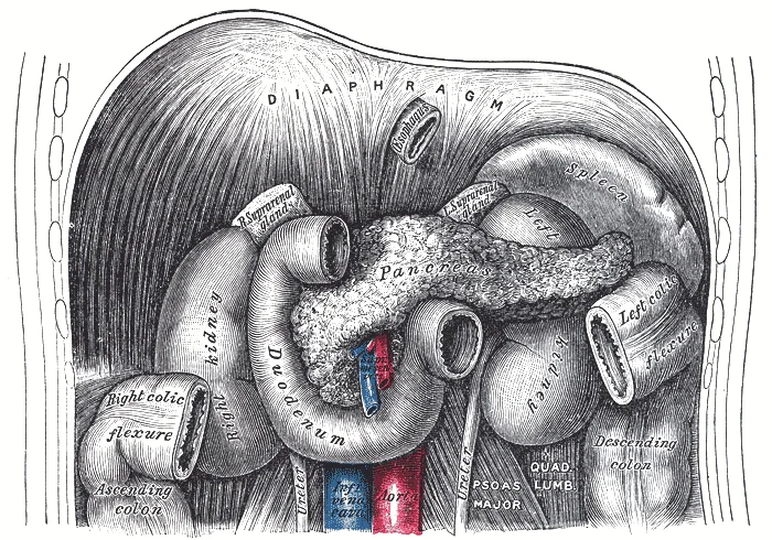
The duodenum and pancreas. Labelled illustration from Wikimedia Commons, used under its open licence.

Figure 6: The duodenum and pancreas.
Veins run parallel to the arteries, following a similar branching pattern. They drain deoxygenated blood and absorbed nutrients from the intestinal wall. This portal system carries nutrient-rich blood directly to the liver for processing. This anatomical arrangement is crucial for the first-pass metabolism of absorbed substances. The liver receives the majority of the blood from the gastrointestinal tract, allowing it to regulate nutrient levels and detoxify harmful substances before they enter the systemic circulation. [1]
The hepatic portal vein is formed by the union of the superior mesenteric vein and the splenic vein. Students should note the parallel course of arteries and veins. This parallelism is a key feature of the mesenteric vasculature. Understanding this anatomy is essential for surgical procedures involving the small intestine. It also aids in interpreting imaging studies such as CT angiography. The parallel course of the vessels is a critical detail to remember. This arrangement facilitates efficient exchange of nutrients and waste products. The caption provides additional context and clarification.
The venous drainage of the small intestine follows a hierarchical pathway that ultimately delivers nutrient-rich blood to the liver for processing. This system is distinct from the arterial supply and relies on the convergence of multiple tributaries into larger trunks.
The primary collector for the small intestine is the superior mesenteric vein. It originates from the union of several smaller veins that drain the jejunum and ileum. These tributaries, known as jejunal and ileal veins, run between the layers of the mesentery. They ascend towards the right side of the abdomen. As they travel, they receive additional blood from the right colic and ileocolic veins. The two vessels are closely related within the root of the mesentery. The superior mesenteric vein joins the splenic vein behind the neck of the pancreas. This union forms the hepatic portal vein.
Branching from its left side are the jejunal and ileal veins. These branches are numerous and converge superiorly. The pancreas lies posterior to this junction.
DefinitionPortal System
The hepatic portal system transports blood from the gastrointestinal tract to the liver before it enters the systemic circulation.
These veins anastomose freely with each other. This creates a rich collateral network within the mesentery. This arrangement allows for efficient drainage. The blood collected is rich in absorbed nutrients. It passes through the liver sinusoids for metabolic regulation.
| Structure | Origin | Termination |
|---|---|---|
| Superior Mesenteric Vein | Union of jejunal/ileal veins | Joins splenic vein |
| Hepatic Portal Vein | Union of SMV and splenic vein | Enters liver at porta hepatis |
| Splenic Vein | Drains spleen and pancreas | Joins SMV behind pancreas |
The anatomical relationship between the superior mesenteric vein and the duodenum is critical. The vein crosses anterior to the third part of the duodenum. This position makes it vulnerable during surgical procedures in this region. Knowledge of this relation prevents iatrogenic injury. The convergence of these veins ensures that all blood from the small intestine is directed to the liver. This is a fundamental principle of abdominal circulation.
Figure 7Gray1060
Long description
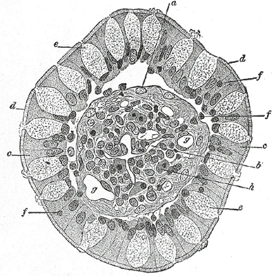
Transverse section of a villus, from the human intestine. (v. Ebner.) X 350. a. Basement membrane, here somewhat shrunken away from the epithelium. b. Lacteal. c. Columnar epithelium. d. Its striated border. e. Goblet cells. f. Leucocytes in epithelium. f’. Leucocytes below epithelium. g. Bloodvesse

Figure 7: Transverse section of a villus, from the human intestine (v.
The lymphatic drainage of the small intestine begins at the microscopic level within the mucosal villi. Each villus contains a central lymphatic capillary known as a lacteal. This structure is distinct from the surrounding capillary bed, which consists of one arteriole and one venule. While breakdown products of carbohydrates and proteins enter the bloodstream directly, lipid breakdown products are absorbed by the lacteals. These lipids are then transported to the bloodstream via the lymphatic system. [1]
DefinitionLacteal
A central lymphatic capillary within each intestinal villus that absorbs dietary lipids.
Moving from the mucosa, lymph vessels converge to form larger collecting trunks. These vessels drain into the mesenteric lymph nodes. The mesenteric nodes are arranged in chains along the branches of the superior mesenteric artery. Lymph from the jejunum and ileum passes through these nodes before reaching the cisterna chyli. From the cisterna chyli, lymph ascends via the thoracic duct to empty into the venous system at the junction of the left internal jugular and left subclavian veins.
Lymphatic Pathway
- VilliLacteals absorb lipids
- Mesenteric NodesFilter lymph from jejunum/ileum
- Cisterna ChyliCollection point in abdomen
- Thoracic DuctAscends to thorax
- Venous JunctionLeft subclavian & internal jugular veins
The immune function of the small intestine is closely linked to its lymphatic architecture. The lamina propria of the small intestine mucosa is studded with mucosa-associated lymphoid tissue (MALT). In addition to solitary lymphatic nodules, aggregations of intestinal MALT are present. These aggregations are typically referred to as Peyer’s patches. Peyer’s patches are concentrated in the distal ileum. They serve to keep bacteria from entering the bloodstream. Peyer’s patches are most prominent in young people. They become less distinct as you age. This change coincides with the general activity of our immune system. [1]
Figure 8Gray1063
Long description
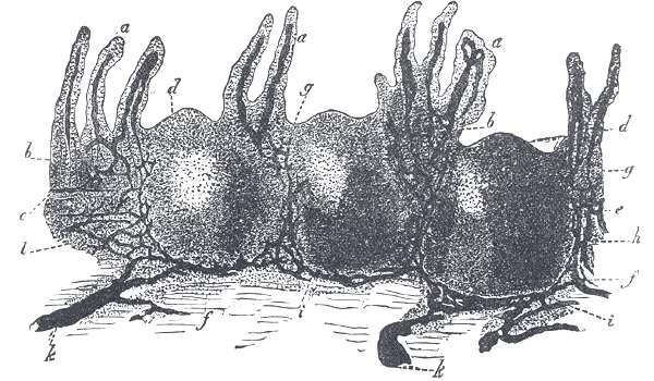
Vertical section of a human aggregated lymphatic nodule, injected through its lymphatic canals. a. Villi with their chyle passages. b. Intestinal glands. c. Muscularis mucosæ. d. Cupola or apex of solitary nodule. e. Mesial zone of nodule. f. Base of nodule. g. Points of exit of the lacteals from th

Figure 8: Vertical section of a human aggregated lymphatic nodule, injected through its lymphatic canals (a).
The submucosa contains larger lymphatic vessels. These vessels travel towards the mesentery.
| Structure | Location | Function |
|---|---|---|
| Lacteal | Central villus | Absorbs dietary lipids |
| Peyer's Patches | Distal ileum mucosa | Immune surveillance |
| Mesenteric Nodes | Along SMA branches | Lymph filtration |
| Thoracic Duct | Thorax | Venous drainage |
Disruption of this drainage can lead to clinical syndromes. Small intestinal bacterial overgrowth is a heterogeneous syndrome characterised by an increased number and/or abnormal type of bacteria in the small bowel. It is a well‐recognised cause of maldigestion and malabsorption. This condition highlights the importance of normal lymphatic and mucosal integrity in preventing bacterial translocation and maintaining nutrient absorption.
The innervation of the small intestine is a complex integration of autonomic and somatic pathways that regulate motility, secretion, and sensation. Understanding this supply requires distinguishing between the sympathetic, parasympathetic, and somatic contributions, as well as recognising the lymphatic drainage which often parallels neural routes.
The autonomic nervous system provides the primary regulatory control. Sympathetic fibres originate from the thoracic spinal cord segments T5 to T9. These preganglionic neurons synapse in the coeliac and superior mesenteric ganglia. Postganglionic fibres then travel along the branches of the superior mesenteric artery to reach the small intestine. These sympathetic inputs generally inhibit smooth muscle contraction and reduce blood flow, preparing the gut for 'fight or flight' responses.
Parasympathetic innervation is primarily mediated by the vagus nerve (cranial nerve X). Preganglionic fibres from the dorsal motor nucleus of the vagus extend down the oesophagus and stomach. They synapse with postganglionic neurons located within the walls of the small intestine, specifically in the myenteric (Auerbach’s) and submucosal (Meissner’s) plexuses. This vagal supply stimulates motility and secretion, promoting digestion and absorption.
Somatic innervation is less direct but crucial for sensation. The afferent sensory fibres that convey pain and stretch information travel back along the sympathetic pathways to the spinal cord. This shared pathway is the basis for referred pain.
Visceral pain from the small intestine is often poorly localised by the patient. This is because the visceral afferent nerves enter the spinal cord at specific segmental levels. When visceral nociceptors are stimulated, the brain may interpret the signal as coming from the somatic structures that share the same spinal segmental entry. Consequently, pain from the small intestine is typically referred to the periumbilical region. This clinical feature is a key diagnostic clue in conditions such as early appendicitis or small bowel obstruction.
The lymphatic drainage of the small intestine is extensive and vital for immune surveillance and fat absorption. Lacteals, which are specialized lymphatic capillaries, are located within the villi of the mucosa. They absorb dietary lipids and fat-soluble vitamins. These lacteals converge into larger lymphatic vessels that run alongside the blood vessels in the mesentery. These vessels drain into the mesenteric lymph nodes, and subsequently into the cisterna chyli via the intestinal trunk.
The nerve supply runs in close anatomical association with these vascular and lymphatic structures within the mesentery. The superior mesenteric plexus, derived from the sympathetic and parasympathetic inputs described above, accompanies the superior mesenteric artery. This plexus distributes fibres to the duodenum, jejunum, and ileum. The close proximity of nerves, arteries, and lymphatics in the mesentery means that pathology affecting one structure, such as lymphadenopathy or vascular compromise, can easily impact the others.
The small intestine is the primary site for nutrient absorption, a function supported by a specialized histological architecture designed to maximize surface area. The mucosa is lined by simple columnar epithelium, which differs significantly from the stratified squamous epithelium found in the oesophagus or the simple columnar epithelium of the stomach. This epithelium contains enterocytes, goblet cells, and enteroendocrine cells. Enterocytes are the dominant cell type, responsible for absorption, and possess a brush border of microvilli on their apical surface. This brush border dramatically increases the absorptive surface area.
These cells differentiate during migration and are exfoliated at the top of the villi exhibiting life spans between days and weeks.
The muscularis mucosae separates the mucosa from the submucosa. The submucosa contains larger blood vessels, lymphatics, and the submucosal plexus (Meissner's plexus), which regulates secretion and blood flow. The muscularis externa consists of an inner circular layer and an outer longitudinal layer, containing the myenteric plexus (Auerbach's plexus) between them, which controls motility.
It is crucial to distinguish the small intestine from the large intestine. Three features are unique to the large intestine: teniae coli, haustra, and epiploic appendages. The teniae coli are three bands of smooth muscle that make up the longitudinal muscle layer of the muscularis of the large intestine, except at its terminal end. Tonic contractions of the teniae coli bunch up the colon into a succession of pouches called haustra (singular = haustrum), which are responsible for the wrinkled appearance of the colon. Attached to the teniae coli are small, fat-filled sacs of visceral peritoneum called epiploic appendages. The purpose of these is unknown. Although the rectum and anal canal have neither teniae coli nor haustra, they do have well-developed layers of muscularis that create the strong contractions needed for defecation. [1]
They have been observed in response to changed nutrition, during diabetes, and after surgical interventions.
DefinitionVillus
A finger-like projection of the mucosa that increases surface area for absorption.
Exam trapLarge vs Small Intestine
Do not confuse haustra and teniae coli with small intestinal features. These are exclusive to the large intestine.
The small intestine originates from the midgut, undergoing complex rotation and fixation to achieve its final anatomical position within the abdominal cavity. This developmental journey ensures the proper arrangement of the duodenum, jejunum, and ileum, establishing the vascular and mesenteric attachments that define adult anatomy. Understanding this embryological origin is crucial for interpreting congenital anomalies such as malrotation, which can compromise blood supply or cause obstruction. The transition from the foregut to the midgut and then to the hindgut marks distinct phases of differentiation, with the small intestine representing the bulk of the midgut derivative.
The mucosal surface undergoes significant expansion through the formation of specialized structures that maximize absorptive capacity. Circular folds, also known as plicae circulares, are deep ridges in the mucosa and submucosa. These structures begin near the proximal part of the duodenum and end near the middle of the ileum. Their presence increases the surface area available for nutrient uptake and alters the flow dynamics of intestinal contents. By causing chyme to spiral rather than move in a straight line, these folds slow the movement of chyme. This deceleration provides the time needed for nutrients to be fully absorbed. [1]
Beneath the circular folds, the mucosa is dotted with deep crevices leading into tubular intestinal glands, or crypts of Lieberkühn. These glands are formed by cells lining the crevices and produce intestinal juice. This secretion is a slightly alkaline mixture of water and mucus with a pH ranging from 7.4 to 7.8. Daily secretion volumes range from 0.95 to 1.9 liters. This output is stimulated by distention of the small intestine or the irritating effects of chyme on the intestinal mucosa. [1]
The muscular layers develop to facilitate coordinated peristalsis and segmentation. The inner circular and outer longitudinal muscle layers allow for the mixing and propulsion of contents. Neural innervation from the enteric nervous system coordinates these movements, responding to local chemical and mechanical stimuli. The vascular supply, derived from the superior mesenteric artery, develops in tandem with the gut tube, ensuring adequate perfusion for metabolic demands. Lymphatic vessels, including lacteals within the villi, develop to absorb dietary lipids. This integrated development of mucosal, muscular, neural, and vascular components creates a highly efficient organ for digestion and absorption.
The central focus is the coiled mass of the small bowel, distinguishable from the large intestine by its narrower lumen and prominent valvulae conniventes, which appear as complete transverse folds crossing the entire width of the bowel wall.
The duodenum is visualised as a C-shaped structure curving around the head of the pancreas. It begins at the pylorus of the stomach, lying to the right of the midline, and descends as the second part. The third part crosses horizontally, before ascending as the fourth part to join the jejunum at the duodenojejunal flexure. This flexure is anchored by the ligament of Treitz, a key landmark for distinguishing the small from the large intestine. The jejunum occupies the upper left quadrant of the abdomen. The valvulae conniventes are dense and closely spaced, giving the jejunum a characteristic 'stack of coins' appearance on cross-sectional imaging. The mesentery attaching the jejunum to the posterior abdominal wall is visible, containing the superior mesenteric vessels.
The ileum constitutes the lower right quadrant and the pelvis. It has a thinner wall and fewer, less prominent valvulae conniventes. The terminal ileum enters the caecum at the ileocaecal valve. The mesentery of the ileum is longer and more fan-shaped than that of the jejunum. The ileocaecal junction is a critical landmark, often appearing as a thickened muscular ring.
The small intestine is suspended from the posterior abdominal wall by the mesentery, which contains the superior mesenteric artery and vein. The superior mesenteric artery arises from the aorta just below the coeliac trunk and supplies the small intestine from the duodenum to the proximal two-thirds of the transverse colon. The venous drainage follows the arterial supply into the superior mesenteric vein, which joins the splenic vein to form the portal vein.
Lymphatic drainage from the small intestine follows the arterial supply to the superior mesenteric lymph nodes, located near the root of the mesentery. These nodes drain into the cisterna chyli, which ascends as the thoracic duct. Understanding this anatomy is essential for interpreting radiological images and planning surgical interventions.
| Segment | Location | Key Radiological Feature |
|---|---|---|
| Duodenum | Right upper quadrant | C-shape around pancreas |
| Jejunum | Left upper quadrant | Thick wall, dense folds |
| Ileum | Right lower quadrant | Thin wall, sparse folds |
The central structure is the small intestine, coiled within the middle and lower abdomen. It is bounded superiorly by the transverse colon and inferiorly by the urinary bladder and reproductive organs. The mesentery fans out from the left upper quadrant, attaching the jejunum and ileum to the posterior abdominal wall, creating the characteristic loops.
The duodenum is the initial segment of the small intestine, receiving chyme from the stomach. It is divided into four parts: superior, descending, horizontal, and ascending. The superior part begins at the pylorus and is intraperitoneal. The descending part runs vertically along the right side of the vertebral column, receiving the common bile duct and pancreatic duct. The horizontal part crosses the midline anterior to the aorta and inferior vena cava. The ascending part ends at the duodenojeju
The jejunum and ileum constitute the majority of the small intestine, suspended by the mesentery. The jejunum is the proximal two-fifths, located in the upper left quadrant. It has a larger diameter, thicker walls, and more prominent circular folds (plicae circulares). The ileum is the distal three-fifths, occupying the lower right quadrant and pelvis. It has a smaller diameter, thinner walls, and fewer folds. The terminal ileum enters the cecum through the ileocaecal valve. The mesentery contains the superior mesenteric vessels, which supply the small intestine.
The cecum is the first part of the large intestine, a sac-like structure suspended inferior to the ileocaecal valve. It is about 6 cm long and receives contents from the ileum. The appendix, or vermiform appendix, is a winding tube attaching to the cecum. Although often considered vestigial, it may serve as a bacterial reservoir during diarrheal illness. Its twisted anatomy allows for the accumulation of enteric bacteria. [1]
Meckel diverticulum is a congenital abnormality of the ileum. It can cause ulceration, bleeding, intussusception, obstruction, and perforation. Understanding these anatomical landmarks is essential for diagnosing and managing these conditions.
Park et al recommended resection if features include male gender, age below 50 years, or length greater than 2 cm. Stone et al recommend against incidental removal of asymptomatic Meckel diverticulum, especially in women.
Clinical pearlAnal Canal Anatomy
The anal canal mucosa is stratified squamous epithelium, unlike the rest of the colon, and contains anal columns with arterial and venous grids.
Exam trapSGLT1 Specificity
SGLT1 transports d-glucose and d-galactose but does not interact with d-fructose.
DefinitionFructose Malabsorption
Functional defects in GLUT5 or decreased transcription are not the predominant cause for fructose malabsorption in IFM.
NoteMeckel Diverticulum Risk
Park et al quantified complication risk as 70%, 42%, 25%, and 17% for all, three, two, or one criteria respectively.
| Feature | Recommendation |
|---|---|
| Male gender | Resect if present |
| Age below 50 years | Resect if present |
| Length > 2 cm | Resect if present |
| Obvious abnormality | Resect if present |
Meckel Diverticulum Management
- Incidental Meckel DiverticulumAssess risk factors
- Question: Symptomatic?
Yes
- Resect
No
- Question: Risk Factors Present?
Yes
- Consider Resection
No
- Do Not Resect
Students frequently confuse the embryological origins of the gastrointestinal tract, leading to errors in understanding innervation and vascular supply. A critical distinction lies at the pectinate line, which marks the junction between the hindgut and the external skin. The mucosa above this line is fairly insensitive, whereas the area below is very sensitive. This resulting difference in pain threshold is due to the fact that the upper region is innervated by visceral sensory fibers, and the lower region is innervated by somatic sensory fibers. [1]
Another frequent error involves the mechanism of glucose absorption. Students often assume water follows glucose via aquaporins exclusively. However, SGLT1-mediated d-glucose uptake into oocytes expressing human SGLT1 precedes water uptake[1]. The uptake generates a local osmotic gradient that is sufficient to drive passive water influx across the BBM[1]. Assuming active water transport mechanisms here ignores the passive osmotic drive demonstrated in experimental models.
Misunderstanding the regulation of transporters is also common. The incorporation of Glut2 into the BBM is not constant but regulated by intracellular conditions. Maximal electrogenic glucose transport by Sglt1 at high intestinal glucose when Sglt1 in the BBM is upregulated and the transporter operates at Vmax is supposed to trigger Glut2 incorporation into the BBM[1]. Students who view Glut2 as a static component miss the dynamic nature of intestinal adaptation to nutrient load.
Exam trapEmbryological Timing
Meckel diverticulum is caused by failure of closure of the vitelline duct at the fifth week of fetal growth.
Students often overlook the clinical significance of congenital malformations. Meckel diverticulum is the most common congenital malformation of the gastrointestinal tract and occurs secondary to persistence of the congenital vitello-intestinal duct. This condition can cause many complications, such as hemorrhage, intussusception, intestinal obstruction and perforation, especially in children. Failing to recognise these potential presentations leads to delayed diagnosis and increased morbidity in paediatric patients.
DefinitionMeckel Diverticulum
A congenital malformation resulting from the persistence of the vitello-intestinal duct, typically presenting with complications like obstruction or hemorrhage in children.
This knowledge is applied at the bedside to interpret symptoms of intestinal obstruction or bleeding. When a child presents with abdominal pain and rectal bleeding, awareness of Meckel diverticulum is crucial because it is rarely observed in children, the condition of remains frequently misdiagnosed during the preoperative period. Early diagnosis allows for curative treatment, whereas delay can lead to perforation or severe haemorrhage.
In the operating theatre, understanding the vascular and neural supply is vital for safe resection. The distinction between visceral and somatic innervation at the pectinate line guides anaesthetic technique. Procedures involving the anal canal below this line require somatic anaesthesia due to the high sensitivity of the somatic sensory fibers. Above the line, visceral anaesthesia may suffice due to the insensitivity of the mucosa.
On imaging, recognising the anatomical landmarks helps differentiate Meckel diverticulum from other pathologies. Since Meckel diverticulum is the most common congenital malformation of the gastrointestinal tract, radiologists must look for signs of inflammation or obstruction in the ileum. The presence of a blind-ending pouch connected to the ileum is diagnostic, often confirmed by a Technetium-99m pertechnetate scan detecting ectopic gastric mucosa.
Diagnostic Approach to Paediatric Abdominal Pain
- StartChild with abdominal pain and rectal bleeding
- Question: Is there a history of intussusception?
Yes
- Consider Meckel diverticulum as cause
No
- Evaluate for other causes
- Perform imagingLook for vitelline duct persistence
In examinations, students must accurately describe the structure and function of the small intestine. The large surface area is necessary for complex processes of digestion and absorption that occur within it. This fundamental principle underpins questions on nutrient uptake and surface area calculations. [1]
NoteSurface Area
The large surface area is necessary for complex processes of digestion and absorption that occur within it.
Understanding the molecular mechanisms of absorption is also tested. The mechanism of glucose-dependent incorporation of Glut2 into the BBM has been investigated in some detail[1]. Students should know that Sglt1-mediated Na+-d-cotransport causes a depolarization of the BBM that activates Ca2+ uptake via Ca2+ channel Cav1.3[1]. This cascade leads to the recruitment of PKC βII to the plasma membrane, promoting Glut2 insertion[1].
| Transporter | Regulation Mechanism | Trigger |
|---|---|---|
| SGLT1 | Upregulated in BBM | High intracellular glucose |
| Glut2 | Incorporated into BBM | Maximal SGLT1 activity |
| Glut1/7/8/12 | Compensatory efflux | Removal of Glut2 |
When Glut2 is removed, Glut1, Glut7, Glut8, or Glut12 is targeted to the BLM when Glut2 is removed and provides a compensatory path for efflux of d-glucose[1]. This ensures continuous glucose exit from the cell even when the primary transporter is downregulated.
Clinical pearlCompensatory Transport
Glut1, Glut7, Glut8, or Glut12 is targeted to the BLM when Glut2 is removed and provides a compensatory path for efflux of d-glucose[1].
Finally, the role of neurotensin in gut function is relevant. The data indicate that d-glucose-mediated stimulation of neurotensin secretion by EEC cells is similar to GLP-1 secretion by L cells[1]. This highlights the endocrine function of the small intestine in regulating metabolism and motility.
The pectinate line marks the junction between hindgut and external skin. Mucosa above the pectinate line is insensitive due to visceral sensory fibers. Mucosa below the pectinate line is sensitive due to somatic sensory fibers. Meckel diverticulum results from failure of vitelline duct closure at week five. SGLT1-mediated glucose uptake creates an osmotic gradient for passive water influx[1]. Glut2 incorporation into BBM is triggered by maximal SGLT1 activity[1]. PKC βII recruitment promotes Glut2 insertion into the plasma membrane[1]. Neurotensin secretion is stimulated by d-glucose in EEC cells[1].
Answer each one in your head first, then open the answer.
What line marks the junction between the hindgut and external skin?
Show answerHide answer
The pectinate line (or dentate line)Why is the mucosa above the pectinate line insensitive?
Show answerHide answer
It is innervated by visceral sensory fibersWhat is the most common congenital malformation of the gastrointestinal tract?
Show answerHide answer
Meckel diverticulumWhen does failure of closure of the vitelline duct occur?
Show answerHide answer
At the fifth week of fetal growthWhat triggers Glut2 incorporation into the BBM?
Show answerHide answer
Maximal electrogenic glucose transport by Sglt1[1]
- Betts JG, Young KA, Wise JA, et al. The Small and Large Intestines. OpenStax, Anatomy and Physiology 2e (CC BY-NC-SA 4.0). Accessed .
Figure credits
- Figure 1: OpenStax College, via Wikimedia Commons, CC BY 3.0, CC BY 3.0.
- Figure 2: Henry Vandyke Carter, via Wikimedia Commons, Public domain, Public domain.
- Figure 3: OpenStax College, via Wikimedia Commons, CC BY 3.0, CC BY 3.0.
- Figure 4: Henry Vandyke Carter, via Wikimedia Commons, Public domain, Public domain.
- Figure 5: Henry Vandyke Carter, via Wikimedia Commons, Public domain, Public domain.
- Figure 6: Henry Vandyke Carter, via Wikimedia Commons, Public domain, Public domain.
- Figure 7: Henry Vandyke Carter, via Wikimedia Commons, Public domain, Public domain.
- Figure 8: Henry Vandyke Carter, via Wikimedia Commons, Public domain, Public domain.
Educational content for medical students and doctors. It is not a substitute for clinical judgement or local guidelines.