Stomach
The stomach is a J-shaped muscular organ that stores food and secretes gastric juice for digestion.
The cardia marks the point where the esophagus connects to the stomach[1].
The oesophagogastric junction is the anatomical point where the oesophagus meets the stomach. This region is functionally critical because it prevents the backflow of gastric contents into the oesophagus. The lower oesophageal sphincter is a functional zone of high pressure located at this junction. It acts as a valve that remains tonically contracted to keep the acidic gastric contents within the stomach. When swallowing occurs, the sphincter relaxes to allow food to pass into the stomach, then immediately contracts again to maintain the barrier. This mechanism is essential for protecting the oesophageal lining from corrosive gastric acid.
The stomach has two distinct borders. The convex lateral surface is known as the greater curvature, while the concave medial border is the lesser curvature[1]. The organ is held in place by two peritoneal folds. The lesser omentum extends from the liver to the lesser curvature, anchoring the stomach to the liver[1]. The greater omentum runs from the greater curvature to the posterior abdominal wall, providing additional support and protection[1]. In the absence of food, the stomach deflates inward, causing the mucosa and submucosa to form large folds called rugae[1]. These folds allow the stomach to expand significantly when food is ingested.
The stomach mucosa is lined by surface mucus cells that secrete a protective alkaline mucus coat[1]. This lining is dotted with gastric pits, which mark the entry points to gastric glands[1]. These glands secrete gastric juice, a complex digestive fluid[1]. The stomach is protected from self-digestion by a mucosal barrier consisting of a thick coating of bicarbonate-rich mucus[1]. This mucus forms a physical barrier and neutralizes acid with bicarbonate ions[1]. Additionally, epithelial cells meet at tight junctions that block gastric juice from penetrating underlying tissues[1]. Stem cells in the gastric glands replace damaged epithelial cells, with the surface epithelium being completely replaced every 3 to 6 days[1].
The stomach participates in virtually all digestive activities except ingestion and defecation[1].
It links the oesophagus to the first part of the small intestine, the duodenum[1].
The organ has two main curvatures. The greater curvature forms the long, convex left border. The lesser curvature forms the short, concave right border. The cardia is the region surrounding the oesophageal opening. The fundus is the dome-shaped portion superior and left of the cardia. The body is the large central region. The pylorus is the distal region connecting to the duodenum. The pyloric sphincter regulates emptying. [1]
The empty stomach is only about the size of your fist. It can stretch to hold as much as 4 liters of food and fluid. This capacity is more than 75 times its empty volume. Contractions provide mechanical assistance to digestion. The stomach vigorously churns food. It stores food as an acidic liquid called chyme. [1]
Relations are key for surgical and clinical understanding. The anterior surface faces the liver and abdominal wall. The posterior surface relates to the pancreas and spleen. The lesser sac lies behind the stomach. The greater omentum hangs from the greater curvature. The lesser omentum connects the lesser curvature to the liver.
DefinitionStomach Capacity
The empty stomach is only about the size of your fist, but can stretch to hold as much as 4 liters of food and fluid, or more than 75 times its empty volume.
The stomach participates in all digestive activities except ingestion and defecation. It secretes gastric juices that break down food. It absorbs certain drugs, including aspirin and some alcohol. Little if any nutrient absorption occurs in the stomach. The exception is the negligible amount of nutrients in alcohol. [1]
Figure 1Gray1048
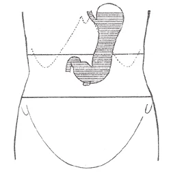
Long description
Diagram showing shape and position of moderately filled stomach. Erect posture. (Hertz.)

Figure 1: Diagram showing shape and position of moderately filled stomach (erect posture).
The stomach continues the digestion of carbohydrates and fats[1].
Gastric emptying is regulated by both the stomach and the duodenum. The contents of the stomach are completely emptied into the duodenum within 2 to 4 hours after you eat a meal. Different types of food take different amounts of time to process. Foods heavy in carbohydrates empty fastest. This is followed by high-protein foods. Meals with a high triglyceride content remain in the stomach the longest. Since enzymes in the small intestine digest fats slowly, food can stay in the stomach for 6 hours or longer when the duodenum is processing fatty chyme. [1]
| Function | Detail |
|---|---|
| Mechanical | Vigorously churns food. |
| Chemical | Breaks down protein with HCl and pepsin. |
| Storage | Stores food as chyme. |
| Absorption | Absorbs aspirin and some alcohol. |
The presence of chyme in the duodenum activates receptors that inhibit gastric secretion. This prevents additional chyme from being released by the stomach before the duodenum is ready to process it. However, it should be noted that the stomach does have a natural means of avoiding excessive acid secretion and potential heartburn. Whenever pH levels drop too low, cells in the stomach react by suspending HCl secretion and increasing mucous secretions. [1]
Figure 2Gray1053
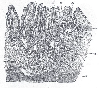
Long description
Section of mucous membrane of human stomach, near the cardiac orifice. (v. Ebner, after J. Schaffer.) X 45. c. Cardiac glands. d. Their ducts. cr. Gland similar to the intestinal glands, with goblet cells. mm. Mucous membrane. m. Muscularis mucosæ. m’. Muscular tissue within the mucous membrane.

Figure 2: Section of mucous membrane of human stomach, near the cardiac orifice ((v).
The oesophagogastric junction marks the entry point. The blood supply arises from the coeliac trunk. Lymph drains to gastric nodes. Nerves include the vagus and sympathetic chains. These structures support the functional anatomy described.
The stomach is a J-shaped hollow viscus situated in the left upper quadrant of the abdomen, occupying the epigastric and left hypochondriac regions. Its anatomical position is defined by its extensive relations with surrounding structures, which are critical for understanding both normal physiology and pathological spread. The organ is primarily covered by peritoneum, making it an intraperitoneal organ, except for the bare area on the posterior surface of the lesser curvature and the pyloric region where it contacts the pancreas and duodenum directly.
Figure 3Gray1049
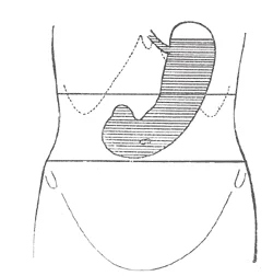
Long description
Diagram showing shape and position of distended stomach. Erect posture. (Hertz.)

Figure 3: Diagram showing shape and position of distended stomach (erect posture).
Figure 4Gray1047
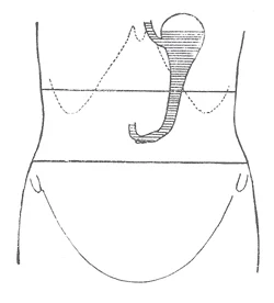
Long description
Diagram showing shape and position of empty stomach. Erect posture. (Hertz.)

Figure 4: Diagram showing shape and position of empty stomach (erect posture).
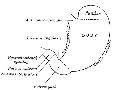
Figure 5Gray1046
Long description
Outline of stomach, showing its anatomical landmarks.

Figure 5: Outline of stomach, showing its anatomical landmarks.
The oesophagus enters the stomach at the cardia, located at the level of the tenth thoracic vertebra, just to the left of the midline. Immediately inferior to the cardia lies the fundus, a dome-shaped region that often contains gas and lies against the diaphragm. The body of the stomach forms the central and largest portion, extending downwards and to the left. The pyloric antrum leads to the pyloric canal, which terminates at the pyloric sphincter, marking the transition to the duodenum. [1]
The anterior surface of the stomach is related to the diaphragm, the left lobe of the liver, and the anterior abdominal wall. The left lobe of the liver lies against the superior part of the anterior surface, while the greater omentum hangs down from the greater curvature, covering the anterior surface of the transverse colon and small intestine. The posterior surface of the stomach forms the anterior wall of the omental bursa (lesser sac). This retrogastric space is bounded by the stomach anteriorly and the pancreas, left kidney, left adrenal gland, spleen, and transverse mesocolon posteriorly. These structures collectively form the stomach bed.
The lesser curvature of the stomach gives attachment to the lesser omentum, which connects the stomach to the liver. The greater curvature gives attachment to the greater omentum, a large apron-like fold of peritoneum. The splenic artery runs along the superior border of the pancreas, close to the posterior aspect of the stomach, and gives off short gastric arteries that supply the fundus. The left gastric artery runs along the lesser curvature, supplying the proximal stomach. The right gastric artery, a branch of the hepatic artery proper, also supplies the lesser curvature. The right gastroepiploic artery, a branch of the gastroduodenal artery, and the left gastroepiploic artery, a branch of the splenic artery, supply the greater curvature.
DefinitionStomach Bed
The structures lying posterior to the stomach, including the pancreas, left kidney, left adrenal gland, spleen, and transverse mesocolon, separated by the omental bursa.
The vascular supply and lymphatic drainage follow the arterial branches. Lymph from the stomach drains into nodes along the lesser and greater curvatures, and then to the coeliac nodes. The innervation is provided by the vagus nerves (parasympathetic) and the sympathetic trunk via the coeliac plexus. The vagus nerves provide motor innervation to the stomach muscles and sensory innervation, while sympathetic fibers mediate vasoconstriction and inhibit motility.
In addition to the typical circular and longitudinal smooth muscle layers, the muscularis has an inner oblique smooth muscle layer[1].
| Layer | Orientation | Function |
|---|---|---|
| Muscularis Externa | Inner Oblique | Churning and mechanical breakdown |
| Muscularis Externa | Middle Circular | Peristalsis and mixing |
| Muscularis Externa | Outer Longitudinal | Propulsion towards pylorus |
The pylorus acts as a valve, regulating the passage of chyme into the duodenum. This distal end has impedance electrodes enclosed in the bag that allow the different measures. Subjects are usually asked to fast at least 8 h before the procedure. The device is positioned in the pylorus either through the endoscope, under endoscopic visualization, or either by fluoroscopy guidance. Pyloric angulation has not been reported as a technical obstacle. The impact of filling in the stomach or using anest [4]
NoteClinical Note
The stomach's ability to churn food is due to the inner oblique muscle layer, a unique adaptation for mechanical digestion. Mixing waves occur every 20 seconds, intensifying towards the pylorus.
The stomach interior is defined by its mucosal lining, which contains specialized glands responsible for secretion and digestion. These glands house distinct cell types, each with a specific role in maintaining the gastric environment.
Parietal cells are located primarily in the middle region of the gastric glands and are among the most highly differentiated epithelial cells in the body. These relatively large cells produce hydrochloric acid (HCl) and intrinsic factor. HCl creates the high acidity of stomach contents, with a pH of 1.5 to 3.5. This acidity is essential to activate the protein-digesting enzyme pepsin and kills ingested bacteria. Intrinsic factor is a glycoprotein required for vitamin B12 absorption in the small intestine. [1]
Gastric glands in the upper part of the stomach contain mucous neck cells. [1]
The secretion of gastric juice is controlled by nerves and hormones, involving stimuli from the brain, stomach, and small intestine. This process occurs in three phases: cephalic, gastric, and intestinal. Once secretion begins, all three phases can occur simultaneously. [1]
The cephalic phase is a brief reflex phase that occurs before food enters the stomach. It is triggered by the smell, taste, sight, or thought of food. For instance, impulses from taste buds or the nose are relayed to the brain, which returns signals to increase gastric secretion. This is a conditioned reflex that can be suppressed by depression or loss of appetite. [1]
The gastric phase lasts 3 to 4 hours and is initiated by local neural and hormonal mechanisms triggered by food entry. Distention of the stomach activates stretch receptors, stimulating parasympathetic neurons to release acetylcholine, which increases gastric juice secretion. Partially digested proteins, caffeine, and rising pH stimulate enteroendocrine G cells to release gastrin. Gastrin induces parietal cells to increase HCl production and activates vigorous smooth muscle contractions. [1]
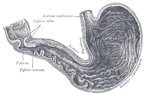
Figure 6Gray's Anatomy - Fig. 1050
Long description
Drawing of the interior of the stomach. Labelled illustration from Wikimedia Commons, used under its open licence.

Figure 6: Drawing of the interior of the stomach.
The upper portion of the gland contains mucous neck cells. The lumen of the gland opens into the gastric pit, which leads to the stomach surface. The surrounding lamina propria supports these structures.
DefinitionGastric Juice Control
Gastric juice secretion is controlled by nerves and hormones, occurring in cephalic, gastric, and intestinal phases, which can happen simultaneously once started.
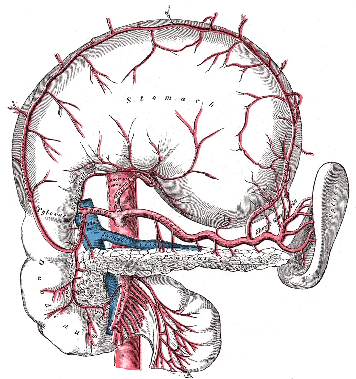
Figure 7Gray533
Long description
The celiac artery and its branches; the stomach has been raised and the peritoneum removed.

Figure 7: The celiac artery and its branches; the stomach has been raised and the peritoneum removed.
The arterial supply of the stomach is derived from the celiac trunk, ensuring a rich vascular network that supports its high metabolic demand for secretion and digestion. The stomach’s blood supply is organized into specific territories corresponding to its anatomical regions, with anastomoses providing collateral circulation.
The pylorus and the lesser curvature of the stomach are primarily supplied by the right and left gastric arteries. The left gastric artery arises directly from the celiac trunk and runs along the lesser curvature. It anastomoses with the right gastric artery, which typically branches from the hepatic artery proper. This arrangement ensures perfusion of the pyloric region and the proximal part of the lesser curvature. [1] [3]
The greater curvature receives blood from the right and left gastro-omental (gastroepiploic) arteries. The left gastro-omental artery arises from the splenic artery, while the right gastro-omental artery branches from the gastroduodenal artery. These vessels run along the greater curvature, anastomosing to form a continuous arterial arcade.
The fundus and the upper body of the stomach are supplied by the short gastric arteries. These vessels arise from the splenic artery or its terminal branches. They pass through the gastrosplenic ligament to reach the fundus, providing essential blood supply to this region, which is critical for the production of gastric juices.
Additionally, the left gastro-omental artery contributes to the supply of the greater curvature, anastomosing with the right gastro-omental artery. This collateral circulation is vital in cases of partial vascular occlusion.
The lymphatic drainage of the stomach follows the arterial supply. Lymph from the pylorus and the lesser curvature drains into the pyloric and left gastric lymph nodes. Lymph from the greater curvature drains into the gastro-omental lymph nodes. Lymph from the fundus drains into the splenic lymph nodes.
These lymph nodes then drain into the coeliac lymph nodes, which finally empty into the intestinal trunk and subsequently the thoracic duct. Understanding this drainage pattern is crucial for staging gastric cancers and planning surgical resections.
The splenic artery gives off the short gastric arteries to the fundus and the left gastro-omental artery along the greater curvature. The common hepatic artery gives rise to the gastroduodenal artery, which becomes the right gastro-omental artery. The right gastric artery, branching from the hepatic artery proper, also runs along the lesser curvature.
DefinitionCeliac Trunk
The primary arterial supply to the foregut, including the stomach, liver, and spleen, arising from the abdominal aorta.
| Region | Artery | Origin |
|---|---|---|
| Lesser Curvature | Left Gastric | Celiac Trunk |
| Lesser Curvature | Right Gastric | Hepatic Artery Proper |
| Greater Curvature | Left Gastro-omental | Splenic Artery |
| Greater Curvature | Right Gastro-omental | Gastroduodenal Artery |
| Fundus | Short Gastric | Splenic Artery |
The venous drainage of the stomach is a critical anatomical pathway that connects the gastric circulation to the portal system. This network ensures that nutrient-rich blood from the digestive tract is directed towards the liver for processing before entering the systemic circulation. Understanding this drainage is essential for comprehending the spread of disease and the physiological regulation of gastric emptying.
The stomach receives its blood supply from branches of the celiac trunk, and its venous return follows a parallel course. Additionally, the short gastric veins drain the fundus, joining the splenic vein. This intricate web of veins allows for collateral circulation, which can become significant in conditions such as portal hypertension.
The pylorus, which holds around 30 mL (1 fluid ounce) of chyme, acts as a filter, permitting only liquids and small food particles to pass through the mostly, but not fully, closed pyloric sphincter. In a process called gastric emptying, rhythmic mixing waves force about 3 mL of chyme at a time through the pyloric sphincter and into the duodenum. Release of a greater amount of chyme at one time would overwhelm the capacity of the small intestine to handle it. The rest of the chyme is pushed back into the body of the stomach, where it continues mixing. This process is repeated when the next mixing waves force more chyme into the duodenum. [1]
The fundus plays an important role, because it stores both undigested food and gases that are released during the process of chemical digestion. Food may sit in the fundus of the stomach for a while before being mixed with the chyme. While the food is in the fundus, the digestive activities of salivary amylase continue until the food begins mixing with the acidic chyme. Ultimately, mixing waves incorporate this food with the chyme, the acidity of which inactivates salivary amylase and activates lingual lipase. Lingual lipase then begins breaking down triglycerides into free fatty acids, and mono- and diglycerides. [1]
The intestinal phase of gastric secretion has both excitatory and inhibitory elements. The duodenum has a major role in regulating the stomach and its emptying. This stimulatory activity is brief, however, because when the intestine distends with chyme, the enterogastric reflex inhibits secretion. One of the effects of this reflex is to close the pyloric sphincter, which blocks additional chyme from entering the duodenum. [1]
The pyloric sphincter is a narrow zone of thickened muscularis and increased luminal pressure. The first one is the circular muscle close to the myenteric plexus, which is dominated by the propagation of the slow wave from the antrum. In this area, pyloric contraction is linked to the propagation of the gastric slow wave. The second area is the deeper circular muscle, which is regulated by motor neurons. Slow waves do not propagate into this area, and the two regions are under independent control. Despite clear anatomic connections between the muscular tissue of the stomach and the duodenum, electrical and mechanical behavior is independent on each side of the pylorus. [4]
Pyloric contractions and relaxations are regulated by both intrinsic and extrinsic innervation. The intrinsic innervation is provided by myenteric plexus of the stomach which extends through the pylorus. Enteric motor neurons regulate pyloric function, through release of neurotransmitters. Transduction of motor signal is then performed through ICC, which conduct the signal to smooth muscle cells. Excitatory response is mediated by enkephalin and acetylcholine, whereas inhibitory response is mediated by nitric oxide (NO) and vasoactive intestinal peptide (VIP). [4]
Pyloric physiology has also been assessed by manometric studies, where it has been described as an area of 1.5 to 2 cm length, with a higher pressure of 5 to 9 mmHg in the fasting period. Fisher and Cohen showed that pyloric pressure increased after duodenal acidification, leading to a diminution of the retrograde movement of duodenal content in stomach. These pyloric contractions also occurred after duodenal triglyceride infusion and seemed to interrupt GE. However, the timing and mechanistic steps of pyloric relaxation and contraction are still not completely understood. Camilleri et al. revealed that antral motility was related to emptying of solid food but that intestinal motility could also impact GE. Studies combining detailed imaging combined with manometry suggested that GE occurred during pyloric relaxation and mostly in periods without gastric contractions. [4]
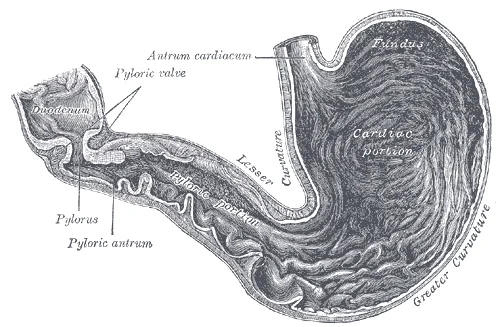
Figure 8Gray1050
Long description
Interior of the stomach. Labelled illustration from Wikimedia Commons, used under its open licence.

Figure 8: Interior of the stomach.
The lymphatic drainage of the stomach is extensive and follows the arterial supply, creating distinct pathways that determine the spread of malignancy. Understanding these routes is essential for surgical planning and staging gastric cancer. The stomach’s lymphatics are grouped into nodes that correspond to specific vascular territories.
Figure 9Gray1051
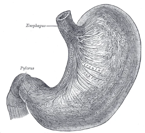
Long description
The longitudinal and circular muscular fibers of the stomach, viewed from above and in front. (Spalteholz.)

Figure 9: The longitudinal and circular muscular fibers of the stomach, viewed from above and in front (Spalteholz.).
DefinitionPerigastric Nodes
Lymph nodes located directly along the greater and lesser curvatures of the stomach, receiving initial drainage from the gastric wall.
The lymphatic vessels from the stomach wall converge into these regional nodes. The left gastric nodes drain the upper part of the lesser curvature and the cardia. These nodes then feed into the celiac nodes. The right gastric nodes drain the lower part of the lesser curvature and the pyloric region. They also drain into the celiac nodes via the hepatic nodes. The pyloric nodes, including suprapyloric and infrapyloric groups, drain the antrum and pylorus. These nodes drain into the pancreaticoduodenal nodes and subsequently the celiac nodes. The splenic nodes drain the fundus and the upper part of the greater curvature. They follow the short gastric and left gastroepiploic vessels. These nodes drain into the celiac nodes via the splenic artery nodes.
Exam trapDrainage Pattern
Do not assume all gastric lymph drains directly into the celiac nodes. Some pathways, particularly from the antrum, may involve the pancreaticoduodenal nodes first.
The celiac nodes are the final common collection point for most gastric lymph. They are located around the origin of the celiac trunk. From here, lymph enters the cisterna chyli and then the thoracic duct. The thoracic duct empties into the junction of the left internal jugular and left subclavian veins. This pathway explains why gastric cancer can metastasize to distant sites, including the supraclavicular nodes (Virchow’s node).
| Node Group | Location | Primary Drainage Area |
|---|---|---|
| Left Gastric | Lesser curvature, near cardia | Upper lesser curvature, cardia |
| Right Gastric | Lesser curvature, near pylorus | Lower lesser curvature |
| Pyloric | Around pylorus | Antrum, pylorus |
| Splenic | Along splenic artery | Fundus, upper greater curvature |
| Celiac | Root of celiac trunk | All above groups |
The clinical significance of this drainage pattern is profound. Surgical resection of gastric cancer must include removal of the relevant lymph node stations. For example, a distal gastrectomy requires dissection of the pyloric and right gastric nodes. A total gastrectomy necessitates removal of all perigastric and celiac nodes. Accurate staging depends on identifying metastatic involvement in these nodes.
The stomach receives a complex innervation that regulates motility, secretion, and sensation. This supply is divided into autonomic and somatic components, working in concert to coordinate gastric function and transmit pain signals.
DefinitionAutonomic Innervation
The autonomic nervous system provides both sympathetic and parasympathetic input to the stomach, regulating smooth muscle tone and glandular secretion.
The parasympathetic supply is predominantly vagal, originating from the dorsal motor nucleus of the vagus nerve. These efferent motor fibers travel via the anterior and posterior vagal trunks to reach the stomach. Sensory nerve fibers are the most frequent and carry afferent signals to the nuclear tractus solitarius (NTS). These fibers convey information regarding stretch and chemical composition. [4]
The efferent motor fibers of the vagus start from dorsal motor nucleus and regulate pyloric response by interacting with the enteric nervous system. Denervation of the pylorus led to a decrease in the compliance and loss of relaxation of pylorus, leading to a decrease in transpyloric outflow. Stimulation of vagal fibers showed consistent results with a decrease in pyloric resistance. The regulation of contractions is [4]
Sympathetic innervation arises from the splanchnic nerves, primarily the greater splanchnic nerve (T5–T9). These fibers travel with the arterial supply to the stomach, providing vasoconstrictor tone and carrying nociceptive (pain) signals. Somatic pain from the stomach is often referred to the epigastric region, corresponding to the T5–T9 dermatomes.
The nerve supply follows the arterial blood supply, which is derived from the celiac trunk. The left and right gastric arteries supply the lesser curvature, while the left and right gastroepiploic arteries supply the greater curvature. The short gastric arteries supply the fundus. Lymphatic drainage follows these vessels to the coeliac nodes.
Figure 10Gray1052
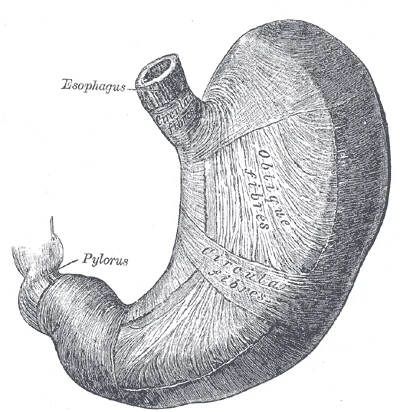
Long description
The oblique muscular fibers of the stomach, viewed from above and in front. (Spalteholz.)

Figure 10: The oblique muscular fibers of the stomach, viewed from above and in front (Spalteholz.).
The anterior trunk gives off the hepatic branch and the anterior gastric branches. The posterior trunk gives off the celiac branch and the posterior gastric branches. These nerves lie on the surface of the stomach, closely related to the lesser and greater curvatures. The hepatic branch runs towards the liver, while the celiac branch extends towards the coeliac plexus. The gastric branches innervate the body and antrum of the stomach. The pyloric region receives specific innervation from the posterior vagal trunk, which is critical for pyloric relaxation.
NoteClinical Pearl
Understanding the vagal innervation is crucial for procedures like G-POEM, where denervation can affect gastric emptying.
The stomach wall exhibits a complex histological architecture designed for secretion, storage, and mechanical processing of food. The mucosa, the innermost layer, is lined by simple columnar epithelium that secretes mucus to protect the underlying tissue from acidic gastric juice. Beneath the epithelium lies the lamina propria, a loose connective tissue containing blood vessels, lymphatics, and immune cells. The muscularis mucosae separates the mucosa from the submucosa.
The submucosa consists of dense irregular connective tissue and houses Meissner's plexus, which regulates glandular secretion and blood flow. This layer also contains larger blood vessels and lymphatic channels that supply the mucosa. The muscularis externa is distinctively thick and comprises three layers of smooth muscle: the inner oblique, the middle circular, and the outer longitudinal. The inner oblique layer is unique to the stomach and aids in the mechanical churning of food. The middle circular layer thickens at the pylorus to form the pyloric sphincter, a crucial valve controlling gastric emptying. The outer longitudinal layer is shorter than the other layers and helps shorten the stomach during contraction.
It provides a smooth, lubricated surface to reduce friction with adjacent organs.
The pylorus plays a key role in the regulation of GE. Postprandially, there is a first brief period of emptying of liquid and small size particles. Then, constriction of the pylorus restricts GE as the gastric peristaltic contraction reaches the distal antrum. Food is then retropulsed to the antrum, where antral contractions will triturate it into smaller particles (2–4 mm). Then, pyloric relaxation in coordination with antro‐pyloro‐duodenal motor activity leads to GE of the food. [4]
DefinitionPyloric Sphincter
A thickening of the circular muscle layer at the distal stomach that regulates the passage of chyme into the duodenum.
| Layer | Orientation | Function |
|---|---|---|
| Inner Oblique | Diagonal | Mechanical churning and mixing |
| Middle Circular | Circular | Forms pyloric sphincter; regulates emptying |
| Outer Longitudinal | Longitudinal | Shortens stomach during contraction |
The histological features of the stomach are adapted to its dual role in chemical digestion and mechanical breakdown. The presence of the inner oblique muscle layer allows for the vigorous mixing required to reduce food particles to the appropriate size for further digestion. The pyloric sphincter acts as a gatekeeper, ensuring that only appropriately processed chyme enters the small intestine.
Clinical pearlClinical Correlation
Understanding the histological layers is essential for interpreting gastric motility disorders and the effects of surgical interventions on gastric emptying.
The mucosal surface contains gastric pits that lead to gastric glands. These glands contain various cell types, including parietal cells that secrete hydrochloric acid and intrinsic factor, chief cells that secrete pepsinogen, and mucous neck cells that secrete mucus. The coordinated activity of these cells ensures the optimal environment for digestion while protecting the stomach wall from autodigestion. [1]
Figure 112415 Histology of StomachN
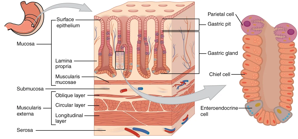
Long description
The stomach wall is adapted for the functions of the stomach. In the epithelium, gastric pits lead to gastric glands that secrete gastric juice. The gastric glands (one gland is shown enlarged on the right) contain different types of cells that secrete a variety of enzymes, including hydrochloride a

Figure 11: The stomach wall is adapted for the functions of the stomach.
The submucosa and muscularis externa work in concert to facilitate the movement of food. Peristaltic waves originate in the body of the stomach and move towards the pylorus, pushing food towards the antrum. The pyloric sphincter opens briefly to allow small particles to pass into the duodenum, while larger particles are retropulsed back into the antrum for further processing. This mechanism ensures that only finely ground chyme enters the small intestine, preventing obstruction and allowing for efficient nutrient absorption.
The stomach is a J-shaped muscular organ located in the upper left quadrant of the abdomen, situated between the oesophagus and the duodenum. Its development begins during the fourth week of embryogenesis as a dilation of the foregut. This initial structure undergoes differential growth, with the dorsal wall expanding more rapidly than the ventral wall to form the greater and lesser curvatures. The dorsal mesogastrium elongates to form the greater omentum, while the ventral mesogastrium contributes to the lesser omentum and falciform ligament. Rotation of the stomach occurs around its longitudinal and anteroposterior axes, positioning the pylorus to the right and the cardia to the left. This rotational movement also determines the final anatomical relations of the organ.
The mucosal layer develops from the endodermal lining of the foregut, giving rise to the gastric glands responsible for secretion. The muscularis externa differentiates into three layers: the inner oblique layer, the middle circular layer, and the outer longitudinal layer. The oblique layer is unique to the stomach and facilitates the churning of food. The serosa, derived from the peritoneum, covers the anterior and posterior surfaces, except at the bare areas where the stomach attaches to surrounding structures.
The blood supply arises from the celiac trunk, a major branch of the abdominal aorta. The left gastric artery supplies the lesser curvature, while the right gastric artery, often a branch of the proper hepatic artery, also contributes to this region. The greater curvature is supplied by the left and right gastroepiploic arteries. Venous drainage follows the arterial pattern, with blood flowing into the portal venous system. Lymphatic drainage is extensive, with nodes along the curvatures and near the pylorus draining into the coeliac nodes.
Nerve supply is provided by the vagus nerves and the sympathetic trunks. The anterior and posterior vagal trunks, derived from the left and right vagus nerves respectively, innervate the stomach and regulate secretion and motility. Sympathetic fibres arise from the greater splanchnic nerves and modulate blood flow and inhibit motility.
The stomach is a J-shaped muscular organ situated in the left upper quadrant, extending from the oesophagogastric junction to the duodenal bulb. Its anatomical divisions include the cardia, fundus, body, antrum, and pylorus. The pylorus plays a key role in the control of gastric content outflow. Impairment of pyloric physiology is a key feature in gastroparesis, particularly when associated with diabetes mellitus or opioid intake. [4]
DefinitionPyloric Function
The pylorus regulates gastric outflow, and its dysfunction is central to conditions like gastroparesis.
The fundus is located superiorly, containing the gas bubble. The body extends inferiorly, leading to the antrum. In cases of hiatus hernia, the gastro-oesophageal junction may be displaced above the diaphragm. Peptic ulcers may appear as niche defects in the mucosal contour, commonly on the lesser curvature or duodenal bulb.
| Structure | Relation |
|---|---|
| Liver | Anterior and right |
| Spleen | Left and posterior |
| Pancreas | Posterior |
| Diaphragm | Superior |
Endoscopic anatomy provides a direct view of the mucosal surface. The cardia surrounds the oesophageal opening. The fundus is the dome-shaped upper region. The body is the main central portion. The antrum is the distal narrowing before the pylorus. The pylorus itself is a muscular sphincter.
NoteEndoscopic Assessment
FLIP is a new tool to identify pyloric dysfunction by measuring impedance planimetry.
The pyloric ring is a distinct anatomical landmark. It appears as a white, circular structure during endoscopy. The mucosa around the pylorus is smooth. The antral mucosa may show folds. The pylorus opens and closes rhythmically. In gastroparesis, the pylorus may fail to relax adequately. This leads to gastric retention. Radiological studies, such as barium meals, can demonstrate delayed emptying. The contrast material remains in the stomach for an extended period. The pylorus appears tight and unyielding. Endoscopic therapy, such as pyloric dilation or myotomy, can relieve this obstruction.
The stomach is a J-shaped muscular organ situated in the left upper quadrant, lying between the oesophagus and the duodenum. Its position is defined by its relations to the diaphragm superiorly, the liver anteriorly, and the pancreas posteriorly. Understanding the anatomical boundaries is crucial for interpreting imaging and surgical approaches, particularly when addressing pathologies such as hiatus hernia and peptic ulcers.
The cardia is located at the superior aspect where the oesophagus enters. The fundus lies superior and posterior to the cardia, often containing gas bubbles on imaging. The body constitutes the main central portion. The antrum is the distal narrowing before the pyloric sphincter. Recognising these landmarks allows for precise localisation of lesions.
DefinitionHiatus Hernia
A condition where part of the stomach protrudes through the oesophageal hiatus of the diaphragm into the thoracic cavity.
Hiatus hernias involve the failure of the gastro-oesophageal junction to remain below the diaphragm. This anatomical defect compromises the anti-reflux barrier. The diaphragmatic crura normally provide a sling effect that supports the lower oesophageal sphincter. When this support is lost, gastric acid can reflux into the oesophagus.
Exam trapPeptic Ulcer Location
Duodenal ulcers are more common than gastric ulcers and typically occur in the first part of the duodenum, not the stomach body.
Peptic ulcers represent mucosal breaks extending into the submucosa. Gastric ulcers often occur on the lesser curvature, an area with relatively poor blood supply compared to the greater curvature. Duodenal ulcers frequently arise in the duodenal bulb. The anatomical proximity of these lesions to major vessels, such as the gastroduodenal artery, poses a risk of significant haemorrhage.
| Structure | Relation to Stomach | Clinical Relevance |
|---|---|---|
| Liver | Anterior and right | Limits spread of inflammation |
| Spleen | Left lateral | Risk during splenic flexure surgery |
| Pancreas | Posterior | Direct contact; source of pain |
| Diaphragm | Superior | Hiatus hernia site |
The blood supply derives from the coeliac trunk. The left gastric artery supplies the lesser curvature, while the right gastric artery reinforces this. The short gastric arteries supply the fundus. Venous drainage follows the arterial pattern into the portal system. Lymphatic drainage is extensive, following the arteries to the coeliac nodes. This anatomical knowledge underpins the management of gastric pathologies.
Gastroparesis is defined as delayed gastric emptying in the absence of mechanical obstruction. The most common causes are diabetes, post-surgical states, or drug-induced effects, particularly by opiates. However, idiopathic gastroparesis remains the most common diagnosis overall. Pyloric dysfunction is a key mechanism, often manifesting as pylorospasm in diabetic patients.
Clinical pearlIdiopathic Prevalence
Idiopathic gastroparesis is the most common diagnosis, despite diabetes and post-surgical causes being frequent aetiologies.
Botulinum toxin injection into the pylorus has been widely evaluated. Symptoms improved include vomiting, nausea, postprandial fullness, and bloating. Most studies noted an acceleration of gastric emptying.
The European and American recommendations do not support the routine use of botulinum toxin. Trials included limited numbers, such as 23 and 32 patients respectively. Recent data suggests P-DI before injection may predict outcomes. Patients with altered P-DI had greater symptom improvement and better gastric emptying.
Satiety, bloating, and nausea improved by a mean reduction of 1.3 points. This was not confirmed in long-term series. Healthcare use reduced, with antiemetic medication reduction in 50% to 70% of responders.
NoteHealthcare Impact
G-POEM reduces hospitalizations and emergency visits, suggesting cost-effectiveness and improved nutritional status via BMI increase.
This leads to an increased post-procedure P-DI index. The improvement is sustained at 3, 6, and 12 months. There was no difference in the adverse event rate between the two techniques.
Symptomatic improvement was confirmed in 27 patients after 17 months.
Clinical pearlGES Augmentation
G-POEM after prior GES therapy is feasible, safe, and leads to clinical response in all patients in one prospective study.
These must address the superiority of performing both techniques over one or the other alone.
Students frequently confuse the anatomical structure of the stomach with its physiological role in digestion, particularly regarding microbial interactions. A common error is assuming that the stomach’s acidity is primarily designed to digest plant cellulose. In reality, the stomach does not digest cellulose; herbivores rely on microbial processes in fermentation chambers such as the rumen or caecum to digest refractory plant material like cellulose and lignin. The stomach’s acidic environment s [1][2]
A significant anatomical and physiological pitfall concerns the pylorus in gastroparesis. Students often attribute pyloric dysfunction solely to neural issues. However, the pylorus in gastroparesis is characterized by a strong reduction in the number of interstitial cells of Cajal (ICCs). This reduction in ICCs leads to a slow-wave free gap, meaning there is no coordination of motor activities in the absence of gastric emptying. After the opening of the pylorus, antral activity propagates aborally and coordinates with duodenal activity. Furthermore, students may misinterpret the mechanism of gastric emptying. It is not merely a peristaltic pump; antral contraction coordinated with pyloric contractions leads to food trituration, and the contribution of a “pressure pump” rather than a “peristaltic pump” controlled by pylorus opening controls gastric emptying.
Another frequent mistake is misinterpreting pyloric distensibility index (P-DI) values. Students might assume a single universal normal value exists. However, normal and pathological values for P-DI still need to be confirmed. For instance, fasting P-DI in non-anesthetized volunteers was 25.2 ± 2.4 mm2/mmHg at 40 ml of bag inflation. In contrast, a study in anesthetized healthy subjects reported a P-DI of 8.4 ± 4.7 mm2/mmHg. The French study set the lower 90th percentile at 10 mm2/mmHg at 40 ml of inflation. Students must recognize that anesthesia and ethnicity may influence these measurements.
Finally, students often confuse the indications for surgical interventions. A common error is assuming Gastric Electrical Stimulation (GES) and G-POEM are interchangeable for all symptoms. However, it has been suggested that GES is performed preferentially in patients with symptoms of nausea and vomiting. In contrast, G-POEM can be performed in patients with other symptoms related to delayed gastric emptying, such as early satiety or postprandial fullness. Whether one is more effective than the other for refractory gastroparesis associated with nausea and/or vomiting remains uninvestigated to date. Additionally, while G-POEM leads to an acceleration of gastric emptying with an acceptable safety profile, further studies are needed to confirm its place.
Knowledge of gastric anatomy and physiology is applied at the bedside to understand the pathophysiology of gastroparesis. The pylorus plays a central role in this condition. Clinicians use the understanding that a reduction in interstitial cells of Cajal (ICCs) leads to uncoordinated motor activities. This insight guides the interpretation of symptoms and the selection of interventions. For example, the measurement of pyloric distensibility index (P-DI) is used as an exploratory tool in the pathophysiology of gastroparesis. The pyloric distensibility index is calculated in the zone with the narrowest cross-sectional area with the corresponding intra-bag pressure. Although normal values vary, a decreased P-DI is associated with gastroparesis symptoms. In the French study, a lower 90th percentile of 10 mm2/mmHg at 40 ml of inflation was used to define decreased P-DI. This threshold helps identify patients with diabetic or idiopathic gastroparesis. [4]
In the operating theatre, this anatomical knowledge informs surgical techniques such as G-POEM. The procedure aims to address pyloric dysfunction by opening the pylorus, allowing antral activity to propagate aborally and coordinate with duodenal activity. This intervention leads to an acceleration of gastric emptying. Surgeons must be aware that the pylorus contributes to a “pressure pump” mechanism rather than just a “peristaltic pump”. Understanding this distinction is vital for post-operative assessment of gastric emptying. Furthermore, the choice between GES and G-POEM depends on the predominant symptoms. GES is preferred for nausea and vomiting, while G-POEM is considered for early satiety or postprandial fullness.
On imaging and endoscopic evaluation, the assessment of gastric emptying and pyloric function is critical. The EndoFlip tool is used to measure cross-sectional area, bag pressure, and distensibility index. This tool provides data on pyloric distensibility, which correlates with gastric emptying and symptoms. However, it is important to note that decreased P-DI has also been observed in patients with chronic nausea and vomiting without delayed gastric emptying. This suggests that pyloric dysfunction can explain “gastroparesis-like” symptoms in some patients. Therefore, imaging and functional tests must be interpreted in the context of the entire clinical picture.
In examinations, students are tested on the specific anatomical and physiological details of the stomach and pylorus. Questions may focus on the role of interstitial cells of Cajal in coordinating motor activities. They may also ask about the differences in P-DI values between anesthetized and non-anesthetized subjects. Understanding that normal values are still being confirmed is a key point. Additionally, students must know the specific indications for GES versus G-POEM. The role of the gastric microbiota in gastric cancer is also a topic, with emphasis on the genetic evidence of bacteria like Lactobacillus and Lachnospiraceae. However, students must remember that viability has not been assessed in these studies. This knowledge is essential for understanding the translational opportunities to reduce gastric cancer morbidity and mortality.
- Herbivores rely on microbial fermentation in the rumen or caecum to digest cellulose, not the stomach itself.
- The stomach’s acidity reduces pathogen risk, but herbivores maintain modest acidity due to low pathogen threat on leaves. [2]
- Genetic evidence of bacteria in gastric cancer does not confirm bacterial viability. [3]
- Gastroparesis pyloric dysfunction involves a reduction in interstitial cells of Cajal (ICCs). [4]
- Lack of ICCs causes a slow-wave free gap, leading to uncoordinated motor activities. [4]
- Gastric emptying is controlled by a “pressure pump” coordinated by pylorus opening, not just peristalsis. [4]
- Pyloric distensibility index (P-DI) values vary; a threshold of 10 mm2/mmHg was used in one study. [4]
- GES is preferred for nausea/vomiting, while G-POEM is used for early satiety or postprandial fullness. [4]
Answer each one in your head first, then open the answer.
What structure is reduced in the pylorus of gastroparesis patients?
Show answerHide answer
Interstitial cells of Cajal (ICCs)What is the mechanism of gastric emptying controlled by pylorus opening?
Show answerHide answer
Pressure pumpWhich symptom profile is GES preferred for?
Show answerHide answer
Nausea and vomitingWhat do herbivores rely on to digest cellulose?
Show answerHide answer
Microbial processes in fermentation chambers
- Betts JG, Young KA, Wise JA, et al. The Stomach. OpenStax, Anatomy and Physiology 2e (CC BY-NC-SA 4.0). Accessed .
- Beasley DE, Koltz AM, Lambert JE, et al. The Evolution of Stomach Acidity and Its Relevance to the Human Microbiome. PloS one 2015;10(7):e0134116. doi:10.1371/journal.pone.0134116. Accessed .
- Noto JM, Peek RM. The gastric microbiome, its interaction with Helicobacter pylori, and its potential role in the progression to stomach cancer. PLoS pathogens 2017;13(10):e1006573. doi:10.1371/journal.ppat.1006573. Accessed .
- Soliman H, Gourcerol G. Targeting the pylorus in gastroparesis: From physiology to endoscopic pyloromyotomy. Neurogastroenterology and motility 2023;35(2):e14529. doi:10.1111/nmo.14529. Accessed .
Figure credits
- Figure 1: Henry Vandyke Carter, via Wikimedia Commons, Public domain, Public domain.
- Figure 2: Henry Vandyke Carter, via Wikimedia Commons, Public domain, Public domain.
- Figure 3: Henry Vandyke Carter, via Wikimedia Commons, Public domain, Public domain.
- Figure 4: Henry Vandyke Carter, via Wikimedia Commons, Public domain, Public domain.
- Figure 5: Henry Vandyke Carter, via Wikimedia Commons, Public domain, Public domain.
- Figure 6: Henry Gray (1825–1861), via Wikimedia Commons, Public domain, Public domain.
- Figure 7: Henry Vandyke Carter, via Wikimedia Commons, Public domain, Public domain.
- Figure 8: Henry Vandyke Carter, via Wikimedia Commons, Public domain, Public domain.
- Figure 9: Henry Vandyke Carter, via Wikimedia Commons, Public domain, Public domain.
- Figure 10: Henry Vandyke Carter, via Wikimedia Commons, Public domain, Public domain.
- Figure 11: OpenStax College, via Wikimedia Commons, CC BY 3.0, CC BY 3.0.
Educational content for medical students and doctors. It is not a substitute for clinical judgement or local guidelines.