Greater and lesser omentum
The greater and lesser omenta are double layers of peritoneum connecting the stomach to adjacent viscera.
The omenta are double folds of peritoneum that serve as anatomical bridges between the stomach and other abdominal structures. They are classified into two distinct parts based on their attachment to the stomach’s curvatures. Understanding their location requires first identifying the stomach’s borders.
The stomach has two primary curvatures. The greater curvature forms the long, convex lateral border of the stomach. The lesser curvature forms the shorter, concave medial border. These curvatures provide the attachment points for the omenta. [3]
The lesser omentum connects the liver to the lesser curvature of the stomach. It is a relatively thin sheet of peritoneum. Anatomically, it extends from the porta hepatis of the liver to the lesser curvature. This structure helps hold the stomach in place within the abdominal cavity. It separates the lesser sac from the greater sac of the peritoneal cavity. [3]
This creates a four-layered apron-like structure. It covers the intestines like an apron, providing protection and insulation. [3]
Surface marking of the greater omentum is significant for clinical examination. When the patient is supine, the greater omentum lies anterior to the small intestines. In thin individuals, the lower edge may be palpable just above the pubic symphysis.
The anatomical relations of the greater omentum are crucial. It lies anterior to the transverse colon and the loops of the small intestine. The greater omentum contains adipose tissue, which provides cushioning. It also contains lymphatic vessels that drain into the gastroepiploic nodes.
The lesser omentum contains important vascular structures. The hepatic artery, portal vein, and bile duct run within its free right edge, known as the hepatoduodenal ligament. This triad is enclosed in the lesser omentum. The left edge of the lesser omentum attaches to the stomach.
Clinical relevance arises when these structures become inflamed. Appendicitis can cause the greater omentum to migrate to the right iliac fossa to wall off infection. This is a protective physiological response. The mobility of the greater omentum allows it to adhere to sites of inflammation.
The embryological development of the omenta involves the dorsal and ventral mesogastrium. The dorsal mesogastrium forms the greater omentum. The ventral mesogastrium forms the lesser omentum. This developmental origin explains their peritoneal nature.
DefinitionOmenta
Double layers of peritoneum connecting the stomach to adjacent organs, providing support and protection.
| Feature | Greater Omentum | Lesser Omentum |
|---|---|---|
| Attachment | Greater curvature to transverse colon | Lesser curvature to liver |
| Layers | Four layers (folded) | Two layers |
| Content | Adipose tissue, lymphatics | Hepatic artery, portal vein, bile duct |
| Function | Protection, insulation | Vascular conduit, support |
These membranous aprons play critical roles in immune defence, fat storage, and limiting the spread of intra-abdominal infection. Understanding their anatomy requires visualising the abdominal cavity in the supine position, where gravity causes these structures to hang inferiorly from the stomach.
The greater and lesser omenta are double layers of peritoneum that connect the stomach to adjacent structures. They are not muscular organs but rather fatty aprons rich in blood vessels, lymphatics, and nerves. Understanding their histology requires looking at the simple squamous mesothelium that lines their surfaces. This mesothelium rests on a thin layer of connective tissue containing adipose cells. The lesser omentum connects the lesser curvature of the stomach to the liver [3]
The lesser omentum consists of two distinct ligaments. The hepatogastric ligament connects the liver to the lesser curvature of the stomach. The hepatoduodenal ligament connects the liver to the duodenum. Within the free right edge of the hepatoduodenal ligament lies the portal triad, containing the hepatic artery proper, the common bile duct, and the portal vein. This structure is clinically significant as it forms the anterior boundary of the epiploic foramen (of Winslow).
The greater omentum is the largest peritoneal fold. It hangs down from the greater curvature of the stomach, covering the transverse colon and small intestines. Its inferior border is often fused with the transverse colon. The greater omentum contains abundant adipose tissue and lymphoid aggregates, earning it the nickname "abdominal policeman" due to its ability to migrate to sites of inflammation and wall off infection.
DefinitionOmental Bursa
A potential space behind the stomach and lesser omentum, accessible via the epiploic foramen.
The vascular supply to the omenta derives from the gastric arteries. The left and right gastric arteries supply the lesser curvature, while the left and right gastroepiploic arteries supply the greater curvature. Venous drainage follows the arterial supply, ultimately reaching the portal system. Lymphatic drainage from the greater omentum flows to the pyloric and pancreaticosplenic nodes. [1]
| Ligament | Attachment 1 | Attachment 2 | Contents |
|---|---|---|---|
| Hepatogastric | Liver (porta hepatis) | Stomach (lesser curvature) | None major |
| Hepatoduodenal | Liver (porta hepatis) | Duodenum (first part) | Portal triad |
Clinical relevance arises when the greater omentum becomes inflamed, as in appendicitis, where it may migrate to the right iliac fossa to contain the infection. Surgical access to the lesser sac is achieved by dividing the gastrocolic ligament, a part of the greater omentum.
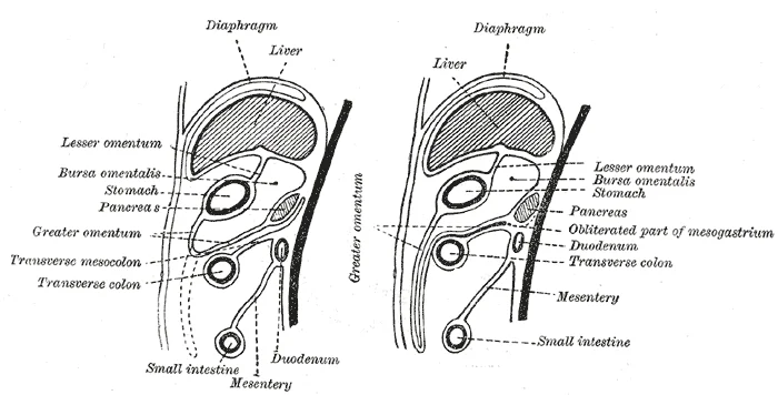
Figure 1Gray990
Long description
Diagrams to illustrate the development of the greater omentum and transverse mesocolon.

Figure 1: Diagrams to illustrate the development of the greater omentum and transverse mesocolon.
The lesser omentum is attached to the lesser curvature of the stomach. It extends superiorly to attach to the porta hepatis of the liver.
The greater and lesser omentum are double-layered folds of peritoneum that suspend the stomach and provide a conduit for vascular and lymphatic structures. They are not solid organs but membranous sheets that create potential spaces and compartments within the abdominal cavity. Understanding their internal architecture requires examining the layers of peritoneum, the vessels they enclose, and the fat distribution within them.
The greater omentum is a large peritoneal fold, hanging from the greater curvature of the stomach. It descends anterior to the intestines like an apron, then folds back upon itself to ascend and attach to the transverse colon. This complex folding creates a multi-layered structure in its distal portion.
DefinitionGreater Omentum
A large, apron-like fold of peritoneum extending from the greater curvature of the stomach to the transverse colon, containing fat and immune cells.
Internally, the greater omentum is rich in adipose tissue, particularly in adults. This fat serves as an energy reserve and provides thermal insulation. Embedded within this fatty matrix are the gastroepiploic vessels. The right and left gastroepiploic arteries run along the greater curvature, anastomosing to supply the stomach wall. These vessels are accompanied by corresponding veins and lymphatic channels. The lymphatics drain into the pyloric and pancreaticosplenic nodes, which are critical in the spread of gastric malignancies.
| Structure | Origin | Course | Termination |
|---|---|---|---|
| Right Gastroepiploic Artery | Gastroduodenal Artery | Runs rightward along greater curvature | Anastomoses with Left Gastroepiploic |
| Left Gastroepiploic Artery | Splenic Artery | Runs leftward along greater curvature | Anastomoses with Right Gastroepiploic |
The lesser omentum connects the lesser curvature of the stomach and the proximal duodenum to the liver. It is divided into two parts: the hepatogastric ligament and the hepatoduodenal ligament. The hepatogastric ligament is thinner and contains the gastric vessels. The hepatoduodenal ligament is thicker and forms the free edge of the lesser omentum. This edge defines the anterior boundary of the epiploic foramen (of Winslow). [2]
DefinitionLesser Omentum
A double layer of peritoneum extending from the liver to the stomach and duodenum, containing the portal triad.
The hepatoduodenal ligament contains the portal triad: the common bile duct, the proper hepatic artery, and the portal vein. These structures are arranged with the bile duct on the right, the hepatic artery on the left, and the portal vein posterior to both. This arrangement is constant and crucial for surgical identification. The lymphatics within the lesser omentum drain into the hepatic nodes, reflecting the liver's role in filtering abdominal contents.
The fat within the omentum appears as yellowish tissue. The common bile duct is on the right, the hepatic artery on the left, and the portal vein posteriorly.
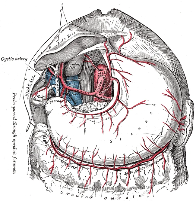
Figure 2Gray532
Long description
The celiac artery and its branches; the liver has been raised, and the lesser omentum and anterior layer of the greater omentum removed.

Figure 2: The celiac artery and its branches; the liver has been raised, and the lesser omentum and anterior layer of the greater omentum removed.
The arterial supply to the greater and lesser omentum is derived from the coeliac trunk, which is the first major anterior branch of the abdominal aorta. This supply is organised into two distinct systems that correspond to the two parts of the omentum. The lesser omentum receives its blood supply primarily from the left and right gastric arteries. The left gastric artery arises directly from the coeliac trunk and runs upwards and to the left towards the cardia of the stomach. It then descends along the lesser curvature of the stomach, anastomosing with the right gastric artery. The right gastric artery usually arises from the proper hepatic artery and runs to the right along the lesser curvature. These vessels lie within the two layers of the lesser omentum, specifically in the hepatogastric ligament. They provide branches to the anterior and posterior surfaces of the stomach and form an arterial arcade along the lesser curvature. This arcade ensures a rich collateral circulation, which is clinically significant in cases of gastric resection or ulcer disease.
The greater omentum hangs inferiorly, while the lesser omentum connects superiorly to the liver. [1]
The greater omentum is supplied by the left and right gastroepiploic arteries. It then enters the greater omentum. The right gastroepiploic artery arises from the gastroduodenal artery, which is a branch of the common hepatic artery. It runs along the greater curvature from right to left. These two arteries anastomose extensively along the greater curvature, forming a continuous arterial arcade. This arcade gives off numerous branches that descend into the greater omentum. These branches supply the four-layered structure of the greater omentum. The greater omentum is often described as the 'abdominal policeman' due to its role in containing infection. Its rich blood supply allows it to migrate and adhere to inflamed areas. The anastomosis between the left and right gastroepiploic arteries is robust, providing redundancy. This is important in surgical procedures involving the stomach. Ligation of one vessel does not necessarily compromise the blood supply to the omentum. The vessels within the greater omentum are relatively large and easily identifiable. They run between the two anterior layers of the greater omentum before penetrating deeper. These supply the fundus of the stomach and the upper part of the greater curvature. The lesser omentum is thinner and contains fewer large vessels. It primarily serves as a conduit for the hepatic artery, portal vein, and bile duct in the hepatoduodenal ligament. The arterial supply is crucial for the metabolic functions of the omentum. It also plays a role in fat storage and immune response. Understanding this supply is vital for surgical interventions such as omentectomy or gastric bypass. The anatomical variations in the origin of the right gastric artery are common. It may arise from the proper hepatic artery or the common hepatic artery. These variations must be recognised during surgery to avoid inadvertent injury. The left gastric artery is the most constant branch of the coeliac trunk. It is often used as a landmark in abdominal surgery. The gastroepiploic arteries are important in flap surgery for reconstruction. This highlights the clinical importance of knowing the arterial anatomy. The anastomotic connections provide a safety net against ischaemia. This is particularly relevant in patients with atherosclerosis. The omentum's vascularity supports its high metabolic activity. It is involved in the production of adipokines and cytokines. The blood supply ensures the delivery of these mediators to the systemic circulation. The lymphatic drainage follows the arterial supply. The nodes along the gastric arteries are important in cancer staging. Knowledge of the arterial supply aids in understanding the spread of disease. The greater omentum's mobility is facilitated by its loose connective tissue and vascular network. This allows it to wrap around inflamed organs. The lesser omentum is more fixed due to its attachment to the liver. The arterial supply reflects these functional differences. The greater omentum has a more extensive network to support its dynamic role. The lesser omentum has a more direct supply to support its structural role. The coeliac trunk is a critical junction in the abdominal circulation. Occlusion of this trunk is a life-threatening emergency. Collateral circulation via the pancreaticoduodenal arteries can sometimes compensate. This anastomosis between the coeliac and superior mesenteric arteries is vital. It involves the gastroduodenal and superior pancreaticoduodenal arteries. The greater omentum is not directly involved in this collateral pathway. However, its blood supply is dependent on the patency of the coeliac branches. The lesser omentum's blood supply is similarly dependent. Any compromise to the coeliac trunk affects both omenta. This underscores the importance of the coeliac axis in abdominal perfusion. The arterial anatomy is consistent across most individuals. Minor variations exist in the branching patterns. These variations are usually clinically insignificant. However, they can complicate surgical dissection. Surgeons must be aware of these possibilities. The arterial supply is a key feature of omental anatomy. It supports the physiological functions of the omentum. It also provides a route for pathological processes. Inflammation, tumours, and infarction all affect the blood vessels. Understanding the supply helps in diagnosis and treatment. The greater omentum's rich supply makes it resistant to ischaemia. This is due to its thinner structure and fewer collaterals. The arterial supply is a fundamental aspect of omental biology. It is essential for medical students to master this knowledge. [1]
The venous drainage of the omenta follows the principle that blood returns to the portal system via tributaries of the superior mesenteric and splenic veins. Understanding this pathway requires tracing the vessels from the capillary beds within the omental aprons back to their confluence.
The greater omentum is a large, fatty apron hanging from the greater curvature of the stomach. Its venous return is primarily managed by the gastroepiploic vessels.
The portal vein is formed by the union of the splenic and superior mesenteric veins.
The lesser omentum connects the lesser curvature of the stomach and the duodenum to the liver. Its venous drainage is more complex due to its proximity to the liver and the porta hepatis.
The left gastric vein drains the lesser curvature of the stomach and the upper part of the lesser omentum. It runs along the lesser curvature, accompanying the left gastric artery. [1]
The right gastric vein drains the lesser curvature of the stomach. The right gastric vein is smaller than the left gastric vein. [1]
In the hepatoduodenal ligament, the portal vein lies posterior to the hepatic artery and the common bile duct. This arrangement is known as the portal triad. [1]
The lymphatic drainage of the omentum is a critical pathway for immune surveillance and potential metastatic spread, particularly in gastrointestinal malignancies. Understanding the distinct routes for the greater and lesser omentum requires an appreciation of their embryological origins and anatomical relationships.
The greater omentum hangs from the greater curvature of the stomach and the proximal duodenum, draping over the intestines like an apron. Its lymphatic vessels follow the gastroepiploic vessels. [1]
DefinitionGastroepiploic Nodes
Lymph nodes located along the greater curvature of the stomach, draining the greater omentum and the anterior wall of the stomach.
The terminal drainage of the greater omentum is into the celiac lymph nodes. These nodes surround the celiac trunk and its branches. From here, lymph enters the intestinal trunk, which then joins the thoracic duct. This pathway is significant because gastric cancers often metastasise to these nodes before spreading distantly.
The lesser omentum connects the lesser curvature of the stomach and the proximal duodenum to the liver. It consists of two parts: the hepatogastric ligament and the hepatoduodenal ligament. The lymphatic drainage of the lesser omentum is more complex due to its proximity to the liver and the porta hepatis.
The greater and lesser omenta are double layers of peritoneum that connect the stomach to adjacent structures. They are not muscular organs but rather fatty aprons rich in blood vessels, lymphatics, and nerves. [1]
Exam trapPortal Hypertension
In portal hypertension, lymphatic drainage from the gut can be compromised, leading to ascites. This is not due to increased production but rather increased pressure in the hepatic sinusoids and lymphatic overflow.
The hepatic nodes receive lymph from the liver, gallbladder, and the lesser omentum. They ultimately drain into the celiac nodes. This convergence means that pathology in the lesser omentum or the liver can affect the same nodal group. Clinically, palpable Virchow’s node (left supraclavicular node) may indicate metastasis from abdominal malignancies, including those involving the omentum, via the thoracic duct.
| Structure | Primary Nodes | Terminal Drainage |
|---|---|---|
| Greater Omentum | Pyloric, Splenic | Celiac Nodes |
| Lesser Omentum | Hepatic | Celiac Nodes |
The autonomic innervation of the greater and lesser omentum is derived from the coeliac plexus, which provides both sympathetic and parasympathetic fibres. Sympathetic fibres originate from the greater splanchnic nerves, arising from the thoracic spinal segments. These fibres travel to the coeliac ganglia, where they synapse. Postganglionic sympathetic fibres then follow the branches of the coeliac artery to reach the omentum. They primarily mediate vasoconstriction of the blood vessels within the omental tissue. Parasympathetic fibres are supplied by the anterior vagal trunk. These fibres tra [2]
Branches extend laterally to form the left and right coeliac ganglia.
Referred pain from the omentum is typically felt in the epigastric region. This occurs because the visceral afferent fibres travel with the sympathetic nerves back to the T5 to T9 spinal segments. The brain interprets these signals as originating from the somatic dermatomes corresponding to these segments, which include the epigastrium. This mechanism explains why inflammation of the omentum, such as in omental infarction, presents with central abdominal pain rather than localised tenderness in the fat itself. Understanding this pathway is essential for differential diagnosis in acute abdominal presentations.
Clinical pearlReferred Pain Mechanism
Visceral pain from the omentum refers to the epigastrium because afferent fibres join sympathetic pathways to T5-T9 spinal levels, where the brain misinterprets the signal as somatic dermatomal pain.
Link to the histology page.
The greater and lesser omenta are double layers of peritoneum that connect the stomach to adjacent structures. They are not muscular organs but rather fatty aprons rich in blood vessels, lymphatics, and nerves. Understanding their histology requires looking at the simple squamous mesothelium that lines their surfaces. This mesothelium rests on a thin layer of connective tissue containing adipose cells. The greater omentum is often described as an 'abdominal policeman' due to its ability to migra
DefinitionOmentum
A double layer of peritoneum connecting the stomach to other abdominal organs, rich in fat and vasculature.
The mesothelial cells lining the omenta secrete a small amount of fluid to reduce friction during movement. This serous fluid allows the omenta to glide smoothly against the intestines and abdominal wall. The connective tissue stroma supports the blood vessels that supply the stomach and liver. Lymphatic vessels are abundant, particularly in the greater omentum, facilitating immune surveillance. Macrophages and other immune cells reside within the connective tissue, ready to respond to infection. The greater omentum can adhere to inflamed areas, such as a perforated ulcer, to wall off the infection. This histological arrangement allows it to perform both mechanical and immunological functions. The lesser omentum, being thinner, is more focused on providing a conduit for neurovascular structures. Its connective tissue is denser to support the tension placed on the hepatoduodenal ligament.
| Structure | Location | Primary Content |
|---|---|---|
| Greater Omentum | Greater curvature of stomach | Fat, lymphatics, immune cells |
| Lesser Omentum | Lesser curvature of stomach | Hepatic artery, bile duct, portal vein |
The histological features reflect the functional demands of each omentum. The greater omentum's fat content allows for energy storage and cushioning. The lesser omentum's dense connective tissue supports the vascular structures it carries. Both omenta are lined by mesothelium, which is continuous with the peritoneal lining of the abdominal cavity. This continuity allows for the spread of disease or fluid throughout the peritoneal cavity. The vascular supply comes from the gastric arteries for the greater omentum and the hepatic artery for the lesser omentum. Lymphatic drainage follows the vascular supply to regional lymph nodes. The innervation is provided by the autonomic nervous system, regulating blood flow and immune response. Understanding these histological details is crucial for interpreting imaging and surgical findings.
The omenta arise from the dorsal mesogastrium, a double layer of peritoneum that suspends the developing stomach from the posterior abdominal wall. During the fourth week of gestation, the stomach begins to rotate, pulling the dorsal mesogastrium with it. This rotation creates the greater sac of the peritoneal cavity. The dorsal mesogastrium elongates significantly to form the greater omentum. It hangs down from the greater curvature of the stomach like an apron. Initially, this structure is thin and transparent. As development proceeds, the layers of the greater omentum fuse together. This fusion creates the characteristic four-layered structure seen in adults. The fusion process is not always complete. In some individuals, small gaps or foramina may persist. These foramina allow communication between the greater and lesser sacs. The lesser omentum forms from the ventral mesogastrium. This structure connects the lesser curvature of the stomach and the proximal duodenum to the liver. The ventral mesogastrium divides into two parts. The hepatogastric ligament connects the liver to the stomach. The hepatoduodenal ligament connects the liver to the duodenum. The hepatoduodenal ligament contains the portal triad. This triad consists of the hepatic artery proper, the common bile duct, and the portal vein. The formation of the omenta is crucial for the positioning of abdominal organs. The greater omentum provides a protective layer for the intestines. It contains fat and immune cells. These cells help fight infection within the abdominal cavity. The lesser omentum provides a pathway for vessels and nerves. It supports the liver and stomach. Understanding the embryological origin helps explain the anatomical relationships. The dorsal mesogastrium gives rise to the spleen as well. The splenic ligaments connect the spleen to the stomach and kidney. The ventral mesogastrium also gives rise to the falciform ligament. This ligament connects the liver to the anterior abdominal wall. The ligamentum teres hepatis is the remnant of the umbilical vein. It runs within the free edge of the falciform ligament. The peritoneal reflections create potential spaces. These spaces can accumulate fluid or pus. Infection can spread through these continuous peritoneal layers. The omenta are highly mobile. They can migrate to sites of inflammation. This movement is a protective response. The greater omentum is often called the 'policeman of the abdomen'. It wraps around infected areas. This action limits the spread of infection. The embryological development explains the vascular supply. The arteries supplying the omenta run within the peritoneal layers. Knowledge of this development aids in surgical procedures. Surgeons must respect these anatomical planes. Dissection along these planes reduces bleeding. The omenta are vital for abdominal homeostasis. They play a role in immunity. Macrophages within the omentum phagocytose debris. This function is essential for healing. The structural integrity of the omenta depends on proper fusion. Abnormal fusion can lead to congenital defects. These defects may cause internal hernias. Internal hernias can lead to bowel obstruction. Therefore, understanding development is clinically relevant. The omenta are not just passive fat pads. They are dynamic structures. They respond to physiological changes. Their position can shift with body habitus. Despite this mobility, their attachments remain constant. The greater curvature attachment is fixed. The lesser curvature attachment is also fixed. These fixed points define the omental boundaries. The variable part is the free edge. This edge can adhere to other organs. Adhesions can form after surgery. These adhesions are fibrous bands. They can cause pain or obstruction. The embryological origin dictates this potential for adhesion. The mesothelial cells allow for sliding and sticking. This duality is key to their function. The omenta are essential for life. They support digestion and immunity. Their development is a complex process. It involves precise genetic regulation. Disruption of this regulation causes anomalies. These anomalies are rare but serious. Medical students must understand this foundation. It underpins all clinical reasoning. The omenta are integral to abdominal anatomy. They connect major organs. They facilitate vascular and neural pathways. They provide immunological surveillance. They are dynamic and responsive. Their study is fundamental to surgery. Their development explains their adult form. Their adult form explains their function. This logical chain is vital for learning. Anatomy is not just memorisation. It is understanding structure and function. The omenta exemplify this principle. They are complex yet elegant. They are essential yet often overlooked. Their importance cannot be overstated. They are the guardians of the abdomen. Their development is the key to understanding them. [1][3] [2]
The greater omentum is a large, apron-like fold of peritoneum. [1]
DefinitionGreater Omentum
A four-layered peritoneal fold extending from the stomach to the transverse colon, rich in adipose tissue and immune cells.
On the axial slice, the greater omentum is situated anterior to the small bowel loops. It often contains visible fat stranding or fluid levels if pathology is present. The structure is highly mobile, which can make its exact boundaries variable between individuals. However, its attachment to the greater curvature of the stomach is consistent. The gastrocolic ligament forms the superior portion of this fold, connecting the stomach to the transverse colon. This ligament is a key landmark for surgeons identifying the plane of dissection.
The lesser omentum is a double layer of peritoneum that extends from the porta hepatis of the liver to the lesser curvature of the stomach and the duodenum. It forms the anterior wall of the omental bursa, also known as the lesser sac. Radiologically, it appears as a thin, linear structure separating the liver from the stomach. [2]
DefinitionLesser Omentum
A peritoneal fold connecting the liver to the stomach and duodenum, forming the anterior boundary of the lesser sac.
The lesser omentum consists of two parts: the hepatogastric ligament and the hepatoduodenal ligament. [1]
| Structure | Position |
|---|---|
| Portal Vein | Posterior and to the right |
| Common Bile Duct | Anterior and to the right |
| Hepatic Artery Proper | Anterior and to the left |
Understanding the radiological appearance of these structures is essential for diagnosing conditions such as cholangitis or portal hypertension. The omenta serve as important barriers to infection, often walled off by inflammatory exudate in cases of perforated peptic ulcers. This can lead to the formation of abscesses within the omental layers, visible as fluid collections on imaging.
The omenta are double layers of peritoneum that connect the stomach to other abdominal viscera. They serve as mobile anatomical bridges, providing pathways for vessels and nerves while offering immunological protection. Understanding their spatial relationships is critical for interpreting abdominal imaging and surgical approaches.
The greater omentum is a large, apron-like fold of peritoneum that hangs down from the greater curvature of the stomach. It extends anterior to the small intestines and folds back upon itself to attach to the transverse colon. This structure is often described as the "abdominal policeman" due to its ability to migrate towards sites of inflammation, such as a perforated appendix, to wall off infection. [1]
DefinitionGreater Omentum
A four-layered peritoneal fold extending from the greater curvature of the stomach to the transverse colon, rich in adipose tissue and lymphoid aggregates.
The greater omentum contains the gastroepiploic vessels. [1]
| Vessel | Origin | Termination |
|---|---|---|
| Right gastroepiploic artery | Gastroduodenal artery | Anastomoses with left gastroepiploic artery |
| Left gastroepiploic artery | Splenic artery | Anastomoses with right gastroepiploic artery |
Clinically, the greater omentum is frequently used in reconstructive surgery due to its robust blood supply and pliability. It can be mobilised as a pedicled flap to cover defects in the chest or abdomen. In cases of acute appendicitis, the omentum may become adherent to the inflamed appendix, limiting the spread of peritonitis.
The lesser omentum is a double layer of peritoneum. [1]
DefinitionLesser Omentum
A peritoneal fold connecting the lesser curvature of the stomach and the first part of the duodenum to the liver.
The hepatoduodenal ligament contains the portal triad, which consists of the common bile duct, the proper hepatic artery, and the portal vein. These structures are enclosed within the free edge of the lesser omentum. The portal triad is a crucial landmark in abdominal surgery, particularly during cholecystectomy, where it must be carefully identified and controlled. [1] [2][3]
| Structure | Type | Function |
|---|---|---|
| Common bile duct | Biliary | Transports bile to the duodenum |
| Proper hepatic artery | Arterial | Supplies oxygenated blood to the liver |
| Portal vein | Venous | Carries nutrient-rich blood to the liver |
The lesser omentum provides a conduit for neurovascular structures entering and exiting the liver. The hepatic plexus of nerves accompanies the vessels, providing autonomic innervation to the liver and biliary tree. Pathologies such as liver cirrhosis can alter the architecture of the lesser omentum, leading to the formation of collateral vessels and varices.
The greater omentum is a large, apron-like fold of peritoneum that hangs from the greater curvature of the stomach and the proximal duodenum. It extends anterior to the small intestines, often described as the "policeman of the abdomen" due to its role in localising infection. Anatomically, it consists of multiple layers of peritoneum fused together. The anterior layers descend from the greater curvature, while the posterior layers ascend to attach to the transverse colon. This complex folding creates a potential space that can accumulate fluid or inflammatory exudate. [1][2]
The lesser omentum is a sheet of peritoneum connecting the liver to the stomach and duodenum. It extends from the liver to the stomach and duodenum. It is divided into parts. The hepatoduodenal ligament contains structures such as the hepatic artery, bile duct, and portal vein. This structure is critical in surgical procedures involving the liver or biliary tree.
DefinitionOmental Foramen
The epiploic foramen is the opening connecting the greater and lesser sacs of the peritoneum, bounded anteriorly by the hepatoduodenal ligament. [2]
Common exam traps involve confusing the attachments of the greater and lesser omenta. The greater omentum attaches to the greater curvature. The lesser omentum attaches to the liver. Students often mistake the contents of the hepatoduodenal ligament; remember the mnemonic for the bile duct, hepatic artery, and portal vein. [2]
| Structure | Attachment | Key Contents |
|---|---|---|
| Greater Omentum | Greater curvature of stomach | Fatty apron, lymph nodes |
| Lesser Omentum | Liver to lesser curvature | Portal triad components |
| Hepatoduodenal Ligament | Porta hepatis to duodenum | Bile duct, hepatic artery, portal vein |
Clinical relevance includes the migration of the greater omentum to site of inflammation, such as in appendicitis. The lesser omentum's thinness makes it susceptible to tearing during rapid decompression of the stomach. Understanding these structures is vital for interpreting abdominal CT scans and planning surgical approaches.
Students frequently confuse the anatomical location and structural composition of various bones and cavities, leading to errors in clinical reasoning and imaging interpretation. A primary error involves misidentifying the components of the hip bone. The ilium is the fan-like, superior region that forms the largest part of the hip bone. It is firmly united to the sacrum at the largely immobile sacroiliac joint. The ischium forms the posteroinferior region of each hip bone. It supports the body when sitting. The pubis forms the anterior portion of the hip bone. The pubis curves medially, where it joins to the pubis of the opposite hip bone at a specialized joint called the pubic symphysis. Confusing these regions can lead to incorrect localisation of pelvic fractures or pain. [1]
Another significant pitfall relates to the nasal cavity and paranasal sinuses. Inside the nasal area of the skull, the nasal cavity is divided into halves by the nasal septum. The upper portion of the nasal septum is formed by the perpendicular plate of the ethmoid bone and the lower portion is the vomer bone. Each side of the nasal cavity is triangular in shape, with a broad inferior space that narrows superiorly. When looking into the nasal cavity from the front of the skull, two bony plates are seen projecting from each lateral wall. The larger of these is the inferior nasal concha, an independent bone of the skull. Located just above the inferior concha is the middle nasal concha, which is part of the ethmoid bone. A third bony plate, also part of the ethmoid bone, is the superior nasal concha. It is much smaller and out of sight, above the middle concha. The superior nasal concha is located just lateral to the perpendicular plate, in the upper nasal cavity. Students often overlook the superior concha due to its position, missing its role in airflow and drainage. [2]
Regarding the temporal bone, the temporal bone is subdivided into several regions. The flattened, upper portion is the squamous portion of the temporal bone. Below this area and projecting anteriorly is the zygomatic process of the temporal bone, which forms the posterior portion of the zygomatic arch. Posteriorly is the mastoid portion of the temporal bone. Projecting inferiorly from this region is a large prominence, the mastoid process, which serves as a muscle attachment site. The mastoid process can easily be felt on the side of the head just behind your earlobe. On the interior of the skull, the petrous portion of each temporal bone forms the prominent, diagonally oriented petrous ridge in the floor of the cranial cavity. Located inside each petrous ridge are small cavities that house the structures of the middle and inner ears. Mistaking the petrous ridge for a fracture line is a common radiological error. [2]
The posterior cranial fossa is the most posterior and deepest portion of the cranial cavity. It contains the cerebellum of the brain. The posterior fossa is bounded anteriorly by the petrous ridges, while the occipital bone forms the floor and posterior wall. It is divided at the midline by the large foramen magnum (“great aperture”), the opening that provides for passage of the spinal cord. Confusion between the fossa boundaries can lead to misinterpretation of posterior fossa masses. [2]
Exam trapSinus Drainage
The maxillary sinuses are most commonly involved during sinus infections. Because their connection to the nasal cavity is located high on their medial wall, they are difficult to drain. This anatomical fact explains why maxillary sinusitis often persists despite standard treatment. [2]
The paranasal sinuses are named for the skull bone that each occupies. The frontal sinus is located just above the eyebrows, within the frontal bone. This irregular space may be divided at the midline into bilateral spaces, or these may be fused into a single sinus space. The frontal sinus is the most anterior of the paranasal sinuses. The largest sinus is the maxillary sinus. These are paired and located within the right and left maxillary bones, where they occupy the area just below the orbits. The sphenoid sinus is a single, midline sinus. [2]
The hyoid bone is an independent bone that does not contact any other bone and thus is not part of the skull. It is a small U-shaped bone located in the upper neck near the level of the inferior mandible, with the tips of the “U” pointing posteriorly. The hyoid serves as the base for the tongue above, and is attached to the larynx below and the pharynx posteriorly. The hyoid is held in position by a series of small muscles that attach to it either from above or below. These muscles act to move t [2]
DefinitionGastric Mucosal Barrier
The stomach is protected from self-digestion by the mucosal barrier[1]. This barrier has several components[1]. First, the stomach wall is covered by a thick coating of bicarbonate-rich mucus[1]. This mucus forms a physical barrier, and its bicarbonate ions neutralize acid[1]. Second, the epithelial cells of the stomach's mucosa meet at tight junctions, which block gastric juice from penetrating the underlying tissue layers[1]. Finally, stem cells located where gastric glands join the gastric pits quickly replace damaged epithelial mucosal cells, when the epithelial cells are shed[1]. In fact, the surface epithelium of the stomach is completely replaced every 3 to 6 days[1]. [3]
There are four main regions in the stomach: the cardia, fundus, body, and pylorus[1]. The cardia (or cardiac region) is the point where the esophagus connects to the stomach and through which food passes into the stomach[1]. Located inferior to the diaphragm, above and to the left of the cardia, is the dome-shaped fundus[1]. Below the fundus is the body, the main part of the stomach[1]. The funnel-shaped pylorus connects the stomach to the duodenum [3] Within a few moments after food enters your stomach, mixing waves begin to occur at intervals of approximately 20 seconds[1]. A mixing wave is a unique type of peristalsis that mixes and softens the food with gastric juices to create chyme[1]. The initial mixing waves are relatively gentle, but these are followed by more intense waves, starting at the body of the stomach and increasing in force as they reach the pylorus[1]. It is fair to say that long before your sushi exits through the pyloric sphincter, it bears little resemblance to the sushi you ate[1]. [3]
Gastric emptying is regulated by both the stomach and the duodenum[1]. The presence of chyme in the duodenum activates receptors that inhibit gastric secretion[1]. This prevents additional chyme from being released by the stomach before the duodenum is ready to process it[1]. [3]
This knowledge is used at the bedside to assess swallowing difficulties, as the hyoid bone's movement is coordinated with the tongue, larynx, and pharynx during swallowing and speaking. In the operating theatre, understanding the sacroiliac joint stability is crucial for pelvic surgery, as the ilium is firmly united to the sacrum at the largely immobile sacroiliac joint. On imaging, recognising the petrous ridge helps distinguish normal anatomy from fractures, as the petrous portion of each temporal bone forms the prominent, diagonally oriented petrous ridge in the floor of the cranial cavity. In examinations, students must identify the superior nasal concha, which is much smaller and out of sight, above the middle concha. [2]
Gastric Emptying Regulation
- Chyme enters duodenum...
- Question: Receptors activated?
Yes
- Inhibit gastric secretion...
No
- Continue normal emptying...
The stomach's mucosal barrier is critical in managing peptic ulcer disease, as the surface epithelium of the stomach is completely replaced every 3 to 6 days[1]. The maxillary sinus's high connection to the nasal cavity explains drainage issues, as their connection to the nasal cavity is located high on their medial wall, they are difficult to drain. The pyloric sphincter's role in controlling emptying is vital in gastroparesis, with the smooth muscle pyloric sphincter located at this latter point of connection and controls stomach emptying[1]. [3]
| Region | Description | Key Feature |
|---|---|---|
| Cardia | Esophagus connection | Food entry point |
| Fundus | Dome-shaped | Above and left of cardia |
| Body | Main part | Below fundus |
| Pylorus | Funnel-shaped | Connects to duodenum |
The nasal septum's composition is relevant in septoplasty, with the upper portion formed by the perpendicular plate of the ethmoid bone and the lower portion is the vomer bone. The mastoid process's muscle attachment role is important in neck surgery, as it serves as a muscle attachment site. The posterior cranial fossa's contents are critical in cerebellar surgery, as it contains the cerebellum of the brain. The foramen magnum's role in spinal cord passage is essential in neurosurgery, as it is the opening that provides for passage of the spinal cord.
Clinical pearlNasal Anatomy
The inferior nasal concha is an independent bone of the skull. This distinguishes it from the middle and superior conchae, which are part of the ethmoid bone.
The frontal sinus's anterior position is key in facial trauma, as it is the most anterior of the paranasal sinuses. The sphenoid sinus's midline location is important in pituitary surgery, as it is a single, midline sinus. The hyoid bone's independence makes it a landmark in airway management, as it does not contact any other bone and thus is not part of the skull. The gastric rugae's role in stomach expansion is evident in endoscopy, as the mucosa and submucosa fall into a large fold called a ruga[1]. [3]
The duodenum's role in regulating gastric emptying is vital in metabolic disorders, as the presence of chyme in the duodenum activates receptors that inhibit gastric secretion[1]. This feedback mechanism ensures the duodenum is ready to process incoming chyme[1].
The ilium is the fan-like, superior region that forms the largest part of the hip bone. The ischium forms the posteroinferior region of each hip bone. The pubis forms the anterior portion of the hip bone. The superior nasal concha is much smaller and out of sight, above the middle concha. The mastoid process can easily be felt on the side of the head just behind your earlobe. The posterior cranial fossa is the most posterior and deepest portion of the cranial cavity. The maxillary sinuses are most commonly involved during sinus infections. The hyoid bone is an independent bone that does not contact any other bone.
Answer each one in your head first, then open the answer.
Which bone forms the upper portion of the nasal septum?
Show answerHide answer
Perpendicular plate of the ethmoid boneWhat is the largest paranasal sinus?
Show answerHide answer
Maxillary sinusWhere is the mastoid process located?
Show answerHide answer
Just behind your earlobeWhat structure divides the posterior cranial fossa at the midline?
Show answerHide answer
Foramen magnumHow often is the stomach surface epithelium replaced?
Show answerHide answer
Every 3 to 6 days
- Betts JG, Young KA, Wise JA, et al. The Pelvic Girdle and Pelvis. OpenStax, Anatomy and Physiology 2e (CC BY-NC-SA 4.0). Accessed .
- Betts JG, Young KA, Wise JA, et al. The Skull. OpenStax, Anatomy and Physiology 2e (CC BY-NC-SA 4.0). Accessed .
- Betts JG, Young KA, Wise JA, et al. The Stomach. OpenStax, Anatomy and Physiology 2e (CC BY-NC-SA 4.0). Accessed .
Figure credits
Educational content for medical students and doctors. It is not a substitute for clinical judgement or local guidelines.