Peritoneum
The peritoneum is a serous membrane lining the abdominal cavity and covering its organs.
Figure 1Gray1038
Long description
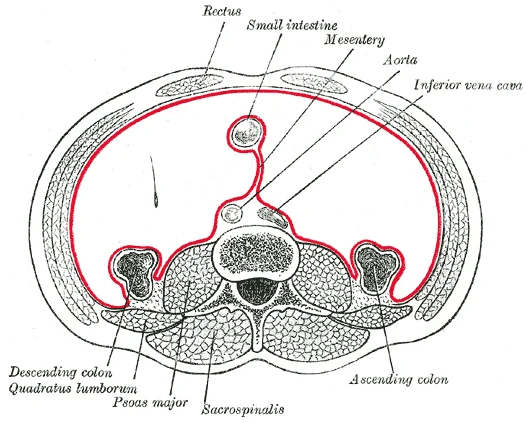
Horizontal disposition of the peritoneum in the lower part of the abdomen.

Figure 1: Horizontal disposition of the peritoneum in the lower part of the abdomen.
It consists of two distinct regions: the parietal peritoneum, which lines the abdominal wall, and the visceral peritoneum, which envelopes the abdominal organs[1].
The digestive organs within the abdominal cavity are held in place by this membrane[1]. Within these folds run blood vessels, lymphatic vessels, and nerves that innervate the associated organs[1]. During fetal development, certain structures such as the duodenum, pancreas, and portions of the large intestine remain posterior to the peritoneum, describing their location as retroperitoneal[1].
Important ligaments include the falciform, hepatoduodenal, hepatogastric, gastrocolic, gastrosplenic, and splenorenal ligaments. The hepatoduodenal ligament connects the liver to the duodenum, while the hepatogastric ligament connects the liver to the stomach[1].
Recesses and paracolic gutters facilitate the spread of fluid and infection. Random peritoneal biopsies may sample normal surfaces including the right hemidiaphragm, bladder reflection, cul-de-sac, right and left paracolic recesses, and pelvic sidewalls. This spread includes the diaphragmatic and liver surfaces. [2]
Intra-abdominal organs are generally intraperitoneal, while retroperitoneal organs lie behind the peritoneum. The alimentary canal forms a continuous tube open to the outside environment at both ends[1]. The wall of the alimentary canal is composed of four basic tissue layers: mucosa, submucosa, muscularis, and serosa[1]. The enteric nervous system provides intrinsic innervation, and the autonomic nervous system provides extrinsic innervation[1].
DefinitionPeritoneal Cavity
The space bounded by the visceral and parietal peritoneal surfaces, containing lubricating fluid.
| Organ | Relation |
|---|---|
| Duodenum | Retroperitoneal |
| Pancreas | Retroperitoneal |
| Ascending Colon | Retroperitoneal |
| Descending Colon | Retroperitoneal |
| Stomach | Intraperitoneal |
| Small Intestine | Intraperitoneal |
It consists of two distinct regions: the parietal peritoneum, which lines the abdominal wall, and the visceral peritoneum, which envelopes the abdominal organs[1].
The parietal peritoneum lines the inner surface of the abdominal wall. The visceral peritoneum covers the stomach, liver, and intestines. The greater omentum hangs down from the stomach, covering the intestines. The mesentery attaches the small intestine to the posterior abdominal wall. These structures create compartments within the cavity.
The serosa is the portion of the alimentary canal superficial to the muscularis. Present only in the region of the alimentary canal within the abdominal cavity, it consists of a layer of visceral peritoneum overlying a layer of loose connective tissue. These tissues serve to hold the alimentary canal in place near the ventral surface of the vertebral column. [1]
The peritoneum creates folds called mesenteries, omenta, and ligaments. Mesenteries suspend organs from the abdominal wall. The greater omentum drapes over the intestines. The lesser omentum connects the liver to the stomach. These structures contain blood vessels, nerves, and lymphatics. They provide a pathway for these structures to reach the organs.
This foramen is located behind the free edge of the lesser omentum.
DefinitionSerosa
The serosa is the portion of the alimentary canal superficial to the muscularis. Present only in the region of the alimentary canal within the abdominal cavity, it consists of a layer of visceral peritoneum overlying a layer of loose connective tissue.
The peritoneum is a serous membrane that lines the abdominal cavity and covers the abdominal viscera. It consists of two continuous layers: the parietal peritoneum, which lines the abdominal wall, and the visceral peritoneum, which invests the organs. Between these layers lies the peritoneal cavity, a potential space containing a small amount of serous fluid that reduces friction during movement. The cavity is divided into the greater sac and the lesser sac (omental bursa). The greater sac is th [1]
Figure 22403 The PeritoneumN
Long description
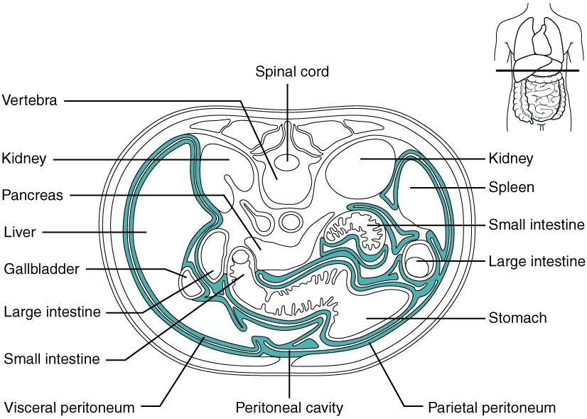
A cross-section of the abdomen shows the relationship between abdominal organs and the peritoneum (darker lines).

Figure 2: A cross-section of the abdomen shows the relationship between abdominal organs and the peritoneum (darker lines).
Figure 3Gray1035
Long description
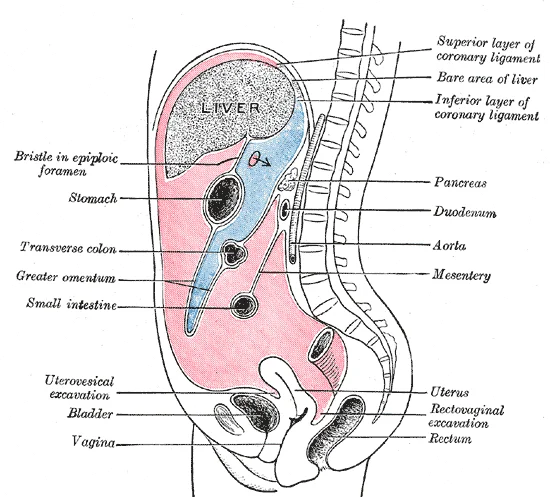
Vertical disposition of the peritoneum. Main cavity, red; omental bursa, blue.

Figure 3: Vertical disposition of the peritoneum.
The visceral peritoneum covers the liver, stomach, and spleen. The greater sac is the large space lateral to the stomach. The lesser sac is the smaller space posterior to the stomach. The falciform ligament attaches the liver to the anterior abdominal wall. The splenorenal ligament connects the spleen to the left kidney. These structures define the boundaries of the peritoneal recesses and compartments.
The peritoneum forms various ligaments, mesenteries, and omenta that suspend organs and convey vessels. The falciform ligament is a broad, thin fold of peritoneum that attaches the liver to the anterior abdominal wall and diaphragm. It contains the ligamentum teres hepatis, the remnant of the umbilical vein. The gastrocolic ligament is part of the greater omentum. It connects the greater curvature of the stomach to the transverse colon. It forms the anterior wall of the lesser sac. The splenorenal ligament connects the hilum of the spleen to the left kidney. It contains the splenic vessels and the tail of the pancreas. These ligaments are double layers of peritoneum. They provide stability to the organs while allowing some movement.
| Structure | Description |
|---|---|
| Parietal Peritoneum | Lines the abdominal wall |
| Visceral Peritoneum | Covers the abdominal organs |
| Falciform Ligament | Attaches liver to anterior wall |
| Gastrocolic Ligament | Connects stomach to transverse colon |
| Splenorenal Ligament | Connects spleen to left kidney |
The mesenteries are double folds of peritoneum that suspend the intestines. They convey blood vessels, nerves, and lymphatics to the gut. The greater omentum hangs from the greater curvature of the stomach. It covers the intestines like an apron. These structures create recesses and spaces. The subphrenic recesses lie between the diaphragm and the liver or spleen. The subhepatic recesses lie between the liver and the transverse colon. Understanding these relations is essential for surgical access and understanding the spread of disease.
The peritoneum forms the serous lining of the abdominal cavity, creating a continuous sac that reflects off the abdominal walls and viscera. It consists of a single layer of simple squamous epithelium, known as mesothelium, resting on a thin layer of connective tissue. This structure allows for the secretion of serous fluid, which reduces friction between moving organs. The cavity is divided into the greater sac and the lesser sac (omental bursa), connected by the epiploic foramen.
Figure 4Gray1036
Long description
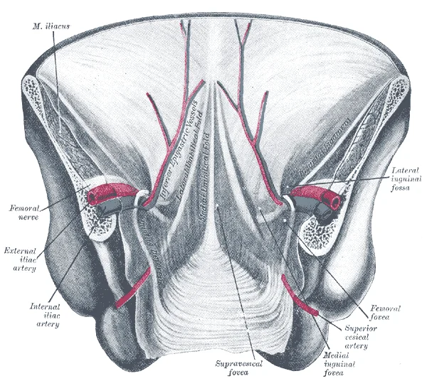
Posterior view of the anterior abdominal wall in its lower half. The peritoneum is in place, and the various cords are shining through. (After Joessel.)

Figure 4: Posterior view of the anterior abdominal wall in its lower half.
The parietal peritoneum lines the inner surface of the abdominal wall. The visceral peritoneum covers the external surface of most abdominal organs. Between these layers lies the peritoneal cavity, containing a small amount of lubricating fluid. The mesentery suspends the small intestine, bringing blood vessels and nerves to the gut. The omentum drapes over the intestines like an apron. The falciform ligament attaches the liver to the anterior abdominal wall. The greater omentum hangs from the greater curvature of the stomach. The lesser omentum connects the liver to the lesser curvature of the stomach. These structures create compartments and pathways for infection or fluid spread. [1]
The peritoneal reflections create potential spaces. The rectouterine pouch (pouch of Douglas) is the most dependent part in females. The rectovesical pouch is the corresponding space in males. These pouches are clinically significant as sites for fluid accumulation. The peritoneum also forms ligaments that stabilize organs. The round ligament of the liver is a remnant of the umbilical vein.
These arteries run within the mesenteries, providing vascular support to the suspended organs. The venous drainage follows the arteries, eventually reaching the portal vein system.
The lymphatic drainage of the peritoneum follows the arterial supply. Lymph from the foregut drains to celiac nodes. Lymph from the midgut drains to superior mesenteric nodes. Lymph from the hindgut drains to inferior mesenteric nodes. These nodes filter lymph before it enters the thoracic duct. The peritoneum plays a crucial role in immune surveillance. Mesothelial cells can phagocytose debris and pathogens. Macrophages within the peritoneal cavity also contribute to immune defence.
The innervation of the parietal peritoneum is somatic. It is supplied by the intercostal and lumbar nerves. This explains why pain from parietal peritoneum is well-localised. The visceral peritoneum is innervated by autonomic nerves. Pain from visceral peritoneum is poorly localised and often referred. This distinction is vital for clinical diagnosis of abdominal conditions.
The blood vessels serving the digestive system have two functions. They transport the protein and carbohydrate nutrients absorbed by mucosal cells after food is digested in the lumen. Lipids are absorbed via lacteals, tiny structures of the lymphatic system. The blood vessels’ second function is to supply the organs of the alimentary canal with the nutrients and oxygen needed to drive their cellular processes. [1]
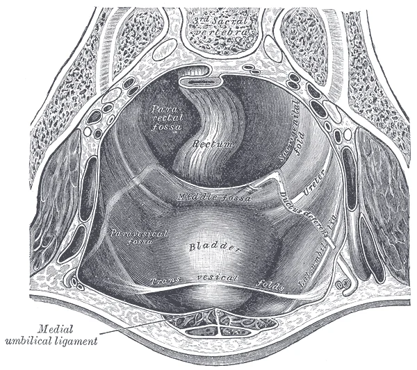
Figure 5Gray1037
Long description
The peritoneum of the male pelvis. (Dixon and Birmingham.)

Figure 5: The peritoneum of the male pelvis (Dixon and birmingham.).
The aorta is central, branching into the coeliac trunk, superior mesenteric artery, and inferior mesenteric artery. These vessels run anterior to the vertebral column and posterior to the peritoneal cavity, supplying the gut loops suspended within.
The coeliac trunk arises from the anterior surface of the abdominal aorta at the level of T12. It supplies the foregut derivatives, including the stomach, liver, and spleen. Its branches include the left gastric, splenic, and common hepatic arteries. The left gastric artery runs to the lesser curvature of the stomach. The splenic artery courses along the superior border of the pancreas to reach the spleen. The common hepatic artery gives rise to the proper hepatic artery, which supplies the liver and gallbladder.
The superior mesenteric artery originates from the anterior aspect of the aorta, just inferior to the coeliac trunk, at the level of L1. It supplies the midgut, which includes the distal duodenum, jejunum, ileum, caecum, ascending colon, and proximal two-thirds of the transverse colon. Its branches include the inferior pancreaticoduodenal artery, jejunal and ileal arteries, ileocolic artery, right colic artery, and middle colic artery. These vessels form arcades within the mesentery to ensure robust blood supply to the long loops of the small intestine.
Figure 6Gray1045
Long description
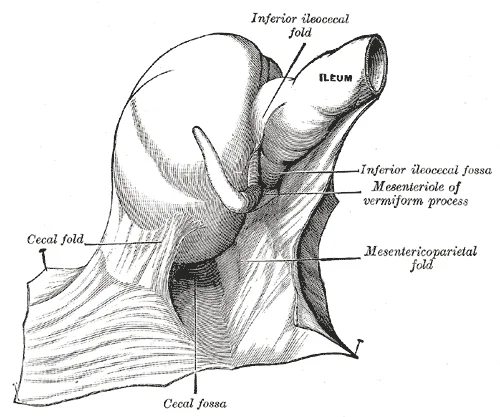
The cecal fossa. The ileum and cecum are drawn backward and upward. (Souligoux.)

Figure 6: The cecal fossa.
Figure 7Gray1044
Long description
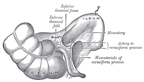
Inferior ileocecal fossa. The cecum and ascending colon have been drawn lateralward and downward, the ileum upward and backward, and the vermiform process downward. (Poirier and Charpy.)

Figure 7: Inferior ileocecal fossa.
Figure 8Gray1042
Long description
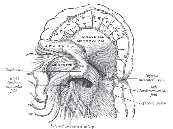
Duodenojejunal fossa. (Poirier and Charpy.)

Figure 8: Duodenojejunal fossa (Poirier and Charpy.).
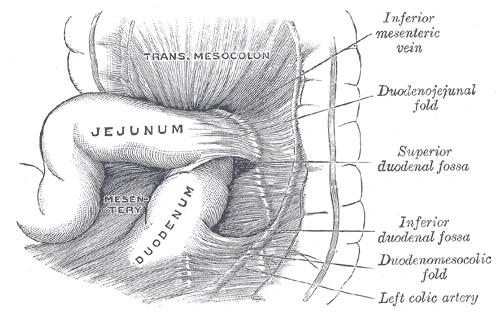
Figure 9Gray1041
Long description
Superior and inferior duodenal fossæ. (Poirier and Charpy.)

Figure 9: Superior and inferior duodenal fossæ (Poirier and Charpy.).
The inferior mesenteric artery arises from the anterior surface of the aorta at the level of L3. It supplies the hindgut, comprising the distal one-third of the transverse colon, descending colon, sigmoid colon, and upper rectum. Its branches are the left colic artery, sigmoid arteries, and superior rectal artery. The superior rectal artery descends into the pelvis to supply the upper part of the rectum.
DefinitionMesenteric Arcades
Complex anastomotic networks formed by the branches of the superior mesenteric artery within the mesentery of the small intestine, ensuring collateral circulation.
The peritoneum itself receives its blood supply from the vessels running within the mesenteries and omenta. The greater omentum is supplied by the gastroepiploic arteries, which are branches of the gastroduodenal and splenic arteries. The lesser omentum receives blood from the left and right gastric arteries and the proper hepatic artery. The mesentery of the small intestine contains the jejunal and ileal arteries, which branch extensively to supply the intestinal wall and the overlying peritoneum.
| Region | Artery | Level of Origin |
|---|---|---|
| Foregut | Coeliac Trunk | T12 |
| Midgut | Superior Mesenteric Artery | L1 |
| Hindgut | Inferior Mesenteric Artery | L3 |
Understanding this vascular arrangement is crucial for interpreting ischaemic patterns in the gut. Occlusion of a specific artery leads to infarction of its corresponding gut segment. For example, superior mesenteric artery occlusion affects the small intestine and right colon. This anatomical knowledge underpins surgical approaches to abdominal emergencies.
Similarly, the marginal artery of Drummond connects the superior and inferior mesenteric arteries along the colon, providing a vital collateral pathway.
NoteClinical Relevance
The marginal artery of Drummond is a critical collateral channel along the colon, connecting the superior and inferior mesenteric arteries.
The parietal peritoneum receives innervation and blood supply from the somatic nerves and vessels of the abdominal wall. This contrasts with the visceral peritoneum, which is supplied by the autonomic nervous system and the visceral arteries. This distinction explains the different pain referral patterns in abdominal pathology.
The venous drainage of the peritoneum is fundamentally linked to the gastrointestinal tract it lines. The peritoneum covering the abdominal viscera drains in parallel with the organs themselves. This arrangement ensures that nutrient-rich blood from the digestive tract is directed to the liver for processing before entering the systemic circulation. This pathway is known as the hepatic portal system.
The portal vein is formed by the union of the superior mesenteric vein and the splenic vein. It lies posterior to the neck of the pancreas. The viewer should note the direction of flow towards the liver.
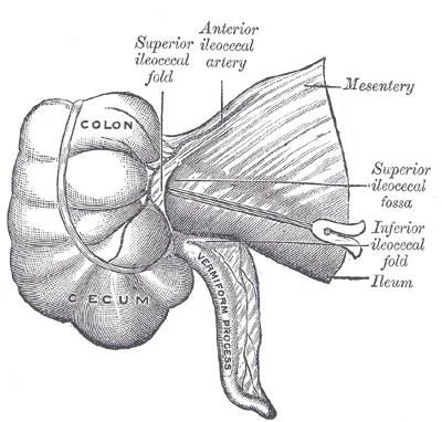
Figure 10Gray1043
Long description
Superior ileocecal fossa. (Poirier and Charpy.)

Figure 10: Superior ileocecal fossa (Poirier and Charpy.).
This venous network takes the blood into the liver where the nutrients are either processed or stored for later use. Only then does the blood drained from the alimentary canal viscera circulate back to the heart. [1]
To appreciate just how demanding the digestive process is on the cardiovascular system, consider that while you are “resting and digesting,” about one-fourth of the blood pumped with each heartbeat enters arteries serving the intestines. This high volume of blood flow necessitates a robust venous drainage system. The peritoneal veins are thin-walled and valveless in many regions. This allows for easy backflow in conditions of portal hypertension. [1]
The drainage can be summarised by structure. The greater omentum drains into the gastroepiploic veins. These empty into the splenic vein and the superior mesenteric vein. The lesser omentum drains into the left gastric vein and the portal vein. The mesentery drains into the superior mesenteric vein. The parietal peritoneum of the abdominal wall drains into the systemic veins. These include the inferior epigastric veins and the lumbar veins. These systemic veins eventually reach the inferior vena cava.
In contrast, the visceral peritoneum follows the portal route. This distinction is critical for understanding the spread of disease. Infections or malignancies in the peritoneum may spread via the portal system to the liver. This is why liver metastases are common in gastrointestinal cancers. The anatomical arrangement dictates the clinical presentation and management of peritoneal conditions.
This system is essential because the alimentary canal is constantly exposed to foodborne bacteria and other foreign matter. To defend against these pathogens, the immune system has evolved specific mechanisms within the gut wall. [1]
This tissue serves a vital immune function by monitoring the contents of the gut lumen. Peyer’s patches are prominent examples of organized lymphoid tissue that facilitate the initiation of immune responses to intestinal antigens. [1]
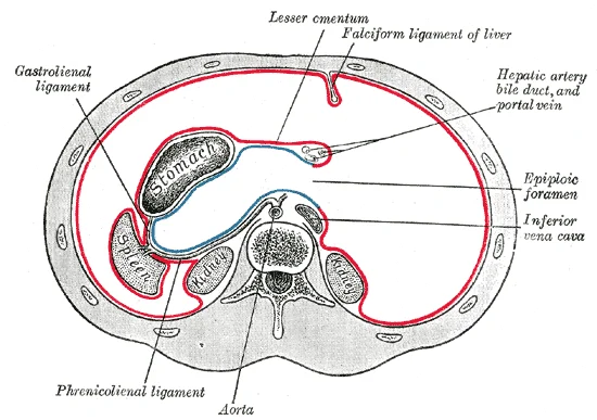
Figure 11Gray1039
Long description
Horizontal disposition of the peritoneum in the upper part of the abdomen.

Figure 11: Horizontal disposition of the peritoneum in the upper part of the abdomen.
The thoracic duct ascends through the aortic hiatus of the diaphragm. It courses posterior to the oesophagus and drains into the junction of the left subclavian and left internal jugular veins. These nodes filter lymph from the small and large intestines before it reaches the cisterna chyli. Understanding this flow is crucial for tracing the spread of infection or malignancy from the gut to the systemic circulation.
The integration of vascular and lymphatic systems in the lamina propria ensures efficient nutrient uptake and immune protection. The presence of MALT and Peyer’s patches highlights the gut's role as a major immune organ. This anatomical arrangement supports both metabolic and defensive functions of the gastrointestinal tract.
The peritoneum receives its innervation from both autonomic and somatic sources, a dual supply that dictates its sensory and motor responses. The autonomic nervous system provides extrinsic innervation to the alimentary canal, comprising both sympathetic and parasympathetic components. Sympathetic activation, associated with the fight-or-flight response, restricts the activity of enteric neurons, thereby decreasing gastrointestinal secretion and motility. In contrast, parasympathetic activation, linked to the rest-and-digest response, increases gastrointestinal secretion and motility by stimulating neurons of the enteric nervous system. [1]
Somatic innervation is distinct and clinically significant. The parietal peritoneum, which lines the abdominal wall, is richly supplied by somatic nerves. These nerves are sensitive to pain, temperature, touch, and pressure. Because these somatic nerves are segmental, pain from irritation of the parietal peritoneum is well-localised. This contrasts with the visceral peritoneum, which covers the organs. The visceral peritoneum is innervated by autonomic nerves and is sensitive to stretch, ischaemia, and chemical irritation, but not to cutting or burning. Pain from visceral peritoneum is poorly localised and often referred to distant somatic areas.
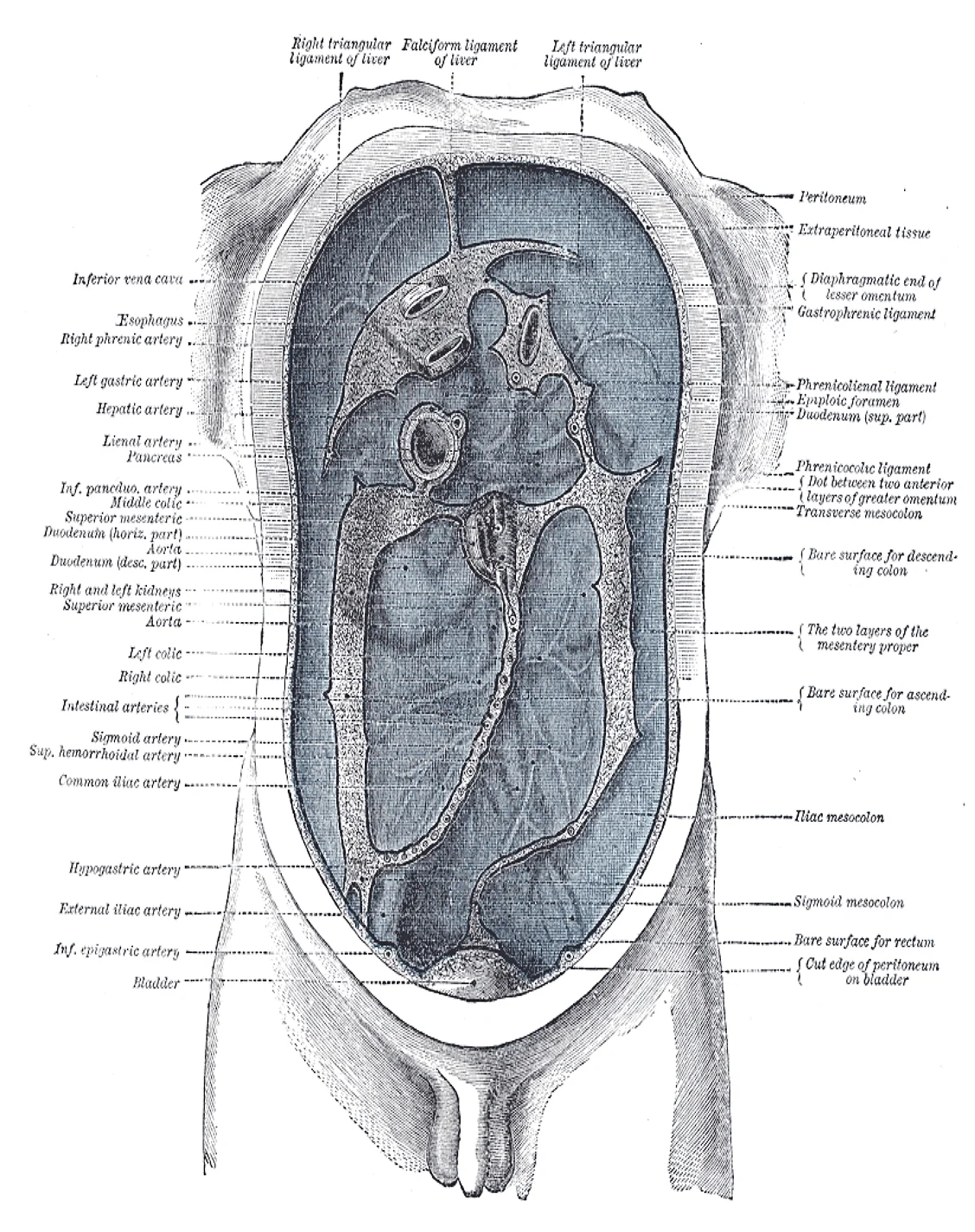
Figure 12Gray1040
Long description
Diagram devised by Delépine to show the lines along which the peritoneum leaves the wall of the abdomen to invest the viscera.

Figure 12: Diagram devised by Delépine to show the lines along which the peritoneum leaves the wall of the abdomen to invest the viscera.
Referred pain is a key clinical consequence of this innervation pattern. Visceral pain is referred to the dermatomes corresponding to the embryonic origin of the organ. For example, pain from the foregut is referred to the epigastrium (T5-T9). Midgut pain is referred to the periumbilical region (T10). Hindgut pain is referred to the suprapubic region (T12-L1). This occurs because visceral afferent fibres enter the spinal cord at the same level as somatic afferent fibres from the skin. The brain interprets the visceral pain as coming from the somatic region.
DefinitionReferred Pain
Pain perceived at a location other than the site of the painful stimulus, due to convergence of visceral and somatic afferent fibres in the spinal cord.
The distinction between parietal and visceral peritoneal pain is crucial for diagnosis. Parietal pain is sharp, well-localised, and exacerbated by movement. Visceral pain is dull, cramping, and poorly localised. Understanding this anatomy helps in localising abdominal pathology.
This structure allows for frictionless movement of abdominal organs. The mesothelial cells secrete a small amount of serous fluid, which lubricates the surfaces. This histological arrangement is fundamental to the peritoneal cavity's function as a potential space.
DefinitionMesothelium
A single layer of simple squamous epithelial cells that lines the serous cavities of the body, including the peritoneum, pleura, and pericardium.
The peritoneum is intimately linked to the embryological development of the gastrointestinal tract. To understand its formation, one must refer to the dedicated embryology page, which details the folding and rotation processes that position the gut within the abdominal cavity. The peritoneum itself arises from the lateral plate mesoderm, splitting into somatic and splanchnic layers. These layers envelop the developing gut tube, creating the primitive coelomic cavity. As the gut elongates and rotates, the peritoneal reflections form the mesenteries, omenta, and ligaments that suspend the visceral organs.
Figure 13Gray1034
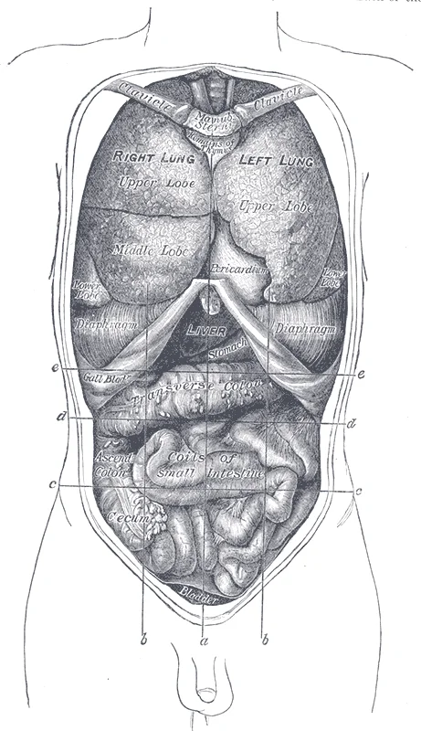
Long description
Front view of the thoracic and abdominal viscera. a. Median plane. b b. Lateral planes. c c. Trans tubercular plane. d d. Subcostal plane. e e. Transpyloric plane.

Figure 13: Front view of the thoracic and abdominal viscera (a).
They maintain a connection to the gut by way of ducts, even after development is complete. [1]
The epithelial lining of the gut also undergoes significant development. In the stomach and intestines, the epithelium is a simple columnar epithelium. This epithelium is in direct contact with the lumen, the space inside the alimentary canal. Interspersed among its epithelial cells are goblet cells, which secrete mucus and fluid into the lumen. Additionally, enteroendocrine cells secrete hormones into the interstitial spaces between cells. This rapid cellular turnover is essential for maintaining the integrity of the peritoneal and gut barriers. [1]
DefinitionPeritoneum
A serous membrane that lines the abdominal cavity and covers the abdominal organs, derived from the lateral plate mesoderm.
The peritoneum serves as a protective and supportive structure. It allows for the movement of organs during digestion and respiration. The mesenteries, formed during development, provide pathways for blood vessels, nerves, and lymphatics to reach the gut.
| Structure | Origin | Function |
|---|---|---|
| Mesentery | Lateral plate mesoderm | Suspends small intestine |
| Greater Omentum | Stomach | Protects abdominal organs |
| Falciform Ligament | Liver | Attaches liver to anterior wall |
Understanding the peritoneum requires knowledge of its embryological roots. This connection ensures that the accessory organs can effectively contribute to digestion. Indeed, you could not live without their vital contributions, and many significant diseases result from their malfunction. [1]
The parietal peritoneum lines the abdominal wall. The visceral peritoneum covers the organs. The mesentery suspends the intestines. The omentum drapes over the stomach. The liver is covered by visceral peritoneum. The spleen is intraperitoneal. The kidneys are retroperitoneal, behind the peritoneum. The bladder is partially covered. Understanding these relations is key to interpreting scans.
The peritoneal cavity is divided into compartments. The greater sac is the main cavity. The lesser sac lies behind the stomach. The omental bursa is another name for the lesser sac. The foramen of Winslow connects them. Radiological anatomy must identify these spaces. Fluid accumulation in the paracolic gutters is common. Ascites is often first seen in the pelvis. The pouch of Douglas is the most dependent part in women. Fluid collects here first. Recognizing these patterns helps in staging cancer.
DefinitionPeritoneal Cavity
The potential space between the parietal and visceral peritoneum, containing a small amount of serous fluid.
NoteImaging Limitation
Radiological scanning does not replace surgical staging with laparotomy.
The space between these layers is the peritoneal cavity, containing a small amount of lubricating fluid. In the female pelvis, the peritoneum reflects from the anterior abdominal wall onto the uterus, forming the vesicouterine pouch anteriorly and the rectouterine pouch (pouch of Douglas) posteriorly. This posterior pouch is the most dependent part of the peritoneal cavity in the upright position, making it a common site for fluid accumulation or abscess formation.
The uterosacral ligaments extend from the cervix to the sacrum, providing structural support. The ovaries are suspended by the mesovarium, a fold of peritoneum. The fallopian tubes lie superior to the ovaries, enclosed within the broad ligament. Understanding this spatial relationship is crucial for interpreting imaging studies, as fluid in the pouch of Douglas may indicate pathology such as haemoperitoneum or infection.
DefinitionPeritoneal Cavity
The potential space between the parietal and visceral peritoneum, containing serous fluid to reduce friction during organ movement.
The greater omentum hangs from the greater curvature of the stomach, draping over the intestines like an apron. It is rich in fat and immune cells, often referred to as the "abdominal policeman" due to its role in containing infections. The lesser omentum connects the liver to the lesser curvature of the stomach and the duodenum. These mesenteries provide pathways for blood vessels, nerves, and lymphatics to reach the organs. The mesentery of the small intestine attaches the jejunum and ileum to the posterior abdominal wall, allowing for mobility while maintaining vascular supply.
| Structure | Origin/Attachment | Course/Relations | Termination/Function |
|---|---|---|---|
| Greater Omentum | Greater curvature of stomach | Drapes over intestines | Attaches to transverse colon |
| Lesser Omentum | Liver hilum | Connects to stomach/duodenum | Forms anterior boundary of omental foramen |
| Mesentery | Posterior abdominal wall | Suspends small intestine | Attaches to jejunum and ileum |
Clinical assessment of pelvic masses requires careful consideration of the peritoneal reflections.
The space between these layers is the peritoneal cavity, which contains a small amount of lubricating fluid. This anatomical arrangement allows for smooth movement of abdominal organs during digestion and respiration.
DefinitionPeritoneal Cavity
The potential space between the parietal and visceral peritoneum, containing serous fluid to reduce friction.
The greater omentum is a large apron-like fold of peritoneum that hangs down from the greater curvature of the stomach. It attaches to the transverse colon and contains fat, lymph nodes, and immune cells. Its primary functions include insulation, fat storage, and immune surveillance within the abdominal cavity.
| Structure | Description |
|---|---|
| Greater Omentum | Apron-like fold from stomach to transverse colon; immune and fat storage. |
| Lesser Omentum | Connects liver to stomach and duodenum; forms anterior lesser sac boundary. |
| Mesentery | Double layer of peritoneum attaching intestines to posterior abdominal wall. |
The peritoneal reflections create several recesses and spaces that are clinically significant. The rectouterine pouch (pouch of Douglas) is the lowest point in the female peritoneal cavity, where fluid or blood may accumulate. In males, the rectovesical pouch serves a similar function. These spaces are common sites for abscess formation or metastatic deposits.
Exam trapPouch of Douglas
Often mistaken for a pathological space; it is a normal anatomical recess in females.
In contrast, the visceral peritoneum is innervated by autonomic nerves, responding primarily to stretch and ischemia. This difference explains why visceral pain is often poorly localized and referred to other areas.
NoteInnervation Difference
Parietal peritoneum: somatic nerves (sharp, localized pain). Visceral peritoneum: autonomic nerves (dull, referred pain).
The peritoneum plays a crucial role in the spread of intra-abdominal infections and malignancies. Pathogens and cancer cells can disseminate through the peritoneal cavity via the lymphatic system or direct extension. Intraperitoneal chemotherapy exploits this anatomy to deliver high concentrations of drugs directly to tumor sites. However, the distribution of chemotherapy agents is uneven due to peritoneal barriers and fluid dynamics.
DefinitionIntraperitoneal Chemotherapy
Delivery of cytotoxic drugs directly into the peritoneal cavity to target surface tumors.
The peritoneal cavity is divided into the greater and lesser sacs. The omental bursa (lesser sac) lies posterior to the stomach and is connected to the greater sac via the epiploic foramen (of Winslow). This foramen is the only communication between the two sacs and is bounded by the inferior vena cava, hepatic artery, and portal vein. Understanding this anatomy is essential for surgical approaches to the pancreas and stomach.
Clinical pearlEpiploic Foramen
The gateway between the greater and lesser sacs; critical for surgical access.
The peritoneum also contributes to the formation of ligaments and mesenteries that support abdominal organs. The falciform ligament attaches the liver to the anterior abdominal wall, while the coronary ligaments attach it to the diaphragm. These structures contain remnants of fetal vessels, such as the ligamentum teres, which is the obliterated umbilical vein.
NoteFetal Remnants
Ligamentum teres is the remnant of the umbilical vein; clinically relevant in portal hypertension.
The peritoneal lining is continuous with the serosal layers of the gastrointestinal tract. This continuity allows for the transmission of peristaltic movements and the protection of underlying tissues.
DefinitionMesentery Proper
Attaches small intestine to posterior wall; contains SMA and SMV.
Peritoneal dialysis utilizes the peritoneum as a semipermeable membrane for waste removal. The procedure involves instilling dialysate into the peritoneal cavity, allowing diffusion of solutes across the peritoneal membrane. The efficiency of this process depends on the surface area and permeability of the peritoneum. Complications include peritonitis, which can damage the peritoneal membrane and reduce dialysis efficacy.
Exam trapPeritonitis Risk
Infection can scar the peritoneum, reducing its effectiveness for dialysis.
Students frequently confuse the anatomical boundaries of the peritoneal cavity with the entire gastrointestinal tract. A critical error is assuming that the contents of the alimentary canal are inside the body. The alimentary canal is a one-way tube about 7.62 meters (25 feet) in length during life and closer to 10.67 meters (35 feet) in length when measured after death, once smooth muscle tone is lost. Because both the mouth and anus are open to the external environment, food and wastes within [1]
Another frequent mistake involves the staging and treatment of ovarian malignancies, particularly regarding screening versus prevention. Students often overestimate the utility of routine screening for high-risk populations. A recent study of multimodal screening using CA125 based on a risk of ovarian cancer algorithm (ROCA) every 4 months and transvaginal ultrasound annually or earlier where indicated by the ROCA in women at high risk of ovarian cancer reported that screening was associated with a low rate of high‐volume disease at primary surgery and very high rates of no residual disease after surgery. However, given that the majority of women with advanced stage ovarian cancer, even with complete resection, will relapse after chemotherapy, this does not seem to be a good alternative to risk‐reducing surgery. The correct clinical approach is that risk‐reducing salpingectomy‐oophorectomy remains the treatment choice for women at high risk of ovarian/fallopian tube cancer. [2]
Misunderstanding the role of opportunistic salpingectomy is also common. While bilateral salpingo-oophorectomy is the gold standard for genetic risk, students may miss the nuance for non-genetic patients. A bulletin from the American College of Obstetricians and Gynecologists (ACOG) has recommended that opportunistic (at the time of a clinically indicated hysterectomy) bilateral salpingectomy be considered in women not at genetic risk who wish to retain their ovaries as a way to reduce their risk of later developing high‐grade serous carcinomas. This highlights a preventive strategy that preserves hormonal function while reducing cancer risk. [2]
Chemotherapy regimens for germ cell tumors present another area of confusion, specifically regarding bleomycin toxicity. Students may not recall the specific dose adjustments required for older patients. Bleomycin/etoposide/cisplatin (BEP) has been widely used to treat patients with metastatic granulosa cell tumors; however, there is significantly increased toxicity of bleomycin in patients over the age of 40 years and there were a number of deaths associated with bleomycin in early GOG trials, which led them to reduce the bleomycin dose to 20 units/m2 intravenously every 3 weeks (x 4) to reduce toxicity. The standard dose is actually Bleomycin (B) 30 IU IV/IM on days 1/8/15 for 12 weeks (optional) (Note: bleomycin is dosed in International Units). Recognizing the toxicity profile is essential for safe prescribing. [2]
Finally, students often confuse the staging systems for different germ cell tumors. Ovarian germ cell tumors are staged similarly to epithelial carcinomas, although the staging system used for male germ cell tumors is probably more useful. The approach to treatment is based on the principles of management of metastatic germ cell tumors of the testis (i.e. low, intermediate, and poor risk). Dysgerminoma is the equivalent of seminoma in testicular cancer. It is exquisitely sensitive to platinum‐based chemotherapy and is radiosensitive. The cure rate is high irrespective of the stage. Confusing these systems can lead to inappropriate treatment planning. [2]
Knowledge of the peritoneum and associated structures is applied at the bedside, in the operating theatre, on imaging, and in examinations. At the bedside, understanding the "outside the body" nature of the gut lumen helps explain why peritoneal infections are distinct from intraluminal issues. This statistic guides prognosis discussions and treatment selection. [2]
In the operating theatre, surgeons perform risk-reducing procedures based on genetic risk profiles. Women at increased genetic risk should be encouraged to consider risk‐reducing bilateral salpingo‐oophorectomy, as this is the most effective way to reduce mortality in this population of women. This procedure is a cornerstone of preventive surgery for BRCA carriers. For those not at genetic risk but undergoing hysterectomy, the surgeon may perform opportunistic salpingectomy to reduce future cancer risk. [2]
On imaging, radiologists look for signs of disease progression or recurrence. The progression‐free interval is defined from the last date of platinum dose until progressive disease is documented. This metric is crucial for interpreting follow-up scans and determining when to initiate second-line therapy. For ovarian germ cell tumors, imaging helps stage the disease, which is similar to epithelial carcinomas. Patients with Stage IA disease may be observed after surgery. A small proportion of patients may recur, but they can be treated successfully at the time of recurrence with a high rate of cure. [2]
In examinations, students must distinguish between different histological subtypes and their treatments. Dysgerminoma is the equivalent of seminoma in testicular cancer. It is exquisitely sensitive to platinum‐based chemotherapy and is radiosensitive. The cure rate is high irrespective of the stage. Other histologic subtypes are equivalent to nonseminomatous testicular cancer. The aggressiveness of the disease is dependent on the type—the most aggressive being endodermal sinus and choriocarcinoma, but with combination chemotherapy, they are highly curable.
Hormonal therapies are also relevant in practice for recurrent granulosa cell tumors. Hormonal therapies have also been widely used to treat patients with recurrent granulosa cell tumors. In contrast, a retrospective study that included 22 patients with evaluable disease reported a response of 18% and 64% had stable disease with an aromatase inhibitor. PARAGON is the only prospective trial and reported a 10.5% partial response rate with anastrozole but a high proportion of patients with stable disease. It is not clear if the stable disease is due to treatment or the indolent biology of granulosa cell tumors. [2]
Management of Ovarian Germ Cell Tumours
- Diagnose Germ Cell TumourConfirm histology (e.g. Dysgerminoma)
- Question: Stage IA?
Yes
- Observe after surgeryHigh cure rate; monitor for recurrence
No
- Adjuvant ChemotherapyPlatinum-based regimen (e.g. BEP)
The alimentary canal is technically outside the body until absorption occurs. The gut is approximately 7.62 meters long in life and 10.67 meters post-mortem. Risk-reducing salpingectomy-oophorectomy is the preferred treatment for high-risk ovarian cancer patients. Opportunistic bilateral salpingectomy is recommended for non-genetic risk women retaining ovaries. Platinum-based chemotherapy has a 63%–80% response rate in advanced disease. [2] Bleomycin dose is reduced to 20 units/m2 in patients over 40 to reduce toxicity. Dysgerminoma is highly sensitive to platinum chemotherapy and radiotherapy. Stage IA ovarian germ cell tumours may be observed after surgery.
Answer each one in your head first, then open the answer.
What is the approximate length of the alimentary canal during life?
Show answerHide answer
7.62 meters (25 feet)What is the recommended treatment for women at high genetic risk of ovarian cancer?
Show answerHide answer
Risk-reducing bilateral salpingo-oophorectomyWhat is the reduced bleomycin dose for patients over 40 years?
Show answerHide answer
20 units/m2 intravenously every 3 weeks (x 4)Which ovarian germ cell tumour is equivalent to seminoma?
Show answerHide answer
DysgerminomaWhat is the overall response rate for platinum-based chemotherapy in advanced disease?
Show answerHide answer
63%–80%
- Betts JG, Young KA, Wise JA, et al. Overview of the Digestive System. OpenStax, Anatomy and Physiology 2e (CC BY-NC-SA 4.0). Accessed .
- Berek JS, Renz M, Kehoe S, et al. Cancer of the ovary, fallopian tube, and peritoneum: 2021 update. International journal of gynaecology and obstetrics: the official organ of the International Federation of Gynaecology and Obstetrics 2021;155 Suppl 1:61-85. doi:10.1002/ijgo.13878. Accessed .
Figure credits
- Figure 1: Henry Vandyke Carter, via Wikimedia Commons, Public domain, Public domain.
- Figure 2: OpenStax College, via Wikimedia Commons, CC BY 3.0, CC BY 3.0.
- Figure 3: Henry Vandyke Carter, via Wikimedia Commons, Public domain, Public domain.
- Figure 4: Henry Vandyke Carter, via Wikimedia Commons, Public domain, Public domain.
- Figure 5: Henry Vandyke Carter, via Wikimedia Commons, Public domain, Public domain.
- Figure 6: Henry Vandyke Carter, via Wikimedia Commons, Public domain, Public domain.
- Figure 7: Henry Vandyke Carter, via Wikimedia Commons, Public domain, Public domain.
- Figure 8: Henry Vandyke Carter, via Wikimedia Commons, Public domain, Public domain.
- Figure 9: Henry Vandyke Carter, via Wikimedia Commons, Public domain, Public domain.
- Figure 10: Henry Vandyke Carter, via Wikimedia Commons, Public domain, Public domain.
- Figure 11: Henry Vandyke Carter, via Wikimedia Commons, Public domain, Public domain.
- Figure 12: Henry Vandyke Carter, via Wikimedia Commons, Public domain, Public domain.
- Figure 13: Henry Vandyke Carter, via Wikimedia Commons, Public domain, Public domain.
Educational content for medical students and doctors. It is not a substitute for clinical judgement or local guidelines.