Inguinal canal
The inguinal canal is a passage in the lower anterior abdominal wall transmitting structures between the abdomen and scrotum or labia.
The inguinal canal is a narrow, oblique passage located in the lower anterior abdominal wall. It serves as a conduit for structures passing between the abdominal cavity and the external genitalia. In males, it transmits the spermatic cord, while in females, it carries the round ligament of the uterus. The canal begins at the deep (internal) inguinal ring and terminates at the superficial (external) inguinal ring. The deep ring is an opening in the transversalis fascia, situated lateral to the inferior epigastric vessels. The superficial ring is a triangular defect in the external oblique aponeurosis, located just superior and lateral to the pubic tubercle. The integrity of the abdominal wall in this region relies on the orientation of the canal and the sphincter-like function of the internal ring. [3]
The walls of the inguinal canal are formed by surrounding muscular and fascial structures. The anterior wall is primarily formed by the external oblique aponeurosis, reinforced laterally by the internal oblique muscle. The posterior wall is supported by the transversalis fascia, with the conjoint tendon (formed by the fusion of internal oblique and transversus abdominis aponeuroses) reinforcing the medial aspect. The roof is formed by the arching fibres of the internal oblique and transversus abdominis muscles. The floor is created by the inguinal ligament and the lacunar ligament.
Hernias in this region are classified based on their relationship to the inferior epigastric vessels. Lateral hernias arise from the internal inguinal ring, often through a patent processus vaginalis, and travel through the canal. Medial hernias protrude through the transversalis fascia in Hesselbach’s triangle, medial to the inferior epigastric vessels. The etiology of these hernias remains uncertain, although the lifetime risk is 27% for men and 3% for women. Lateral hernias are more frequent, [3]
The femoral region lies inferior to the inguinal ligament and is separated from the inguinal canal by this strong fibrous band. The myopectineal orifice, also known as the femoral ring area, is a potential weak point in the pelvic floor. It is bounded by the inguinal ligament anteriorly, the pectineal ligament posteriorly, the iliopubic tract superiorly, and the lacunar ligament medially. This area is critical in hernia surgery, as both inguinal and femoral hernias can protrude through defects in this region. Understanding the boundaries and contents of the inguinal canal is essential for diagnosing and managing groin pathologies.
The inguinal canal itself does not contain traditional joints, but it is a passageway through which several structures pass, including elements that connect to joints. In men, the spermatic cord traverses the inguinal canal, containing the vas deferens, blood vessels, nerves, and the cremaster muscle. The cremaster muscle is a thin muscle derived from the internal oblique that covers the spermatic cord and can reflexively elevate the testis, which is connected to the spermatic cord. This reflex is part of the cremasteric reflex, which involves the genitofemoral nerve and can be tested clinically.
In women, the round ligament of the uterus passes through the inguinal canal, extending from the uterus to the labia majora. The round ligament is a remnant of the gubernaculum and helps maintain the position of the uterus. While not directly forming a joint, these structures contribute to the stability and function of the inguinal region.
The inguinal canal is bounded by several ligamentous structures that provide support and define its boundaries. The inguinal ligament (Poupart's ligament) forms the floor, running from the anterior superior iliac spine to the pubic tubercle. The conjoint tendon (formed by the fusion of the internal oblique and transversus abdominus aponeuroses) forms the medial wall, while the inferior epigastric vessels mark the lateral boundary. These ligaments help maintain the integrity of the canal while allowing passage of structures.
Movements within the inguinal canal are primarily related to the passage of structures rather than joint articulation. The canal allows for the descent of the testis in males and the round ligament in females during development. In adults, the canal permits the passage of structures while maintaining a barrier against herniation. The cremaster muscle can cause movement of the testis within the canal, and changes in intra-abdominal pressure during activities like Valsalva can temporarily increase
NoteClinical Relevance
The inguinal canal's boundaries are important clinically for differentiating direct and indirect inguinal hernias. Indirect hernias protrude lateral to the inferior epigastric vessels, while direct hernias medial to them.
The inguinal canal is an oblique passage within the anterior abdominal wall, situated above the medial half of the inguinal ligament. It transmits the spermatic cord in males and the round ligament of the uterus in females. To locate it clinically, identify the pubic tubercle and the anterior superior iliac spine. The inguinal ligament runs between these two bony landmarks. The canal lies approximately 1.5 cm above the inguinal ligament. Its length is roughly 4 cm in adults. [3] [1]
Figure 1Gray1227
Long description
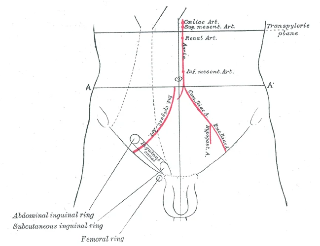
Gray1227.png. Labelled illustration from Wikimedia Commons, used under its open licence.

Figure 1: Gray1227.png.
png. The inguinal ligament is drawn as a thick band extending from the anterior superior iliac spine to the pubic tubercle. The superficial inguinal ring is marked at the medial end, near the pubic tubercle. The deep inguinal ring is indicated laterally, midway between the anterior superior iliac spine and the pubic symphysis. The external oblique aponeurosis forms the anterior wall.
The anterior wall is formed by the aponeurosis of the external oblique muscle. The posterior wall is formed by the transversalis fascia. The roof is formed by the arching fibres of the internal oblique and transversus abdominis muscles. The floor is formed by the inguinal ligament and the lacunar ligament. The superficial inguinal ring is a triangular opening in the external oblique aponeurosis. It lies just superior and lateral to the pubic tubercle. The deep inguinal ring is an opening in the transversalis fascia. It lies lateral to the inferior epigastric vessels.
Figure 2Gray394
Long description
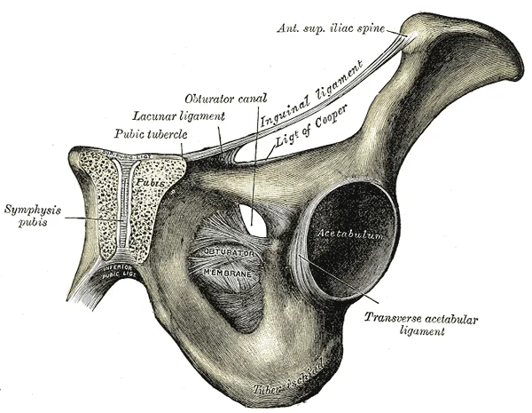
The inguinal and lacunar ligaments. Labelled illustration from Wikimedia Commons, used under its open licence.

Figure 2: The inguinal and lacunar ligaments.
Palpation of the inguinal canal involves examining the superficial inguinal ring. The examiner places fingers over the ring and asks the patient to cough. A bulge indicates a hernia. The spermatic cord can be palpated within the canal. In males, the vas deferens feels like a firm, cord-like structure. The testis and epididymis are palpable in the scrotum. Routine examination includes checking for masses in the inguinal area. A hard mobile mass may suggest a lymph node or other pathology. Ultrasound imaging can clarify the nature of such masses. [3]
DefinitionInguinal Canal
An oblique passage in the anterior abdominal wall transmitting the spermatic cord or round ligament, bounded by the inguinal ligament inferiorly and the arching abdominal muscles superiorly.
The inguinal canal is a oblique passage located in the lower anterior abdominal wall. It is not a true cavity but a potential space formed by the aponeuroses and muscles of the abdominal wall. Its boundaries are defined by specific bony landmarks and ligaments.
The roof of the inguinal canal is formed by the arching fibres of the internal oblique and transversus abdominis muscles. These muscles originate from the inguinal ligament and the anterior two-thirds of the iliac crest. They arch over the canal, providing superior coverage. The floor of the canal is formed by the inguinal ligament, which is the thickened lower border of the external oblique aponeurosis. The inguinal ligament runs from the anterior superior iliac spine to the pubic tubercle.
The anterior wall is primarily formed by the external oblique aponeurosis. In the lateral part, it is reinforced by the internal oblique muscle. The posterior wall is formed by the transversalis fascia. This fascia is reinforced medially by the conjoint tendon, which is the fused insertion of the internal oblique and transversus abdominis muscles into the pubic crest.
The medial boundary is the lateral edge of the rectus abdominis muscle. These vessels arise from the external iliac artery and run upwards and medially along the posterior aspect of the anterior abdominal wall. They serve as a crucial landmark for distinguishing direct from indirect inguinal hernias.
The inguinal ligament attaches medially to the pubic tubercle. This bony prominence is palpable and serves as a key surface landmark. The pubic symphysis is the midline joint between the two pubic bones. It lies medial to the inguinal canals.
The anterior superior iliac spine is the lateral attachment of the inguinal ligament. It is a prominent bony projection on the ilium. It is easily palpable and serves as a reference point for locating the inguinal ligament.
The iliac crest forms the superior boundary of the pelvis. The inguinal ligament originates from the anterior two-thirds of this crest. The pubic crest is the superior border of the pubic bone. The conjoint tendon inserts into this crest.
The spermatic cord passes through the inguinal canal. It contains the vas deferens, testicular artery, and pampiniform plexus. In females, the round ligament of the uterus passes through the canal. The canal transmits these structures from the abdomen to the scrotum or labia majora.
The deep inguinal ring is an opening in the transversalis fascia. It is located lateral to the inferior epigastric vessels. It serves as the entrance to the inguinal canal. The superficial inguinal ring is a triangular opening in the external oblique aponeurosis. It is located just superior and lateral to the pubic tubercle. It serves as the exit of the inguinal canal.
The inguinal canal is approximately 4 cm long in adults. It runs obliquely downwards and medially. It is present in both males and females. Its primary function is to transmit the spermatic cord or round ligament. It also provides a pathway for the genital branch of the genitofemoral nerve and the ilioinguinal nerve.
The bony landmarks are essential for surgical approaches to the inguinal region. Knowledge of these structures prevents injury to the neurovascular bundles. The inferior epigastric vessels are particularly important to identify during hernia repair.
The pubic tubercle is a small bony projection on the superior ramus of the pubis. It is the medial attachment of the inguinal ligament. It is palpable in the groin region. The anterior superior iliac spine is the lateral attachment. It is used to locate the inguinal ligament.
The inguinal ligament is a thick band of fibrous tissue. It supports the abdominal wall. It prevents herniation of abdominal contents. The conjoint tendon reinforces the posterior wall. It is formed by the fusion of the internal oblique and transversus abdominis muscles.
The transversalis fascia is a layer of connective tissue. It lines the inner surface of the abdominal muscles. It forms the posterior wall of the inguinal canal. It is reinforced by the conjoint tendon medially.
The external oblique aponeurosis forms the anterior wall. It is a broad, flat tendon. It covers the lateral part of the rectus abdominis muscle. It forms the inguinal ligament inferiorly.
The internal oblique muscle forms the roof. It originates from the inguinal ligament and iliac crest. It arches over the inguinal canal. It contributes to the conjoint tendon.
The transversus abdominis muscle forms the roof. It originates from the inguinal ligament and iliac crest. It arches over the inguinal canal. It contributes to the conjoint tendon.
The rectus abdominis muscle forms the medial boundary. It is a vertical muscle. It lies on either side of the midline. It is enclosed in the rectus sheath.
The inferior epigastric vessels form the lateral boundary. They arise from the external iliac artery. They run upwards and medially. They are important landmarks for hernia classification.
The deep inguinal ring is the entrance. It is located lateral to the inferior epigastric vessels. It is an opening in the transversalis fascia. It is not a true ring but a slit.
The superficial inguinal ring is the exit. It is located superior and lateral to the pubic tubercle. It is an opening in the external oblique aponeurosis. It is triangular in shape.
The spermatic cord contains the vas deferens. It also contains the testicular artery. It contains the pampiniform plexus. It contains the genital branch of the genitofemoral nerve.
The round ligament of the uterus passes through the canal. It is a fibrous cord. It connects the uterus to the labia majora. It is the female equivalent of the spermatic cord.
The ilioinguinal nerve passes through the canal. It provides sensory innervation to the skin of the upper medial thigh. It also innervates the skin of the root of the penis and anterior scrotum in males. In females, it innervates the skin of the mons pubis and labia majora.
The genital branch of the genitofemoral nerve passes through the canal. It provides motor innervation to the cremaster muscle. It also provides sensory innervation to the skin of the scrotum or labia majora.
The inguinal canal is a potential space. It is formed by the aponeuroses and muscles of the abdominal wall. It transmits the spermatic cord or round ligament. It is located in the lower anterior abdominal wall.
The boundaries are defined by bony landmarks and ligaments. The roof is formed by the internal oblique and transversus abdominis muscles. The floor is formed by the inguinal ligament. The anterior wall is formed by the external oblique aponeurosis. The posterior wall is formed by the transversalis fascia.
The medial boundary is the lateral edge of the rectus abdominis muscle. The deep inguinal ring is the entrance. The superficial inguinal ring is the exit.
The inguinal canal is approximately 4 cm long. It runs obliquely downwards and medially. It is present in both males and females. Its primary function is to transmit the spermatic cord or round ligament.
The bony landmarks are essential for surgical approaches. Knowledge of these structures prevents injury to the neurovascular bundles. The inferior epigastric vessels are particularly important to identify during hernia repair.
The pubic tubercle is a small bony projection. It is the medial attachment of the inguinal ligament. It is palpable in the groin region. The anterior superior iliac spine is the lateral attachment. It is used to locate the inguinal ligament.
The inguinal ligament is a thick band of fibrous tissue. It supports the abdominal wall. It prevents herniation of abdominal contents. The conjoint tendon reinforces the posterior wall. It is formed by the fusion of the internal oblique and transversus abdominis muscles.
The transversalis fascia is a layer of connective tissue. It lines the inner surface of the abdominal muscles. It forms the posterior wall of the inguinal canal. It is reinforced by the conjoint tendon medially.
The external oblique aponeurosis forms the anterior wall. It is a broad, flat tendon. It covers the lateral part of the rectus abdominis muscle. It forms the inguinal ligament inferiorly.
The internal oblique muscle forms the roof. It originates from the inguinal ligament and iliac crest. It arches over the inguinal canal. It contributes to the conjoint tendon.
The transversus abdominis muscle forms the roof. It originates from the inguinal ligament and iliac crest. It arches over the inguinal canal. It contributes to the conjoint tendon.
The rectus abdominis muscle forms the medial boundary. It is a vertical muscle. It lies on either side of the midline. It is enclosed in the rectus sheath.
The inferior epigastric vessels form the lateral boundary. They arise from the external iliac artery. They run upwards and medially. They are important landmarks for hernia classification.
The deep inguinal ring is the entrance. It is located lateral to the inferior epigastric vessels. It is an opening in the transversalis fascia. It is not a true ring but a slit.
The superficial inguinal ring is the exit. It is located superior and lateral to the pubic tubercle. It is an opening in the external oblique aponeurosis. It is triangular in shape.
The spermatic cord contains the vas deferens. It also contains the testicular artery. It contains the pampiniform plexus. It contains the genital branch of the genitofemoral nerve.
The round ligament of the uterus passes through the canal. It is a fibrous cord. It connects the uterus to the labia majora. It is the female equivalent of the spermatic cord.
The ilioinguinal nerve passes through the canal. It provides sensory innervation to the skin of the upper medial thigh. It also innervates the skin of the root of the penis and anterior scrotum in males. In females, it innervates the skin of the mons pubis and labia majora.
The genital branch of the genitofemoral nerve passes through the canal. It provides motor innervation to the cremaster muscle. It also provides sensory innervation to the skin of the scrotum or labia majora.
The inguinal canal is an oblique passage in the lower anterior abdominal wall, transmitting the spermatic cord in males and the round ligament in females. It lies above the inguinal ligament and is bounded by specific fascial layers that provide structural integrity. [2]
The inguinal ligament forms the inferior boundary, running from the anterior superior iliac spine to the pubic tubercle. The cremasteric fascia, derived from the internal oblique, surrounds the spermatic cord. The transversalis fascia lines the posterior aspect, deep to the muscles. The conjoint tendon, formed by the fusion of the internal oblique and transversus abdominis aponeuroses, inserts onto the pubic crest and pectineal line, reinforcing the posterior wall medially. The deep inguinal ring is an opening in the transversalis fascia, located lateral to the inferior epigastric vessels. The superficial inguinal ring is a triangular defect in the external oblique aponeurosis, situated above and lateral to the pubic tubercle. The canal itself lies between these two rings.
Figure 3Gray393
Long description
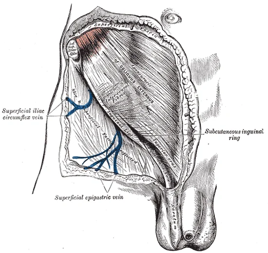
The subcutaneous inguinal ring. Labelled illustration from Wikimedia Commons, used under its open licence.

Figure 3: The subcutaneous inguinal ring.
The boundaries are defined by four walls. The anterior wall is formed primarily by the external oblique aponeurosis, reinforced laterally by the internal oblique muscle. The posterior wall is formed by the transversalis fascia, reinforced medially by the conjoint tendon. The roof is formed by the arching fibres of the internal oblique and transversus abdominis muscles. The floor is formed by the inguinal ligament and the lacunar ligament.
Risk factors for developing a primary inguinal hernia include male gender and old age. A patent processus vaginalis is a key factor. Systemic connective tissue disorders also increase risk. High body mass index increases intraabdominal pressure. Constipation does not appear to be a risk factor. [3]
DefinitionInguinal Canal
An oblique passage in the lower anterior abdominal wall, transmitting the spermatic cord or round ligament, bounded by the inguinal ligament inferiorly and the aponeuroses of the abdominal muscles.
| Muscle | Role in Canal |
|---|---|
| External Oblique | Forms anterior wall via aponeurosis |
| Internal Oblique | Reinforces anterior wall and forms roof |
| Transversus Abdominis | Contributes to roof and posterior reinforcement |
| Transversalis Fascia | Forms posterior wall |
The inguinal canal is a oblique passage in the lower anterior abdominal wall, transmitting the spermatic cord in males and the round ligament in females. Its boundaries are defined by specific muscular and fascial structures. The anterior wall is formed primarily by the aponeurosis of the external oblique muscle, reinforced laterally by the internal oblique muscle. The posterior wall is supported by the transversalis fascia and, medially, by the conjoint tendon. The roof is formed by the arching fibres of the internal oblique and transversus abdominis muscles, while the floor is created by the inguinal ligament and the lacunar ligament.
Figure 4Gray547
Long description
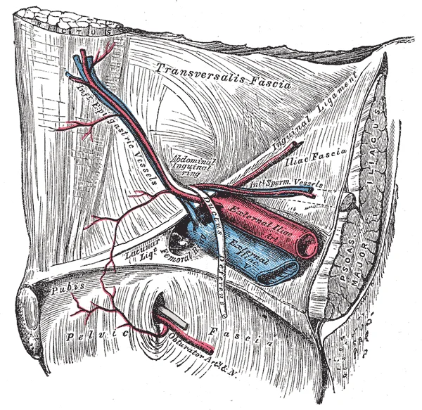
Blick von innen auf die Leistengegend der rechten Seite mit Darstellung der aus- und eintretenden Gefäße und anderen Strukturen.

Figure 4: Blick von innen auf die Leistengegend der rechten Seite mit Darstellung der aus- und eintretenden Gefäße und anderen Strukturen.
The external oblique aponeurosis forms the most superficial layer. Deep to this lies the internal oblique muscle, whose fibres arch over the canal. The transversus abdominis is the deepest muscle layer. The inguinal ligament runs along the inferior border, formed by the folded inferior edge of the external oblique aponeurosis. The conjoint tendon, formed by the fusion of the internal oblique and transversus abdominis aponeuroses, attaches to the pubic crest. The spermatic cord passes through the canal, lying anterior to the transversalis fascia. Lymphatic vessels accompany the spermatic cord towards the external iliac nodes.
The external oblique muscle originates from the external surfaces of the lower eight ribs. Its fibres run inferomedially, ending in an aponeurosis that contributes to the anterior wall of the inguinal canal. It inserts into the pubic tubercle, pectineal line, and anterior half of the inguinal ligament. Its action includes compression of the abdominal contents and rotation of the trunk. It is supplied by the lower six intercostal nerves and the iliohypogastric nerve.
The internal oblique muscle originates from the thoracolumbar fascia, the anterior two-thirds of the iliac crest, and the lateral half of the inguinal ligament. Its fibres run superomedially. The lower fibres contribute to the roof of the inguinal canal and form the conjoint tendon. It inserts into the costal cartilages of the lower three ribs and the linea alba. It compresses the abdomen and assists in trunk rotation.
The transversus abdominis muscle originates from the lower six costal cartilages, the thoracolumbar fascia, the iliac crest, and the lateral third of the inguinal ligament. Its fibres run horizontally. The lower fibres contribute to the conjoint tendon and the posterior wall of the inguinal canal. It inserts into the linea alba and the pubic crest. It compresses the abdominal viscera.
The inguinal ligament is a thickened inferior border of the external oblique aponeurosis. It runs from the anterior superior iliac spine to the pubic tubercle. It forms the floor of the inguinal canal. The lacunar ligament is a medial expansion of the inguinal ligament, attaching to the pectineal line.
The conjoint tendon is formed by the fusion of the lower fibres of the internal oblique and transversus abdominis aponeuroses. It inserts into the pubic crest and pectineal line. It reinforces the posterior wall of the inguinal canal medially.
Nerve supply to the anterior abdominal wall muscles includes the lower six thoracic nerves (T7-T12) and the first lumbar nerve (L1). The iliohypogastric and ilioinguinal nerves run between the internal oblique and transversus abdominis. The genitofemoral nerve supplies the cremaster muscle.
Lymphatic drainage from the anterior abdominal wall above the umbilicus goes to the axillary nodes. Below the umbilicus, it drains to the superficial inguinal nodes. The deep inguinal nodes receive lymph from the glans penis and the distal urethra.
| Muscle | Origin | Insertion | Action | Nerve Supply |
|---|---|---|---|---|
| External Oblique | Lower 8 ribs | Pubic tubercle, inguinal ligament, linea alba | Compress abdomen, rotate trunk | Lower 6 intercostal nerves, iliohypogastric, ilioinguinal |
| Internal Oblique | Thoracolumbar fascia, iliac crest, inguinal ligament | Costal cartilages 10-12, linea alba, pubic crest | Compress abdomen, rotate trunk | Lower intercostal nerves, iliohypogastric, ilioinguinal |
| Transversus Abdominis | Costal cartilages 7-12, thoracolumbar fascia, iliac crest, inguinal ligament | Linea alba, pubic crest | Compress abdomen | Lower intercostal nerves, iliohypogastric, ilioinguinal |
Figure 5Iliac artery bifurcation
Long description
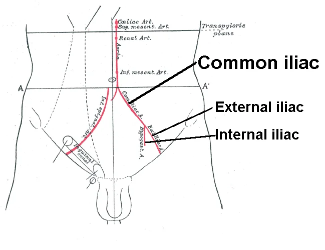
Front of abdomen, showing surface markings for arteries and inguinal canal.

Figure 5: Front of abdomen, showing surface markings for arteries and inguinal canal.
The arterial supply to the inguinal canal is derived primarily from the external iliac artery and its branches. The most significant vessel is the cremasteric artery, a branch of the inferior epigastric artery. This vessel descends along the spermatic cord, supplying the cremaster muscle and the coverings of the testis and spermatic cord. It anastomoses with the testicular artery and the artery to the ductus deferens. [1]
The inferior epigastric artery arises from the external iliac artery just above the inguinal ligament. It courses superiorly and medially, passing posterior to the rectus abdominis muscle. This vessel serves as a crucial landmark for the medial boundary of the deep inguinal ring. The deep circumflex iliac artery, another branch of the external iliac, runs along the inguinal ligament and contributes to the lateral supply.
DefinitionCremasteric Artery
A branch of the inferior epigastric artery that supplies the cremaster muscle and spermatic cord coverings.
The testicular artery, arising directly from the abdominal aorta, enters the inguinal canal through the deep inguinal ring. It travels within the spermatic cord, accompanied by the pampiniform plexus and the vas deferens. This artery provides the primary blood supply to the testis and epididymis.
The artery to the ductus deferens, a branch of the superior vesical artery, accompanies the vas deferens through the inguinal canal. It anastomoses with the cremasteric and testicular arteries, ensuring collateral circulation.
| Vessel | Origin | Course |
|---|---|---|
| Cremasteric | Inferior epigastric | Descends along spermatic cord |
| Testicular | Abdominal aorta | Enters deep ring, travels in cord |
| Artery to ductus deferens | Superior vesical | Accompanies vas deferens |
Figure 6Iliac artery bifurcation and aorta
Long description
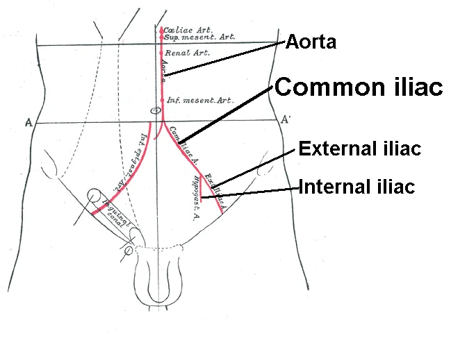
Front of abdomen, showing surface markings for arteries and inguinal canal.

Figure 6: Front of abdomen, showing surface markings for arteries and inguinal canal.
The cremasteric artery branches off and descends along the spermatic cord. The artery to the ductus deferens accompanies the vas deferens.
NoteClinical Relevance
The inferior epigastric artery is a key landmark for distinguishing direct from indirect inguinal hernias.
The blood supply is vital for the function of the cremaster muscle and the viability of the testicular structures. Any compromise to these vessels can lead to ischemia or atrophy. Understanding this vascular anatomy is essential for surgical procedures in the inguinal region.
The venous drainage of the inguinal canal is a critical anatomical consideration, particularly when evaluating pelvic and abdominal pathology. The primary vessels responsible for draining this region are the external pudendal veins and the cremasteric veins. These vessels accompany their arterial counterparts and ultimately drain into the great saphenous vein or the femoral vein. Understanding this drainage pathway is essential for interpreting clinical signs such as varicoceles or pelvic congestion.
Figure 7Gray1146
Long description
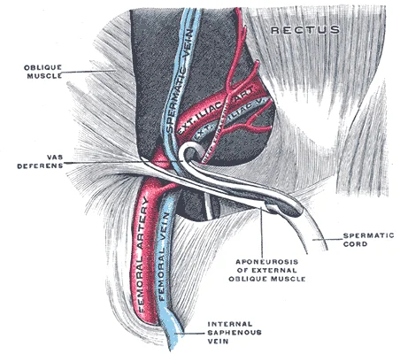
The spermatic cord in the inguinal canal. (Poirier and Charpy.)

Figure 7: The spermatic cord in the inguinal canal (Poirier and charpy.).
The external pudendal veins drain the skin of the anterior abdominal wall and the external genitalia. They run superficial to the fascia lata and empty into the great saphenous vein. This connection is clinically significant because it provides a collateral pathway for blood flow in cases of femoral vein obstruction. The great saphenous vein itself is a long superficial vein that ascends along the medial aspect of the leg and thigh. It pierces the cribriform fascia to join the femoral vein.
On the right side, the testicular vein drains directly into the inferior vena cava. On the left side, it drains into the left renal vein. This anatomical difference explains why left-sided varicoceles are more common than right-sided ones. The left renal vein passes between the superior mesenteric artery and the aorta, creating a potential site of compression. [3]
In clinical practice, knowledge of these venous pathways aids in the diagnosis of pelvic inflammatory disease or thrombosis. For instance, obstruction of the femoral vein can lead to engorgement of the external pudendal veins. This may present as swelling or discomfort in the groin region. Surgeons must be aware of these vessels during inguinal hernia repairs to avoid inadvertent injury. Ligation of these veins can lead to venous congestion and chronic pain.
Clinical pearlClinical Pearl
The left testicular vein drains into the left renal vein, while the right drains into the inferior vena cava, explaining the higher incidence of left-sided varicoceles.
| Structure | Drains Into | Clinical Significance |
|---|---|---|
| External Pudendal Veins | Great Saphenous Vein | Collateral flow in femoral obstruction |
| Cremasteric Veins | Internal Iliac/Inferior Epigastric | Part of pampiniform plexus |
| Testicular Vein (Left) | Left Renal Vein | Site of varicocele formation |
| Testicular Vein (Right) | Inferior Vena Cava | Direct drainage |
The innervation of the inguinal canal is critical for both motor function and sensory perception in the anterior thigh and genital region. The primary nerve supplying this region is the ilioinguinal nerve, which emerges from the lateral border of the psoas major muscle. It courses obliquely across the quadratus lumborum and iliacus muscles before piercing the transversus abdominis muscle near the anterior superior iliac spine. It then travels through the inguinal canal, lying anterior to the spermatic cord in males or the round ligament in females. Finally, it exits the superficial inguinal ring to supply the skin of the upper medial thigh and the root of the penis or mons pubis.
The genitofemoral nerve also plays a significant role in this area. It arises from the anterior rami of L1 and L2. The genital branch descends on the psoas major muscle and enters the deep inguinal ring. It travels within the spermatic cord, supplying the cremaster muscle and the skin of the anterior scrotum or labia majora. The femoral branch does not enter the canal but descends lateral to the external iliac artery to supply the skin of the upper anterior thigh.
DefinitionIlioinguinal Nerve
A branch of the L1 nerve root that supplies the skin of the upper medial thigh and the root of the penis or mons pubis. [1]
DefinitionGenitofemoral Nerve
A nerve arising from L1-L2 that divides into a genital branch supplying the cremaster muscle and a femoral branch supplying the upper anterior thigh.
| Nerve | Motor Supply | Cutaneous Supply |
|---|---|---|
| Ilioinguinal | None | Skin of upper medial thigh, root of penis, anterior scrotum or mons pubis |
| Genitofemoral (Genital branch) | Cremaster muscle | Skin of anterior scrotum or labia majora |
| Genitofemoral (Femoral branch) | None | Skin of upper anterior thigh |
Lesions of these nerves can result in significant sensory deficits. Damage to the ilioinguinal nerve may cause numbness in the distribution area, often occurring after inguinal hernia repair. Injury to the genitofemoral nerve can lead to loss of the cremasteric reflex and sensory changes in the genital region. Understanding the precise anatomical course of these nerves is essential for avoiding iatrogenic injury during surgical procedures in the inguinal region.
The lymphatic drainage of the inguinal canal is a critical pathway for immune surveillance and potential metastatic spread, particularly from the lower abdominal wall, perineum, and external genitalia. Understanding this network requires distinguishing between superficial and deep systems, as their drainage patterns diverge based on anatomical boundaries.
The superficial inguinal lymph nodes are situated in the subcutaneous tissue, just below the inguinal ligament. They are arranged in two groups: a horizontal group and a vertical group. The horizontal group lies parallel to the inguinal ligament, receiving lymph from the lower anterior abdominal wall, the buttocks, the perineum, and the external genitalia. These nodes filt [1]
They receive lymph from the glans penis or clitoris, the deep structures of the lower limb, and the superficial inguinal nodes. This pathway is significant in the staging of cancers affecting the external genitalia. [1]
Obstruction or infiltration of these lymph nodes can lead to lymphoedema of the lower limb or genitalia. Infections such as chancroid or syphilis may present with tender inguinal lymphadenopathy. Malignancies of the scrotum, penis, or vulva often metastasize to these nodes, making their examination essential in clinical practice.
Clinical pearlKey Point
Superficial inguinal nodes drain the skin of the lower limb and perineum, while deep nodes drain the glans and superficial nodes.
| Structure | Drainage Area | Primary Nodes |
|---|---|---|
| Lower Limb | Skin and superficial tissues | Superficial inguinal |
| Perineum | External genitalia | Superficial inguinal |
| Glans Penis/Clitoris | Deep structures | Deep inguinal |
| Superficial Nodes | Efferent vessels | Deep inguinal |
The inguinal canal is an oblique passage situated within the lower anterior abdominal wall, serving as a conduit for structures connecting the abdomen to the external genitalia. It extends approximately four centimetres in length, running from the deep inguinal ring to the superficial inguinal ring. The canal is bounded by four walls, each formed by distinct anatomical layers that provide structural integrity and protection to the passing contents.
The anterior wall is formed primarily by the aponeurosis of the external oblique muscle, reinforced laterally by the internal oblique muscle. This arrangement ensures that the canal remains closed during increased intra-abdominal pressure, a mechanism critical for preventing herniation. The posterior wall is composed of the transversalis fascia, strengthened medially by the conjoint tendon, which is the fused aponeuroses of the internal oblique and transversus abdominis muscles. This posterior reinforcement is particularly important in the region of Hesselbach’s triangle, a clinically significant area where direct inguinal hernias may protrude.
The roof of the canal is formed by the arching fibres of the internal oblique and transversus abdominis muscles, while the floor is supported by the inguinal ligament and the lacunar ligament. These boundaries create a protected tunnel that accommodates the spermatic cord in males or the round ligament of the uterus in females. The spermatic cord itself is a complex structure comprising several layers and components. It is enclosed by three coverings derived from the abdominal wall layers: the external spermatic fascia from the external oblique aponeurosis, the cremasteric muscle and fascia from the internal oblique, and the internal spermatic fascia from the transversalis fascia.
Inside the spermatic cord, the vas deferens, testicular artery, pampiniform plexus, and genital branch of the genitofemoral nerve are arranged in a specific spatial relationship. The vas deferens is palpable as a firm, cord-like structure, while the pampiniform plexus of veins provides a counter-current heat exchange mechanism to protect spermatogenesis. The testicular artery supplies oxygenated blood to the testis, ensuring optimal temperature regulation for sperm production.
Within the cord, the vas deferens is identified as a thick-walled tube, distinct from the softer, convoluted pampiniform plexus. The testicular artery runs alongside these structures, providing a vital blood supply. Understanding these spatial relationships is essential for surgical procedures and diagnosing hernias.
| Structure | Origin | Insertion | Action | Nerve Supply |
|---|---|---|---|---|
| External Oblique | Lower 8 ribs | Iliac crest, pubic tubercle | Compress abdomen | Lower intercostal nerves |
| Internal Oblique | Inguinal ligament | Inguinal ligament | Compress abdomen | Lower intercostal nerves |
| Transversus Abdominis | Inguinal ligament | Inguinal ligament | Compress abdomen | Lower intercostal nerves |
The inguinal canal is a oblique passage in the lower anterior abdominal wall, transmitting the spermatic cord in males and the round ligament in females. Its radiological assessment relies on multiple modalities to define its boundaries and contents.
The inguinal ligament forms the floor, appearing as a dense band extending from the anterior superior iliac spine to the pubic tubercle. The conjoint tendon forms the roof posteriorly. The external spermatic fascia covers the cord superficially. The ilioinguinal nerve lies anterior to the spermatic cord. The genital branch of the genitofemoral nerve runs within the spermatic cord. The vas deferens is a thick-walled tubular structure within the cord. The testicular vessels accompany the vas deferens.
Ultrasound is the primary modality for dynamic assessment. It visualises the inguinal canal in real-time during Valsalva manoeuvres. A normal canal contains only the spermatic cord or round ligament. The cord appears as a hypoechoic structure with internal echogenic strands representing vessels and nerves. The external oblique aponeurosis forms the anterior wall. The pubic tubercle is the medial landmark. The anterior superior iliac spine is the lateral landmark. Ultrasound can detect indirect hernias as bowel loops protruding through the deep inguinal ring. Direct hernias appear as bulging of the posterior wall through Hesselbach's triangle.
CT provides detailed anatomical context. Axial CT scans show the relationship of the inguinal canal to the pelvic bones. The superficial inguinal ring is a triangular defect in the external oblique aponeurosis. CT is useful for identifying complications such as strangulated hernias. It shows bowel wall thickening and fat stranding.
MRI offers superior soft tissue contrast. T2-weighted images highlight fluid and oedema. The inguinal canal is well-defined by the high signal of surrounding fat. MRI is sensitive for detecting occult hernias. It can distinguish between different types of hernias based on their content and location. The iliopubic tract is visible as a low-signal band reinforcing the posterior wall.
DefinitionInguinal Canal
An oblique passage in the lower anterior abdominal wall, transmitting the spermatic cord or round ligament.
| Muscle | Origin | Insertion | Action | Nerve Supply |
|---|---|---|---|---|
| External Oblique | Lower 8 ribs | Iliac crest, inguinal ligament, pubic tubercle | Compress abdomen, flex trunk | Lower intercostal nerves |
| Internal Oblique | Thoracolumbar fascia, inguinal ligament, iliac crest | Lower 3 ribs, linea alba, pubic crest | Compress abdomen, flex trunk | Lower intercostal nerves, iliohypogastric, ilioinguinal |
| Transversus Abdominis | Lower 6 costal cartilages, thoracolumbar fascia, inguinal ligament, iliac crest | Linea alba, pubic crest, pectineal line | Compress abdomen | Lower intercostal nerves, iliohypogastric, ilioinguinal |
The inguinal canal is a four-centimetre oblique passage in the lower anterior abdominal wall, transmitting the spermatic cord in males and the round ligament in females. It runs from the deep inguinal ring to the superficial inguinal ring. Clinically, this region is a common site for herniation and nerve injury. Understanding the surface landmarks is vital for physical examination and surgical planning. The deep inguinal ring lies approximately 1.5 cm above the mid-inguinal point, which is halfway between the pubic symphysis and the anterior superior iliac spine. The superficial ring is palpable just above and lateral to the pubic tubercle.
Beneath it, the internal oblique and transversus abdominis muscles contribute to the roof and posterior wall. The cremasteric muscle, derived from the internal oblique, surrounds the spermatic cord. The hernia sac pushes directly through the posterior wall, medial to the inferior epigastric vessels. This contrasts with indirect hernias, which pass through the deep ring. Injury to this nerve during surgery can cause sensory loss over the upper medial thigh and scrotum.
Direct inguinal hernias are acquired defects resulting from weakness in the transversalis fascia. They are more common in older men due to age-related tissue degeneration. Medial hernias, such as direct inguinal hernias, appear to have more profound connective tissue alteration than lateral hernias. The hernia sac emerges medial to the inferior epigastric artery, distinguishing it from indirect hernias. Clinically, a direct hernia presents as a bulge in the groin that may increase with straining. It rarely descends into the scrotum. Examination involves asking the patient to cough while the examiner’s finger is placed over the superficial ring. A direct hernia will push against the finger tip directly. [3]
Nerve palsies are a known complication of inguinal hernia repair. The iliohypogastric and ilioinguinal nerves are at risk during dissection. Damage leads to chronic groin pain or sensory deficits. The genitofemoral nerve may also be injured, causing sensory loss on the anterior scrotum or labia majora. Surgeons must identify these nerves to avoid iatrogenic injury. Surface landmarks guide the incision and help locate these structures. The mid-inguinal point is a key reference for locating the femoral artery and the deep inguinal ring. Accurate identification prevents complications and ensures effective repair.
The inguinal canal is an oblique passage, approximately 4 cm long, located above the medial half of the inguinal ligament. It transmits the spermatic cord in males and the round ligament of the uterus in females. The canal has four walls: anterior, posterior, roof, and floor. The anterior wall is formed primarily by the aponeurosis of the external oblique muscle, reinforced laterally by the internal oblique muscle. The posterior wall is formed by the transversalis fascia, reinforced medially by the conjoint tendon. The roof is formed by the arching fibres of the internal oblique and transversus abdominis muscles. The floor is formed by the inguinal ligament and the lacunar ligament.
DefinitionInguinal Canal Boundaries
The anterior wall is the external oblique aponeurosis; the posterior wall is the transversalis fascia and conjoint tendon; the roof is the internal oblique and transversus abdominis; the floor is the inguinal ligament.
The deep inguinal ring is an opening in the transversalis fascia, located lateral to the inferior epigastric vessels. It serves as the entrance to the canal. The superficial inguinal ring is a triangular opening in the external oblique aponeurosis, located just superior and lateral to the pubic tubercle. It serves as the exit of the canal. The spermatic cord passes through both rings.
Exam trapHernia Differentiation
Indirect inguinal hernias pass through the deep ring, lateral to the inferior epigastric vessels. Direct inguinal hernias protrude through Hesselbach's triangle, medial to the inferior epigastric vessels, and do not enter the deep ring.
Hesselbach's triangle is bounded by the rectus abdominis muscle medially, the inferior epigastric vessels laterally, and the inguinal ligament inferiorly. Direct hernias occur here due to weakness in the transversalis fascia. Indirect hernias are congenital, resulting from a patent processus vaginalis. They are more common than direct hernias. The cord structures within the spermatic cord include the vas deferens, testicular artery, pampiniform plexus, and genital branch of the genitofemoral nerve.
| Muscle | Origin | Insertion | Action | Nerve Supply |
|---|---|---|---|---|
| External Oblique | Lower 8 ribs | Iliac crest, pubic tubercle | Compress abdomen, rotate trunk | Lower intercostal nerves |
| Internal Oblique | Thoracolumbar fascia, inguinal ligament | Lower 3 ribs, linea alba | Compress abdomen, rotate trunk | Lower intercostal, iliohypogastric, ilioinguinal |
| Transversus Abdominis | Inguinal ligament, lower ribs | Linea alba, pubic crest | Compress abdomen | Lower intercostal, iliohypogastric, ilioinguinal |
Clinical pearlNerve Supply
The ilioinguinal nerve (L1) provides sensory innervation to the skin of the upper medial thigh and root of the penis or mons pubis. It travels through the inguinal canal but is not part of the spermatic cord.
Clinical examination involves inspecting for bulges at the superficial ring. Patients are asked to cough or strain to increase intra-abdominal pressure. A finger is inserted into the scrotum through the scrotal skin, and the tip is guided up to the superficial ring. The examiner then asks the patient to cough again. A bulge felt at the tip indicates a hernia. To distinguish indirect from direct, the examiner occludes the deep ring by pressing laterally to the mid-inguinal point. If the hernia does not reappear, it is indirect. If it does, it is direct.
NoteVascular Relations
The inferior epigastric vessels are the key landmark. They run on the posterior aspect of the rectus abdominis. Hernias lateral to these vessels are indirect; hernias medial are direct.
Complications include incarceration and strangulation. Incarceration occurs when the hernia contents cannot be reduced. Strangulation occurs when blood supply is compromised, leading to ischaemia and necrosis. This is a surgical emergency. Signs include severe pain, tenderness, erythema, and systemic signs of sepsis. Immediate surgical intervention is required to prevent bowel perforation and peritonitis.
DefinitionSpermatic Cord Contents
The spermatic cord contains the vas deferens, testicular artery, pampiniform plexus, genital branch of the genitofemoral nerve, sympathetic nerves, and lymphatics.
Anatomy variations exist. The conjoint tendon may be weak or absent in some individuals, predisposing to direct hernias. Understanding these variations is crucial for surgical repair. Mesh repair is the standard treatment for inguinal hernias. The Lichtenstein tension-free repair uses a mesh to reinforce the posterior wall. Laparoscopic approaches, such as TAPP and TEP, are also common. They involve placing mesh behind the defect. Recurrence rates are low with proper technique. Chronic pain is a potential complication, affecting a small percentage of patients. Nerve entrapment or injury can cause neuropathic pain. Careful dissection around the ilioinguinal and iliohypogastric nerves is essential.
Exam trapNerve Injury
The iliohypogastric nerve runs above the inguinal canal. The ilioinguinal nerve runs through the canal. The genitofemoral nerve genital branch runs within the spermatic cord. Injury to any can cause sensory loss or pain.
In summary, the inguinal canal is a critical anatomical region. Its boundaries, contents, and relations are fundamental for clinical practice. Mastery of this anatomy allows for accurate diagnosis and effective management of inguinal hernias. The distinction between indirect and direct hernias guides surgical approach. Awareness of nerve anatomy prevents postoperative complications. The inguinal canal remains a high-yield topic for medical examinations.
Students frequently confuse the anatomical boundaries of the inguinal canal with the clinical landmarks of hernias. A primary error is assuming the inguinal ligament forms the roof of the canal. In reality, the inguinal ligament forms the floor. The roof is formed by the arching fibres of the internal oblique and transversus abdominis muscles. Confusing these structures leads to incorrect identification of hernia types during physical examination.
Another frequent mistake involves the contents of the canal. Students often list the round ligament of the uterus as a standard content for both sexes. This is incorrect. The round ligament of the uterus is present only in females. In males, the corresponding structure is the spermatic cord. The spermatic cord contains the vas deferens, testicular vessels, and nerves. The vas deferens travels through the deep inguinal ring, enters the canal, and exits via the superficial inguinal ring.
A third pitfall relates to the nerves. Many students forget that the ilioinguinal nerve does not pass through the deep inguinal ring. It pierces the internal oblique muscle and travels within the canal, exiting via the superficial inguinal ring. It does not accompany the spermatic cord through the deep ring. This distinction is crucial for understanding sensory innervation of the anterior scrotum or labia majora.
Students also often misidentify the boundaries of Hesselbach’s triangle. They may confuse the inferior epigastric vessels with the external iliac vessels. The lateral boundary of Hesselbach’s triangle is the inferior epigastric vessels. Direct inguinal hernias protrude through this triangle, medial to these vessels. Indirect inguinal hernias protrude lateral to the inferior epigastric vessels, through the deep inguinal ring.
A further error is neglecting the layers of the spermatic cord. Students may list only the vas deferens and vessels. The cord is covered by three layers derived from the abdominal wall layers. These are the external spermatic fascia, cremasteric fascia, and internal spermatic fascia. Understanding these layers helps in identifying hernia sacs during surgery.
Finally, students often confuse the superficial and deep inguinal rings. The deep ring is an opening in the transversalis fascia. The superficial ring is a defect in the external oblique aponeurosis. The deep ring is located midway between the anterior superior iliac spine and the pubic symphysis. The superficial ring is located just superior and lateral to the pubic tubercle.
In the operating theatre, knowledge of the inguinal canal is vital for hernia repair. Surgeons must identify the deep and superficial rings to reduce hernia sacs. They must also identify the ilioinguinal nerve to avoid injury. Injury to this nerve causes chronic postoperative pain. The canal is also accessed for orchidopexy in undescended testes. The surgeon follows the spermatic cord through the canal to bring the testis into the scrotum.
On imaging, radiologists use the anatomy of the canal to diagnose hernias. Ultrasound and CT scans show the relationship of the hernia sac to the inferior epigastric vessels. Direct hernias appear medial to these vessels. Indirect hernias appear lateral. This distinction guides surgical planning. Radiologists also look for the presence of the vas deferens and testicular vessels within the cord structures.
At the bedside, clinicians perform physical examinations to detect hernias. They ask the patient to cough or strain. This increases intra-abdominal pressure. A bulge at the superficial inguinal ring suggests a hernia. The clinician checks if the bulge reduces when pressure is applied. They also check if the bulge emerges lateral or medial to the pubic tubercle. This helps distinguish between direct and indirect hernias.
In examinations, students are tested on the boundaries and contents of the canal. They must list the muscles forming the roof and floor. They must identify the nerves passing through the canal. They must distinguish between the contents of the spermatic cord and the round ligament. Questions often involve clinical scenarios of hernias. Students must apply anatomical knowledge to diagnose the type of hernia.
The canal is also relevant in paediatric surgery. Congenital indirect hernias result from a patent processus vaginalis. The surgeon must ligate the processus vaginalis at the deep ring. Knowledge of the canal anatomy ensures complete ligation and prevents recurrence.
| Structure | Role in Canal | Nerve Supply |
|---|---|---|
| External Oblique | Forms anterior wall | Ilioinguinal nerve |
| Internal Oblique | Forms roof and part of anterior wall | Ilioinguinal and genitofemoral nerves |
| Transversus Abdominis | Forms roof | Ilioinguinal and genitofemoral nerves |
| Transversalis Fascia | Forms posterior wall | Ilioinguinal and genitofemoral nerves |
Hernia Diagnosis
- Palpate groinCheck for bulge
- Question: Bulge present?
Yes
- Question: Relation to pubic tubercle?
Lateral
- Indirect Hernia
Medial
- Direct Hernia
No
- No Hernia
The inguinal canal is an oblique passage in the lower anterior abdominal wall. It transmits the spermatic cord in males and the round ligament in females. The floor is formed by the inguinal ligament. The roof is formed by the internal oblique and transversus abdominis muscles. The anterior wall is formed by the external oblique aponeurosis. The posterior wall is formed by the transversalis fascia. The deep inguinal ring is an opening in the transversalis fascia. The superficial inguinal ring is a defect in the external oblique aponeurosis.
Answer each one in your head first, then open the answer.
Which structure forms the floor of the inguinal canal?
Show answerHide answer
Inguinal ligamentWhich nerve pierces the internal oblique muscle to enter the inguinal canal?
Show answerHide answer
Ilioinguinal nerveWhat is the lateral boundary of Hesselbach’s triangle?
Show answerHide answer
Inferior epigastric vesselsWhich structure is contained in the spermatic cord but not the round ligament?
Show answerHide answer
Vas deferensWhere is the deep inguinal ring located relative to the inferior epigastric vessels?
Show answerHide answer
Lateral
- Kim JY, Lee YT, Kang HJ, Lee CH. Primary mucinous cystadenoma of the spermatic cord within the inguinal canal. Diagnostic pathology 2012;7:139. doi:10.1186/1746-1596-7-139. Accessed .
- Ward BK, Carey JP, Minor LB. Superior Canal Dehiscence Syndrome: Lessons from the First 20 Years. Frontiers in neurology 2017;8:177. doi:10.3389/fneur.2017.00177. Accessed .
- Öberg S, Andresen K, Rosenberg J. Etiology of Inguinal Hernias: A Comprehensive Review. Frontiers in surgery 2017;4:52. doi:10.3389/fsurg.2017.00052. Accessed .
Figure credits
- Figure 1: Henry Vandyke Carter, via Wikimedia Commons, Public domain, Public domain.
- Figure 2: Henry Vandyke Carter, via Wikimedia Commons, Public domain, Public domain.
- Figure 3: Henry Vandyke Carter, via Wikimedia Commons, Public domain, Public domain.
- Figure 4: Henry Vandyke Carter, via Wikimedia Commons, Public domain, Public domain.
- Figure 5: User:Mikael Häggström, via Wikimedia Commons, Public domain, Public domain.
- Figure 6: Mikael Häggström. When using this image in external works, it may be cited as: Häggström, Mikael (2014). "Medical gallery of Mikael Häggström 2014". WikiJournal of Medicine 1 (2). DOI:10.15347/wjm/2014.008. ISSN 2002-4436. Public Domain. or By Mikael Häggström, used with permission., via Wikimedia Commons, Public domain, Public domain.
- Figure 7: Henry Vandyke Carter, via Wikimedia Commons, Public domain, Public domain.
Educational content for medical students and doctors. It is not a substitute for clinical judgement or local guidelines.