Anterior abdominal wall
Anatomy of the anterior abdominal wall muscles, vessels, and clinical relevance of ventral defects.
The anterior abdominal wall is a complex structure formed by layers of muscle, fascia, and skin that protect the abdominal viscera and maintain intra-abdominal pressure. It is bounded superiorly by the costal margin and xiphoid process, inferiorly by the pubic symphysis and inguinal ligaments, and laterally by the flanks. The wall consists of four pairs of muscles that cover the anterior and lateral abdominal region and meet at the anterior midline[1]. These muscles of the anterolateral abdominal wall can be divided into four groups: the external obliques, the internal obliques, the transversus abdominis, and the rectus abdominis[1].
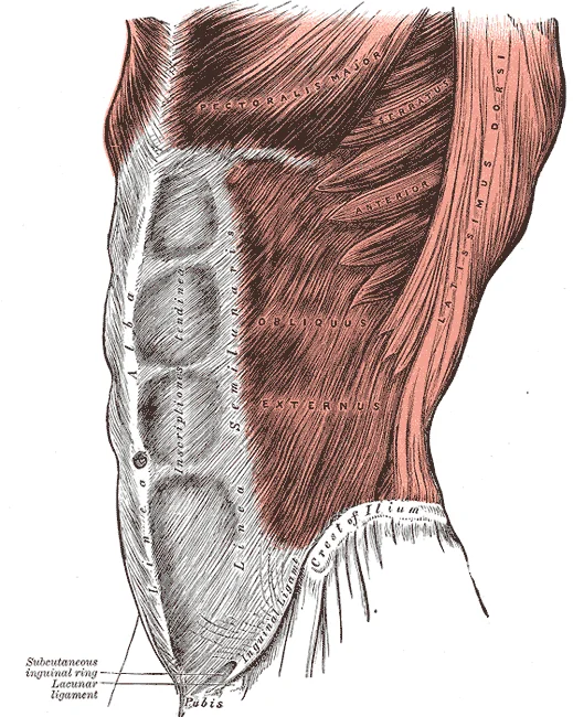
Figure 1Gray392
Long description
The Obliquus externus abdominis. Labelled illustration from Wikimedia Commons, used under its open licence.

Figure 1: The Obliquus externus abdominis.
The rectus abdominis is a paired muscle running vertically on each side of the anterior wall. It is enclosed within the rectus sheath, a fibrous compartment formed by the aponeuroses of the lateral abdominal muscles. The inferior epigastric artery (IEA) is a major blood vessel that supplies the anterior abdominal wall. It arises from the external iliac artery, proximal to the inguinal ligament. Soon thereafter it ascends on the anterior abdominal wall medial to the deep inguinal ring. After piercing the transversalis fascia, the IEA enters the rectus sheath ascending posterior to the rectus abdominis muscle. [5]
Ventral wall defects represent a significant clinical challenge, ranging from small incisional hernias to large midline defects. Entry into the abdomen is performed per the surgeon’s preference and skill. [2]
NoteClinical Pearl
The inferior epigastric artery is a key landmark for distinguishing direct from indirect inguinal hernias and is at risk during lower abdominal incisions.
| Muscle Group | General Orientation | Primary Action |
|---|---|---|
| External Oblique | Inferomedial | Trunk flexion and rotation |
| Internal Oblique | Superomedial | Trunk flexion and rotation |
| Transversus Abdominis | Horizontal | Compression of abdominal viscera |
| Rectus Abdominis | Vertical | Trunk flexion |
The umbilicus lies at the level of the third lumbar vertebra. It serves as a central landmark for dividing the abdomen into nine regions. These regions are defined by two vertical midclavicular lines and two horizontal planes: the subcostal plane and the transtubercular plane. The subcostal plane passes through the inferior margin of the tenth costal cartilage. The transtubercular plane passes through the tubercles of the iliac crests.
The anterior abdominal wall is composed of skin, fascia, and four pairs of muscles. These muscles protect the abdominal organs and move the vertebral column. The muscles include the rectus abdominis, external oblique, internal oblique, and transversus abdominis. The rectus abdominis extends through the entire length of the trunk. It is enclosed within the rectus sheath. The sheath is formed by the aponeuroses of the lateral muscles. The quadratus lumborum forms the posterior abdominal wall, not the anterior. [1]
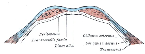
Figure 2Gray400
Long description
Diagram of a transverse section through the anterior abdomina wall, below the linea semicircularis.

Figure 2: Diagram of a transverse section through the anterior abdomina wall, below the linea semicircularis.
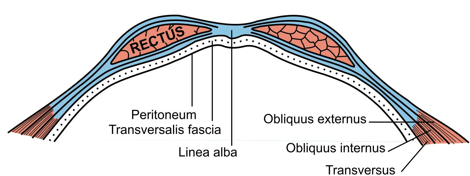
Figure 3Gray399
Long description
Diagram of sheath of Rectus. Labelled illustration from Wikimedia Commons, used under its open licence.

Figure 3: Diagram of sheath of Rectus.
Palpable landmarks include the xiphoid process, the costal margin, the anterior superior iliac spines, and the pubic symphysis. The mid-inguinal point lies midway between the anterior superior iliac spine and the pubic symphysis. This point is crucial for locating the femoral artery. The inferior epigastric artery runs vertically in the anterior abdominal wall. Its course is relevant to invasive procedures. Studies show the mean distance of the inferior epigastric artery from the midline is 5.17 ± 0.93 cm at the level of the mid-inguinal point. At the level of the anterior superior iliac spine, the distance is 4.57 ± 1.05 cm. At the level of the umbilicus, the distance is 5.27 ± 1.17 cm. [5]
Incisions must respect the anatomy of the muscles and nerves. Lower abdominal wall surgical incisions can injure the inferior epigastric artery. This injury may result in abdominal wall haematomas. Haematomas may expand considerably due to lack of tissue resistance. Safe areas for invasive procedures should be determined by localising the artery's course relative to standard anatomic landmarks. [5]
| Muscle | Origin | Insertion | Action | Nerve Supply |
|---|---|---|---|---|
| Rectus abdominis | Pubic symphysis and crest | Xiphoid process and costal cartilages | Flexes trunk | Intercostal nerves |
| External oblique | Lower eight ribs | Iliac crest and linea alba | Flexes and rotates trunk | Intercostal nerves |
| Internal oblique | Thoracolumbar fascia and iliac crest | Lower three ribs and linea alba | Flexes and rotates trunk | Intercostal and iliohypogastric nerves |
| Transversus abdominis | Inguinal ligament, iliac crest, thoracolumbar fascia | Linea alba and pubic crest | Compresses abdominal contents | Intercostal and iliohypogastric nerves |
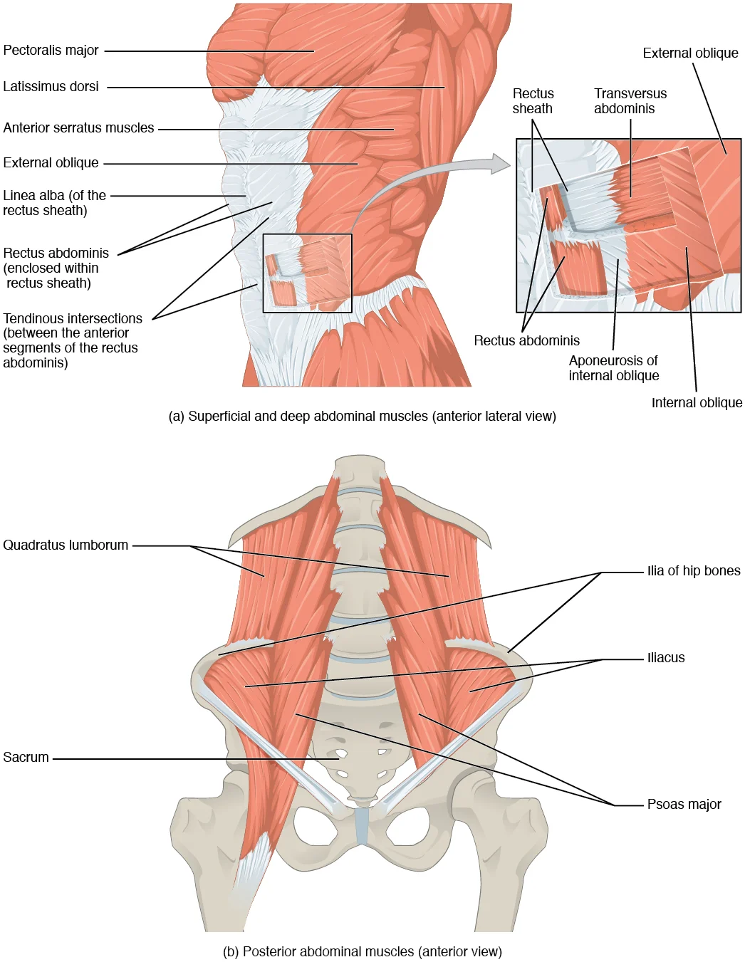
The posterior abdominal wall is formed by the lumbar vertebrae, parts of the ilia of the hip bones, psoas major and iliacus muscles, and quadratus lumborum muscle. This part of the core plays a key role in stabilizing the rest of the body and maintaining posture. [1]
The most superior landmark visible is the xiphoid process of the sternum. Although primarily part of the thoracic skeleton, it serves as the superior attachment point for the linea alba. Pathological conditions such as a chronic infective osteomyelitis of the xiphoid process of the sternum can mimic abdominal wall hernias, necessitating careful differential diagnosis. [2]
Moving inferiorly, the costal margins are formed by the cartilages of ribs seven to ten. These margins define the superior boundary of the abdominal cavity and protect the underlying liver and spleen. The inferior boundary is marked by the pubic symphysis and the pubic crests. The linea alba, a fibrous structure running vertically between the rectus abdominis muscles, extends from the xiphoid process to the pubic symphysis. In primiparous women, the inter-rectus distance (IRD) may be considered "normal" at wider values than in nulliparous women, reflecting changes in the linea alba during p [2]
The iliac crests form the lateral and inferior boundaries of the posterior abdominal wall structures. The psoas major and iliacus muscles, which originate from the lumbar vertebrae and ilia respectively, converge to form the iliopsoas tendon, passing deep to the inguinal ligament. The quadratus lumborum muscle attaches to the iliac crest and the twelfth rib, providing lateral stability.
The anterior abdominal wall lacks synovial joints in the traditional sense. Instead, structural integrity relies on fibrous connections and fascial continuity. The primary articulation occurs at the symphysis pubis, a secondary cartilaginous joint. This joint connects the pubic bones via a fibrocartilaginous disc. It allows minimal movement, providing stability for the pelvic girdle. The anterior abdominal wall muscles attach to the pubic crest and pectineal line. These attachments are secured by strong fibrous bands.
The symphysis pubis is central. It lies anterior to the bladder. The fibrocartilaginous disc fills the gap between them. Superior to the joint, the rectus abdominis muscles attach. The linea alba runs vertically between them. Lateral to the rectus sheaths, the external oblique aponeurosis contributes to the anterior rectus sheath. Inferiorly, the inguinal ligament runs from the anterior superior iliac spine to the pubic tubercle. This ligament forms the inferior boundary of the anterior abdominal wall. It supports the structures passing beneath it. The joint itself is reinforced by anterior and posterior ligaments. These ligaments prevent excessive separation of the pubic bones. [1]
The costal cartilages of ribs seven to ten form synchondroses with the sternum or each other. These are cartilaginous joints. They allow slight expansion during respiration. The xiphisternal joint is also a synchondrosis. It connects the body of the sternum to the xiphoid process. This joint may ossify in adulthood. The interchondral joints connect the cartilages of ribs six to nine. These are plane synovial joints. They permit gliding movements.
The lumbar vertebrae articulate with the psoas major and quadratus lumborum muscles. These muscles originate from the vertebral bodies and transverse processes. They do not form joints with the abdominal wall proper. However, their attachments define the posterior boundary. The anterior abdominal wall is essentially a continuous sheet of fascia and muscle. The linea alba serves as the midline fibrous raphe. It extends from the xiphoid process to the pubic symphysis. This structure is formed by the interlacing aponeuroses of the lateral abdominal muscles. It provides attachment for the rectus abdominis muscles. The strength of the wall depends on the integrity of these fascial connections.
DefinitionSymphysis Pubis
A secondary cartilaginous joint connecting the pubic bones, allowing minimal movement for pelvic stability.
| Structure | Type | Function |
|---|---|---|
| Symphysis Pubis | Cartilaginous | Connects pubic bones, stabilises pelvis |
| Linea Alba | Fibrous Raphe | Midline attachment for rectus muscles |
| Inguinal Ligament | Fibrous | Inferior boundary, supports pelvic structures |
| Xiphisternal Joint | Cartilaginous | Connects sternum to xiphoid process |
The fatty layer is variable in thickness and contains adipose tissue and superficial veins.
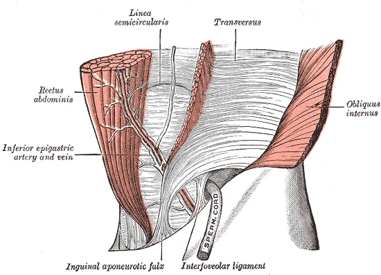
Figure 4Gray398
Long description
The interfoveolar ligament, seen from in front. (Modified from Braune.)

Figure 4: The interfoveolar ligament, seen from in front (Modified from Braune.).
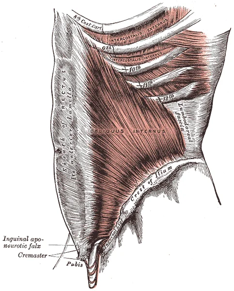
Figure 5Gray395
Long description
The Obliquus internus abdominis. Labelled illustration from Wikimedia Commons, used under its open licence.

Figure 5: The Obliquus internus abdominis.
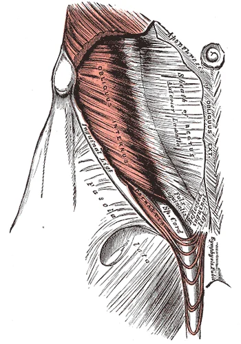
Figure 6Gray396
Long description
The Cremaster. Labelled illustration from Wikimedia Commons, used under its open licence.

Figure 6: The Cremaster.
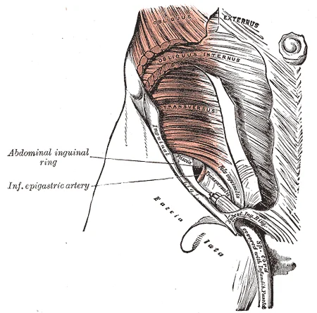
Figure 7Gray401
Long description
The abdominal inguinal ring. Labelled illustration from Wikimedia Commons, used under its open licence.

Figure 7: The abdominal inguinal ring.
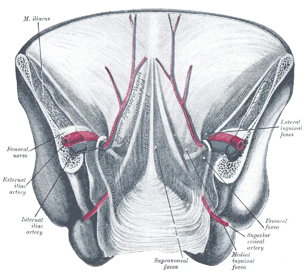
Figure 8Gray1036
Long description
Posterior view of the anterior abdominal wall in its lower half. The peritoneum is in place, and the various cords are shining through. (After Joessel.)

Figure 8: Posterior view of the anterior abdominal wall in its lower half.
Figure 91112 Muscles of the Abdomen
Long description
(a) The anterior abdominal muscles include the medially located rectus abdominis, which is covered by a sheet of connective tissue called the rectus sheath. On the flanks of the body, medial to the rectus abdominis, the abdominal wall is composed of three layers. The external oblique muscles form th

Figure 9: (a) The anterior abdominal muscles include the medially located rectus abdominis, which is covered by a sheet of connective tissue called the rectus sheath.
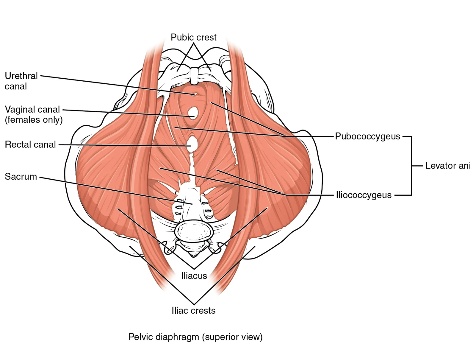
Figure 101115 Muscles of the Pelvic Floor
Long description
The pelvic floor muscles support the pelvic organs, resist intra-abdominal pressure, and work as sphincters for the urethra, rectum, and vagina.

Figure 10: The pelvic floor muscles support the pelvic organs, resist intra-abdominal pressure, and work as sphincters for the urethra, rectum, and vagina.
Each muscle is segmented by three transverse bands of collagen fibers called the tendinous intersections. These segments create the appearance of "six-pack abs" when hypertrophied. The linea alba is a white, fibrous band formed by the bilateral rectus sheaths joining at the anterior midline. The nerve and vessel supply run in the plane between the internal oblique and transversus abdominis muscles. [1]
The muscles of the vertebral column, thorax, and abdominal wall extend, flex, and stabilize different parts of the body’s trunk. The deep muscles of the core help maintain posture. The brain sends out electrical impulses to these various muscle groups to control posture by alternate contraction and relaxation. This is necessary so that no single muscle group becomes fatigued too quickly. If any one group fails to function, body posture will be compromised. [1]
Motor innervation of the abdominal muscles is segmental, through the intercostal nerves (Th7–Th11), subcostal nerve (Th12) and L1 branches in the plane between the internal oblique and transversus abdominis. Lateral, subcostal or lumbar incisions can injure these bundles. Prolonged denervation is associated with fibro-fatty replacement. [4]
| Muscle | Origin | Insertion | Action | Nerve Supply |
|---|---|---|---|---|
| Rectus Abdominis | Pubic crest and symphysis | Xiphoid process and costal cartilages of ribs 5-7 | Flexes trunk, compresses abdomen | Intercostal nerves (Th7-11), subcostal (Th12) |
| External Oblique | Lower 8 ribs | Linea alba, pubic tubercle, anterior iliac crest | Flexes and rotates trunk | Intercostal nerves (Th7-11), iliohypogastric, ilioinguinal |
| Internal Oblique | Inguinal ligament, iliac crest, thoracolumbar fascia | Lower 3 ribs, linea alba, pubic crest | Flexes and rotates trunk | Intercostal nerves (Th7-11), iliohypogastric, ilioinguinal |
| Transversus Abdominis | Inguinal ligament, iliac crest, thoracolumbar fascia, costal cartilages | Linea alba, pubic crest | Compresses abdominal contents | Intercostal nerves (Th7-11), iliohypogastric, ilioinguinal |
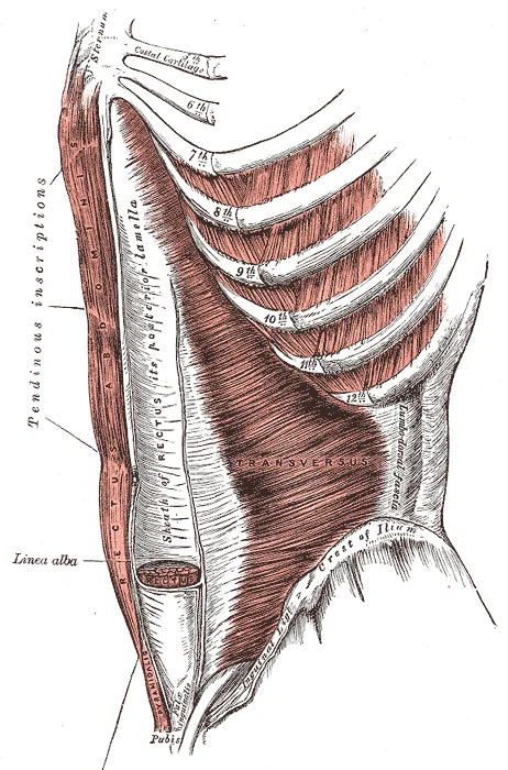
The rectus abdominis muscles are commonly called the “sit-up” muscles. The linea alba is a white, fibrous band that is made of the bilateral rectus sheaths that join at the anterior midline of the body. These enclose the rectus abdominis muscles. Each muscle is segmented by three transverse bands of collagen fibers called the tendinous intersections. [1]
NoteClinical Note
Lateral, subcostal or lumbar incisions can injure these bundles.
The arterial supply to the anterior abdominal wall is derived from three main sources: the superior epigastric, inferior epigastric, and lower intercostal arteries. These vessels anastomose extensively, providing collateral circulation. The image shows the layers including the rectus sheath, nerves, and vessels, with the umbilicus centrally located. The superior epigastric artery descends within the rectus sheath, while the inferior epigastric artery ascends from the external iliac region. The intercostal arteries run along the costal margins. Understanding these relations is crucial for surgical planning.
The superior epigastric artery is the terminal branch of the internal thoracic artery. It enters the rectus sheath behind the rectus abdominis muscle at the level of the sixth costal cartilage. It descends between the rectus abdominis and the posterior layer of the rectus sheath. It anastomoses with the inferior epigastric artery. This vessel supplies the upper part of the rectus abdominis muscle.
The inferior epigastric artery arises from the external iliac artery just above the inguinal ligament. It ascends obliquely along the medial side of the deep inguinal ring. It enters the rectus sheath by passing behind the rectus abdominis muscle. The artery gives off several branches, including the pubic branch and the cremasteric artery in males. It anastomoses with the superior epigastric artery. The course of the inferior epigastric artery is a key landmark for distinguishing direct from indirect inguinal hernias. Understanding the course of IEA would also help in minimising the likelihood of injury. [5]
The lower six intercostal arteries and the subcostal arteries contribute to the lateral supply. They run between the internal oblique and transversus abdominis muscles. They pierce the posterior layer of the rectus sheath to supply the rectus abdominis muscle. The lumbar arteries also provide collateral supply from the posterior aspect.
DefinitionClinical Pearl
The inferior epigastric artery serves as the lateral boundary of Hesselbach's triangle, a critical landmark for inguinal hernia repair.
| Vessel | Origin | Course | Termination |
|---|---|---|---|
| Superior Epigastric | Internal Thoracic | Descends in rectus sheath | Anastomoses with inferior epigastric |
| Inferior Epigastric | External Iliac | Ascends behind rectus abdominis | Anastomoses with superior epigastric |
| Lower Intercostals | Thoracic Aorta | Run between oblique muscles | Pierce posterior rectus sheath |
The venous drainage of the anterior abdominal wall is a critical anatomical consideration, particularly when evaluating surgical approaches or complications such as hernias, which affect millions of patients worldwide. Understanding these vessels is essential for safe dissection and avoiding significant haemorrhage during abdominal wall reconstruction. [2]
Lateral to the rectus muscles, the superficial epigastric vein runs in the subcutaneous tissue. It drains into the great saphenous vein near the saphenofemoral junction.
This connection is vital for collateral flow between the superior and inferior vena cava systems. It ascends along the posterior aspect of the rectus abdominis muscle. It terminates by anastomosing with the superior epigastric vein. This pathway is often compromised in patients with increased intra-abdominal pressure, such as those with large ventral hernias.
This vessel drains the skin and subcutaneous tissues of the lower anterior abdominal wall. It is often used in venous bypass procedures due to its accessibility. This vessel is important for drainage of the upper lateral abdominal region.
In patients undergoing abdominal wall repair, knowledge of these veins prevents inadvertent injury. The complexity of abdominal wall hernia disease means that surgical plans must account for vascular variations. For instance, in laparoscopic approaches, trocars must be placed carefully to avoid the inferior epigastric vessels. Bowel injury is also a concern during such procedures. Therefore, precise anatomical knowledge is paramount. [2]
The venous system also plays a role in postoperative recovery. Abdominal binders may reduce pain and improve physical function after major abdominal surgery. This support helps manage swelling and may influence venous return. In cases of infected mesh, replacement strategies must consider vascular integrity. The choice of surgical approach, whether open, laparoscopic, or endoscopic, depends on the specific vascular and muscular anatomy. [2] [3]
DefinitionSuperior Epigastric Vein
Drains the upper rectus sheath and anastomoses with the inferior epigastric vein, connecting the internal thoracic and external iliac systems.
DefinitionInferior Epigastric Vein
Ascends within the rectus sheath to join the superior epigastric vein, providing a key collateral pathway between the caval systems.
| Vein | Origin | Termination | Drainage Area |
|---|---|---|---|
| Superior Epigastric | Internal Thoracic | Inferior Epigastric | Upper Rectus Sheath |
| Inferior Epigastric | External Iliac | Superior Epigastric | Lower Rectus Sheath |
| Superficial Epigastric | Subcutaneous Tissue | Great Saphenous | Lower Abdominal Skin |
| Lateral Thoracic | Lateral Chest Wall | Axillary | Lateral Abdominal Wall |
These nerves run in the neurovascular plane between the internal oblique and transversus abdominis muscles, providing both motor supply to the muscles and sensory supply to the overlying skin.
This arrangement allows for coordinated contraction of the abdominal wall. [1]
| Structure | Motor Supply | Cutaneous Supply |
|---|---|---|
| External Oblique | T7-T11, T12, L1 | T7-T12, L1 |
| Internal Oblique | T7-T11, T12, L1 | T7-T12, L1 |
| Transversus Abdominis | T7-T11, T12, L1 | T7-T12, L1 |
| Rectus Abdominis | T7-T11 | T7-T11 |
| Skin (Upper) | T7-T9 | T7-T9 |
| Skin (Middle) | T10-T11 | T10-T11 |
| Skin (Lower) | T12, L1 | T12, L1 |
The cutaneous supply follows a dermatomal pattern. The upper abdomen is supplied by T7-T9, the middle abdomen by T10-T11, and the lower abdomen by T12 and L1. This segmental innervation is clinically important for localising lesions and planning surgical incisions to avoid nerve damage. The iliohypogastric and ilioinguinal nerves also supply the skin of the suprapubic and inguinal regions.
Clinical pearlClinical Pearl
Damage to the lower intercostal nerves during surgery can lead to weakness of the abdominal wall and sensory loss in the corresponding dermatomes.
The cooperation between the diaphragm and abdominal muscles, known as the Valsalva maneuver, involves the contraction of these innervated muscles to increase intra-abdominal pressure. This is essential for defecation, urination, and childbirth. The nerves ensure that the muscles contract in a coordinated manner to achieve this effect. [1]
Above the umbilicus, the lymphatics follow the superficial epigastric and thoracoepigastric veins towards the axilla. Below the umbilicus, they drain into the superficial inguinal nodes.
The lymphatic drainage of the anterior abdominal wall is divided into two main regions by the umbilicus. This anatomical landmark serves as a crucial boundary for clinical assessment.
The lymphatics from the skin and subcutaneous tissue above the umbilicus drain primarily into the axillary lymph nodes. Specifically, the vessels follow the course of the thoracoepigastric vein and the lateral thoracic vein. These vessels travel laterally towards the axilla. This pathway is significant because pathology in the upper abdominal wall may present with axillary lymphadenopathy.
The lymphatics from the skin and subcutaneous tissue below the umbilicus drain into the superficial inguinal lymph nodes. The vessels accompany the superficial epigastric vein and the external pudendal vessels. They travel inferiorly towards the groin. The superficial inguinal nodes are located just below the inguinal ligament. This drainage pattern is clinically important for understanding the spread of infections or malignancies from the lower abdominal wall and perineum.
The umbilicus acts as a watershed area. Lesions above the umbilicus typically drain to the axilla, while lesions below drain to the inguinal nodes. However, there can be some anastomosis between these systems. Understanding this drainage is essential for surgical planning and staging of diseases. For instance, a melanoma above the umbilicus may require axillary node evaluation, whereas one below may require inguinal node assessment.
Lateral to these muscles are the flat muscles of the abdominal wall: the external oblique, internal oblique, and transversus abdominis.
The spaces within the abdominal wall are defined by these fascial layers. The extraperitoneal space lies between the transversalis fascia and the peritoneum, containing fat and the inferior epigastric vessels. These vessels arise from the external iliac artery and ascend posterior to the rectus abdominis muscle, piercing the transversalis fascia to enter the rectus sheath. They anastomose with the superior epigastric artery, a terminal branch of the internal thoracic artery, which descends within the sheath. This vascular arrangement provides collateral circulation but also poses a risk of significant haemorrhage if injured during trocar insertion. These findings may be useful not only for laparoscopic procedures but also for image-guided biopsy, abdominal paracentesis, and placement of abdominal drains. [5]
Figure 11Gray397
Long description
The Transversus abdominis, Rectus abdominis, and Pyramidalis.

Figure 11: The Transversus abdominis, Rectus abdominis, and Pyramidalis.
The contents of the anterior abdominal wall include the rectus abdominis muscles, the pyramidalis muscle, the neurovascular bundles, and the aponeuroses of the lateral muscles. The anterior layer of the rectus sheath receives contributions from the external oblique aponeurosis and the anterior leaf of the internal oblique aponeurosis. The posterior layer, present only above the arcuate line, receives the posterior leaf of the internal oblique aponeurosis and the transversus abdominis aponeurosis. The arcuate line itself is typically located halfway between the umbilicus and the pubic symphysis. Understanding these relations is essential for safe surgical entry and repair of ventral hernias, where the integrity of the rectus sheath and the position of the inferior epigastric vessels dictate the choice of incision and mesh placement.
The outermost layer is the skin, followed by the subcutaneous tissue containing fat and the superficial fascia. Deep to this lies the rectus abdominis muscle, enclosed within the rectus sheath. The linea alba runs vertically in the midline, separating the two rectus muscles. Lateral to the rectus sheath are the three flat muscles: the external oblique, internal oblique, and transversus abdominis. Their aponeuroses contribute to the rectus sheath anteriorly and posteriorly, depending on the level. The peritoneum lines the inner surface. Understanding these layers is critical for interpreting pathology such as hernias or diastasis. [1]
Computed tomography (CT) and magnetic resonance imaging (MRI) are not routinely recommended for diagnosing ventral hernias before laparoscopic repair. However, CT is frequently used to assess abdominal wall closure after component separation techniques. On CT, the anterior abdominal wall appears as distinct soft-tissue layers. Muscle cross-sectional area, mean attenuation, and intermuscular fat can be measured at the L3–L4 level. No anterior abdominal wall-specific diagnostic cut-off is available for these measurements. [2][4]
Magnetic resonance imaging provides detailed soft-tissue contrast, useful for evaluating muscle integrity and hernia defects. Ultrasound is a non-invasive diagnostic tool that allows differentiation of hernia from other abdominal swellings. Ultrasound and calipers are adequate methods to assess diastasis of the rectus abdominal muscle. Ultrasound was the most evaluated method in systematic reviews. [2]
X-ray has limited utility for soft-tissue anatomy of the anterior abdominal wall. It may show gas patterns or foreign bodies but does not delineate muscle layers or hernia defects effectively. Therefore, cross-sectional imaging or ultrasound is preferred for detailed anatomical assessment.
| Modality | Primary Use | Key Limitation |
|---|---|---|
| CT | Assess muscle mass, hernia size | No AAW-specific cut-offs |
| MRI | Soft tissue detail | Not routine for diagnosis |
| Ultrasound | Hernia vs swelling, diastasis | Operator dependent |
| X-ray | Indirect signs | Poor soft tissue resolution |
NoteImaging Context
CT measurements at L3–L4 are standard for sarcopenia but lack anterior abdominal wall-specific diagnostic cut-offs.
The anterior abdominal wall serves as a dynamic barrier, separating the abdominal cavity from the external environment while facilitating core stability and respiration. Clinically, understanding its layered architecture is essential for interpreting surface landmarks, managing trauma, and performing surgical interventions. The wall’s integrity relies on the coordinated action of its musculature and the innervation provided by the lower thoracic and upper lumbar nerves.
The sheath is formed by the aponeuroses of the lateral abdominal muscles. Superior to this line, the posterior rectus sheath is present; inferior to it, it is absent. Understanding these relations helps in identifying potential injury sites during laparoscopic procedures and in assessing hernia defects.
Injuries to the anterior abdominal wall often involve penetrating trauma or blunt force, potentially damaging the underlying viscera or causing muscle tears. Nerve palsies, particularly of the iliohypogastric and ilioinguinal nerves, can result from surgical incisions, leading to chronic pain or sensory deficits in the suprapubic and inguinal regions. These nerves provide motor supply to the internal oblique and transversus abdominis muscles and sensory innervation to the skin of the lower abdomen and groin.
Procedures such as laparoscopic ventral hernia repair require precise knowledge of the abdominal wall layers. Minimally invasive techniques aim to reduce morbidity by avoiding large incisions. Standard uncoated permanent synthetic meshes, such as polypropylene, PVDF, or polyester, may be used for reinforcement. The choice of mesh and placement technique depends on the defect size and location. For complex defects, component separation techniques may be employed to achieve midline closure. [3]
Surface landmarks are vital for clinical examination and procedural guidance. The midline linea alba, formed by the fusion of aponeuroses, is a common site for incisions. The lateral borders of the rectus abdominis, the linea semilunaris, mark the transition to the flat abdominal muscles. These landmarks guide the placement of trocars and the assessment of hernia boundaries.
| Muscle | Origin | Insertion | Action | Nerve Supply |
|---|---|---|---|---|
| Rectus abdominis | Pubic symphysis and crest | Xiphoid process and costal cartilages 5-7 | Flexes trunk, compresses abdomen | Lower intercostal nerves (T7-T12) |
| External oblique | Lower 8 ribs | Iliac crest, pubic tubercle, linea alba | Compresses abdomen, rotates trunk | Lower intercostal nerves, iliohypogastric, ilioinguinal |
| Internal oblique | Thoracolumbar fascia, iliac crest, inguinal ligament | Lower 3 ribs, linea alba, pubic crest | Compresses abdomen, rotates trunk | Lower intercostal nerves, iliohypogastric, ilioinguinal |
| Transversus abdominis | Lower 6 costal cartilages, thoracolumbar fascia, iliac crest, inguinal ligament | Linea alba, pubic crest | Compresses abdomen, stabilizes trunk | Lower intercostal nerves, iliohypogastric, ilioinguinal |
Clinical pearlClinical Pearl
The arcuate line marks the inferior limit of the posterior rectus sheath, making the area below it more susceptible to herniation and hematoma formation.
Below this line, the posterior sheath is absent, leaving only the transversalis fascia and peritoneum posterior to the muscle.
DefinitionArcuate Line
The arcuate line marks the inferior limit of the posterior rectus sheath, typically located halfway between the umbilicus and the pubic symphysis.
The neurovascular supply follows a segmental pattern. The lower six intercostal nerves (T7–T11) and the subcostal nerve (T12) supply the anterolateral wall. These nerves run between the internal oblique and transversus abdominis muscles, forming the neurovascular plane. This plane is critical during open hernia repairs to avoid nerve injury. The iliohypogastric and ilioinguinal nerves (L1) supply the suprapubic region and groin. [1]
Exam trapNerve Injury
Injury to the iliohypogastric nerve during lower abdominal incisions can lead to sensory loss over the suprapubic region and weakness of the abdominal muscles.
The linea alba, a fibrous structure running down the midline, results from the interlacing of the aponeuroses. It is avascular and a common site for midline incisions.
| Muscle | Origin | Insertion | Action | Nerve Supply |
|---|---|---|---|---|
| Rectus Abdominis | Pubic symphysis and crest | Xiphoid process and costal cartilages 5–7 | Flexes trunk, compresses abdomen | Lower intercostal nerves (T7–T12) |
| External Oblique | Lower 8 ribs | Linea alba, pubic tubercle, ASIS | Compresses abdomen, rotates trunk | Lower intercostal nerves, iliohypogastric |
| Internal Oblique | Inguinal ligament, iliac crest | Lower 3 ribs, linea alba | Compresses abdomen, rotates trunk | Lower intercostal nerves, iliohypogastric, ilioinguinal |
| Transversus Abdominis | Inguinal ligament, iliac crest, lower 6 costal cartilages | Linea alba, pubic crest | Compresses abdomen | Lower intercostal nerves, iliohypogastric, ilioinguinal |
Clinical relevance includes the formation of hernias. Weaknesses in the abdominal wall, such as the inguinal canal or the femoral ring, allow protrusion of intra-abdominal contents.
Clinical pearlInguinal Ligament
The inguinal ligament is the thickened inferior border of the external oblique aponeurosis and serves as a key landmark for hernia repair.
The blood supply arises from the superior and inferior epigastric arteries. The superior epigastric artery is a terminal branch of the internal thoracic artery. The inferior epigastric artery arises from the external iliac artery. These vessels anastomose within the rectus sheath, providing collateral circulation. Knowledge of this vascular pattern is essential to prevent ischaemia during surgery.
NoteVascular Anatomy
The inferior epigastric vessels serve as a lateral boundary for direct inguinal hernias, which occur medial to these vessels.
Surgical approaches must respect these anatomical layers. Open repairs involve incising the skin, subcutaneous tissue, and aponeuroses in the correct plane. Laparoscopic repairs enter the preperitoneal space, avoiding the neurovascular plane. Understanding the origin, course, relations, branches, and termination of each structure ensures safe and effective surgical outcomes.
Students frequently confuse the anatomical definition of diastasis recti with simple abdominal obesity or generalised laxity. Diastasis recti is specifically defined as the bulging of the linea alba under increased intra-abdominal pressure, not merely a cosmetic concern. A critical error is assuming that all cases resolve spontaneously. While women during pregnancy typically develop this condition which regresses within a year after child birth, in some patients, the diastasis persists. [3]
Another frequent mistake involves the measurement technique for diagnosis. The width of the linea alba is measured at 2 reference points. The first is at 3 cm superior to the umbilicus and the second is 2 cm inferior. A patient is considered to have diastasis if the width between the 2 medial edges of the recti is greater than 22 mm at the first reference point and greater than 16 mm at the second point. Students often measure at arbitrary points or fail to distinguish between the medial edges of the rectus abdominis muscles. [3]
In surgical contexts, a major pitfall is combining primary and incisional ventral hernias in analysis or treatment planning. Studies have consistently shown that the outcomes of these two types of ventral hernias are different, so it would be inappropriate to combine them when attempting to study ventral hernia repair. Furthermore, assuming that primary hernias always have better outcomes than incisional ones is incorrect. Some sub-populations of primary hernias will have more risk/worse outcomes than some sub-populations of incisional/secondary hernias. [2]
Regarding repair techniques, students may overlook the specific risks associated with laparoscopic repair of large incisional hernias. Abdominal bulging is a specific problem associated with laparoscopic repair of large incisional hernias. This wide range may be attributable in part to surgical technique differences between surgeons whether close hernia defect, or in part to interpretation by the examiner and the opinion of the patient, but evidence for this is limited. [2]
Finally, when considering component separation, it is erroneous to assume a single technique is universally superior. New topics included in this update are: In which patient group is a component separation indicated? Should the component separation be done open or endoscopically? Is an anterior component separation better than the posterior one?. We are well aware that with respect to these innovations the evidence is not yet strong enough to give valuable statements or recommendations, however, the guidelines should inform the surgical community and stimulate further studies to gain more knowledge in the coming years.
At the bedside, the diagnosis of diastasis recti is managed on a regular basis by surgeons. It is simply described as bulging of the linea alba with increased intra–intra-abdominal pressure. Clinicians must differentiate between transient postpartum changes and persistent defects requiring intervention.
In the operating theatre, understanding the anatomy of the anterior abdominal wall is crucial for hernia repair. The search was performed in October 2011 and a total of two unique publications were returned from this search. Both were clinical studies. A secondary search revealed additional 10 publications pertinent to this topic. Additional information on this topic was searched for on UpToDate. This highlights the need for robust evidence when selecting techniques like component separation.
On imaging, ultrasound is the gold standard for non-invasive IRD measurement in parous women when investigating diastasis recti; however, its use is limited when IRD is large. Another Reliability and validity study promotes the value of extended field of view (FOV) technique: Ultrasound imaging is the gold standard for non-invasive IRD measurement in parous women when investigating diastasis recti; however, its use is limited when IRD is large. Extended FOV techniques (panoramic USI or using acoustic standoff pads) allow complete visualization of the linea alba when the IRD is large and conventional imaging is not sufficient. FOV techniques were highly correlated with those acquired using conventional imaging (r > 0.95, p < 0.0001). [2]
In examinations, students must recall the classification of myofascial deformities. Nahas proposed a classification that included 4 types of “myofascial deformities” with a tailored treatment for each type. Additionally, knowledge of hernia recurrence risks is tested. Hope et al. [14] reported in this review that the most common techniques in the literature is a combination of suture and tack fixation and the double-crown technique. The risk of recurrence is most likely found in the inferior/pubic region. Additional suture fixation may reduce this complication. [2] [3]
DefinitionDiastasis Recti
Bulging of the linea alba with increased intra-abdominal pressure, diagnosed when the width between the 2 medial edges of the recti is greater than 22 mm at 3 cm superior to the umbilicus and greater than 16 mm at 2 cm inferior.
| Reference Point | Location | Threshold for Diagnosis |
|---|---|---|
| First | 3 cm superior to the umbilicus | > 22 mm |
| Second | 2 cm inferior to the umbilicus | > 16 mm |
Diastasis Recti Management
- Assess Abdominal WallCheck for bulging of the linea alba with increased intra-abdominal pressure
- Question: Measure IRD
Yes, >22mm/16mm
- Diagnose DiastasisPersistent in 1/3 of postpartum women
- Question: Symptomatic?
Yes
- Consider RepairTailored treatment based on Nahas classification
No
- Conservative ManagementMonitor regression
No, <22mm/16mm
- Normal AnatomyNo diastasis recti
Diastasis recti is bulging of the linea alba with increased intra-abdominal pressure. It typically develops in women during pregnancy and regresses within a year after child birth. In a third of those patients, the diastasis persists beyond the postpartum period. The width of the linea alba is measured at 2 reference points for diagnosis. The first reference point is at 3 cm superior to the umbilicus. The second reference point is 2 cm inferior to the umbilicus. A diagnosis requires width >22 mm at the first point and >16 mm at the second. Ultrasound is the gold standard for non-invasive IRD measurement in parous women.
Answer each one in your head first, then open the answer.
What is the diagnostic threshold for diastasis recti at 3 cm superior to the umbilicus?
Show answerHide answer
Greater than 22 mmWhat is the diagnostic threshold for diastasis recti at 2 cm inferior to the umbilicus?
Show answerHide answer
Greater than 16 mmWhat percentage of postpartum women experience persistent diastasis recti?
Show answerHide answer
One thirdWhat is the gold standard imaging for non-invasive IRD measurement in parous women?
Show answerHide answer
UltrasoundWhere is the risk of recurrence most likely found in ventral hernia repair?
Show answerHide answer
In the inferior/pubic region
- Betts JG, Young KA, Wise JA, et al. Axial Muscles of the Abdominal Wall, and Thorax. OpenStax, Anatomy and Physiology 2e (CC BY-NC-SA 4.0). Accessed .
- Bittner R, Bain K, Bansal VK, et al. Update of Guidelines for laparoscopic treatment of ventral and incisional abdominal wall hernias (International Endohernia Society (IEHS))-Part A. Surgical endoscopy 2019;33(10):3069-3139. doi:10.1007/s00464-019-06907-7. Accessed .
- Bittner R, Bain K, Bansal VK, et al. Update of Guidelines for laparoscopic treatment of ventral and incisional abdominal wall hernias (International Endohernia Society (IEHS)): Part B. Surgical endoscopy 2019;33(11):3511-3549. doi:10.1007/s00464-019-06908-6. Accessed .
- Muraviev SY, Akulov ZA, Sukhanova MA, et al. Metabolism, Morphology and Function of the Anterior Abdominal Wall Musculature in Ventral Hernias and After Repair: A Narrative Review and Research Agenda. Journal of personalized medicine 2026;16(8):419. doi:10.3390/jpm16080419. Accessed .
- Joy P, Prithishkumar IJ, Isaac B. Clinical anatomy of the inferior epigastric artery with special relevance to invasive procedures of the anterior abdominal wall. Journal of minimal access surgery 2017;13(1):18-21. doi:10.4103/0972-9941.181331. Accessed .
Figure credits
- Figure 1: Henry Vandyke Carter, via Wikimedia Commons, Public domain, Public domain.
- Figure 2: Henry Vandyke Carter, via Wikimedia Commons, Public domain, Public domain.
- Figure 3: Henry Vandyke Carter, via Wikimedia Commons, Public domain, Public domain.
- Figure 4: Henry Vandyke Carter, via Wikimedia Commons, Public domain, Public domain.
- Figure 5: Henry Vandyke Carter, via Wikimedia Commons, Public domain, Public domain.
- Figure 6: Henry Vandyke Carter, via Wikimedia Commons, Public domain, Public domain.
- Figure 7: Henry Vandyke Carter, via Wikimedia Commons, Public domain, Public domain.
- Figure 8: Henry Vandyke Carter, via Wikimedia Commons, Public domain, Public domain.
- Figure 9: OpenStax, via Wikimedia Commons, CC BY 4.0, CC BY 4.0.
- Figure 10: OpenStax, via Wikimedia Commons, CC BY 4.0, CC BY 4.0. resized.
- Figure 11: Henry Vandyke Carter, via Wikimedia Commons, Public domain, Public domain.
Educational content for medical students and doctors. It is not a substitute for clinical judgement or local guidelines.