Applied thoracic anatomy
Applied thoracic anatomy covering cardiac, lymphatic, spinal and CSF structures.
The thoracic cavity houses the heart, lungs, and major vessels, bounded by the thoracic wall. The heart shape resembles a pinecone, broad superiorly and tapering to the apex. A typical heart measures 12 cm in length, 8 cm wide, and 6 cm thick. Female hearts weigh 250–300 grams, while male hearts weigh 300–350 grams. Athletes may exhibit hypertrophy, increasing cell size without increasing cell numbers. [1]
Auscultation points correspond to valve locations, though specific points are not detailed in the provided texts. CT and MRI sections visualise the heart, lungs, and mediastinal structures. Echocardiographic windows assess cardiac function and valve integrity. Normal variants include size differences between sexes and athletic hypertrophy.
Valves ensure one-way flow: atrioventricular valves between atria and ventricles, and semilunar valves leading to the pulmonary trunk and aorta. The interventricular septum separates the ventricles. [1]
Coronary circulation supplies the heart, peaking during relaxation and ceasing during contraction. This cycling is critical due to the heart's ceaseless activity. [1]
The lymphatic system drains interstitial fluid. Capillary filtration releases 20 liters of plasma daily into interstitial space. Seventeen liters are reabsorbed by blood vessels. The remaining three liters enter the lymphatic system as lymph. Lymph is not pumped by the heart but moves via body movement, skeletal muscle contraction, and breathing. One-way valves prevent backflow. Lymph empties into the bloodstream at the junction of the jugular and subclavian veins. [2]
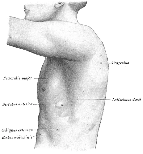
Figure 1Gray1215
Long description
The left side of the thorax. Labelled illustration from Wikimedia Commons, used under its open licence.

Figure 1: The left side of the thorax.
The spleen is a secondary lymphoid organ, 12 cm long, attached to the stomach. It filters blood, removing microbes and dying red blood cells. [2]
Thoracic spine anatomy involves costovertebral, costotransverse, and facet joints[1]. Thoracic nerve roots traverse the radicular canal, divided into retrodiscal, parapedicular, and foraminal sections[1]. The intervertebral foramen borders include the vertebral body, disc, ligaments, and articular processes[1]. Rib heads can obscure neural elements[1]. [3]
Cerebrospinal fluid (CSF) volume is 150 mL, produced at 500 mL daily[1]. Production is 60% choroid plexus and 40% extrachoroidal[1]. Absorption begins at 68 mm H2O and rises linearly to 250 mm H2O[1]. Equilibrium occurs at 112 mm H2O[1]. [3]
CSF leaks post-chest operation are diagnosed by CT myelography or radionuclide cisternography[1]. CT myelography aids surgical planning but has a high false-negative rate[1]. Nuclear cisternography with 111In-DTPA is more sensitive but lacks anatomical detail[1]. [3]
DefinitionHypertrophy
Exercise increases cardiomyocyte size by adding protein myofilaments, without increasing cell numbers. [1]
| Structure | Function/Detail |
|---|---|
| Heart | Pinecone shape, 12x8x6 cm |
| Lymph | Drains 3L excess interstitial fluid daily |
| Spleen | 12 cm long, filters blood |
| CSF | 150 mL volume, 500 mL/day production[1] |
CSF Dynamics
- CSF Production500 mL/day (60% choroid, 40% interstitial)
- Question: Pressure < 68 mm H2O?
Yes
- Low Absorption
No
- Absorption Begins
- Equilibrium at 112 mm H2OFormation equals absorption
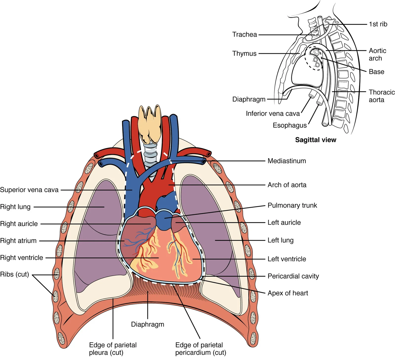
Figure 22001 Heart Position in ThoraxN
Long description
The heart is located within the thoracic cavity, medially between the lungs in the mediastinum. It is about the size of a fist, is broad at the top (called the base), and tapers toward the apex.

Figure 2: The heart is located within the thoracic cavity, medially between the lungs in the mediastinum.
The heart lies medially within the thoracic cavity, separated from other mediastinal structures by the pericardium[1]. Its anterior surface sits deep to the sternum and costal cartilages, while the dorsal surface lies near the vertebral bodies[1]. The base of the heart, where the great vessels attach, is located at the level of the third costal cartilage[1]. This position is critical for placing a stethoscope to listen for heart sounds[1]. The slight deviation of the apex to the left creates a depression in the medial surface of the superior lobe of the left lung, known as the cardiac notch[1].
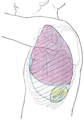
The heart’s apex is marked at the left fifth intercostal space, mid-clavicular line. The pleural lines are marked on the chest wall, indicating the inferior extent of the pleura. These points guide auscultation and help correlate surface anatomy with internal structures.
Surface markings are essential for safe thoracocentesis and chest drain insertion. The pleura extends further down the chest wall than the lung tissue. These levels guide needle insertion to avoid lung puncture. For chest drains, the insertion site is usually in the fourth or fifth intercostal space, anterior to the mid-axillary line, to ensure drainage of pleural fluid or air. The surface markings on a chest radiograph help correlate clinical findings with internal anatomy. Auscultation points for the heart valves are derived from the surface projection of the valve planes. The tricuspid valve, which separates the right atrium and ventricle, is best heard at the lower left sternal border[1]. The mitral valve is heard at the apex. The aortic and pulmonary valves are heard at the second intercostal spaces. Understanding these surface markings allows clinicians to accurately assess cardiac and pleural pathology.
Figure 3Gray1217
Long description
Side of thorax, showing surface markings for bones, lungs (purple), pleura (blue), and spleen (green).

Figure 3: Side of thorax, showing surface markings for bones, lungs (purple), pleura (blue), and spleen (green).
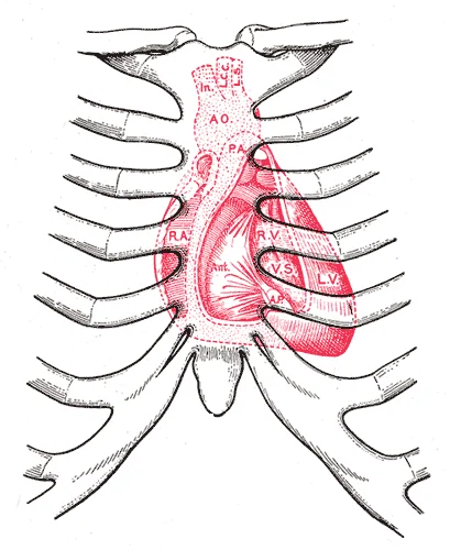
Figure 4Gray1218
Long description
Diagram showing relations of opened heart to front of thoracic wall. Ant. Anterior segment of tricuspid valve. A O. Aorta. A.P. Anterior papillary muscle. In. Innominate artery. L.C.C. Left common carotid artery. L.S. Left subclavian artery. L.V. Left ventricle. P.A. Pulmonary artery. R.A. Right atr

Figure 4: Diagram showing relations of opened heart to front of thoracic wall (ant).
The head articulates with the vertebral body, while the tubercle connects to the transverse process. Understanding these landmarks is critical for safe surgical access and avoiding neural injury.
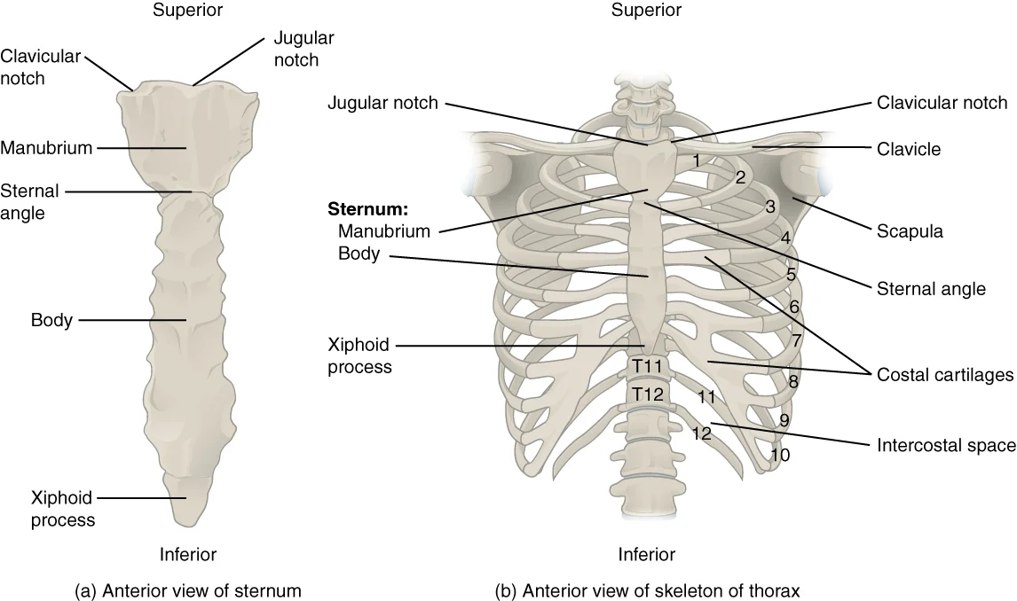
The costovertebral joint connects the head of the rib to the vertebral body. The rib head possesses a facet that articulates in planar joints with a single costal facet on T1, T10, T11, and T12. With all other levels having articulations with the costal facet of their numbered level and additionally the inferior costal demifacet of the vertebral body one level above and with the associated intervertebral disc. The vertebral body costal facets are found dorsally near the root of the pedicle, with more rostral location in the upper thoracic spine and more caudal location in the lower thoracic spine. The ribs that have two articulations have two rib head facets with an intervening interarticular crest that has ligamentous attachments to the intervertebral disc. The capsular, radiate, and intraarticular ligaments are associated with this joint. [3]
Figure 5721 Rib Cage
Long description
The thoracic cage is formed by the (a) sternum and (b) 12 pairs of ribs with their costal cartilages. The ribs are anchored posteriorly to the 12 thoracic vertebrae. The sternum consists of the manubrium, body, and xiphoid process. The ribs are classified as true ribs (1–7) and false ribs (8–12). Th

Figure 5: The thoracic cage is formed by the (a) sternum and (b) 12 pairs of ribs with their costal cartilages.
The costotransverse joint links the rib tubercle to the transverse process. An articular region on the tubercle of the first ten ribs articulates with a facet on the corresponding transverse process. These synovial joints transition from a convex rib portion in the upper thoracic spine to planar in the lower thoracic spine. The costotransverse, superior costotransverse, and lateral costotransverse ligaments are associated with this joint. The costotransverse ligament attaches the rib to the transverse process, occupying the costotransverse foramen. The superior costotransverse ligament has anterior and posterior layers—the anterior layer attaches the crest of the rib neck to the transverse process and is continuous laterally with the internal intercostal membrane, crossed by the intercostal vessels and nerves. [3]
Foraminal ligaments modify the intervertebral foramen. There are various foraminal ligaments closely related to the exiting nerve root that may be present in the thoracic spine intervertebral foramen. The ligamentous structures vary in width and thickness from 2 to 5 mm and substantially decrease the aperture of the intervertebral foramen—while no data is reported specific to the thoracic spine, the actual aperture size in thoracolumbar spine among 49 human nonpathological intervertebral foramen was on average 31.5% less than the size of the osseous foramen.
Understanding these relationships is crucial for interpreting thoracic trauma and surgical approaches.
The costovertebral joints are synovial joints where the head of the rib articulates with the demifacets of the vertebral bodies. The superior fibers of the joint capsule extend through the intervertebral foramen to attach to the posterior aspect of the intervertebral disc. The posterior capsular fibers are continuous with the fibers of the costotransverse ligament. The radiate ligaments fan out from the anterior aspect of the rib to the anterior vertebral body—superiorly to the vertebra above, laterally to the intervertebral disc, and inferiorly to the corresponding vertebra. Costovertebral joints where the rib articulates with two vertebrae also have an extrasynovial, intra-articular ligament that attaches from the crest on the rib head that lies between the two costal demifacets to the intervertebral disc. These ligaments provide stability against rotational forces. [3]
The costotransverse joints are synovial joints between the tubercle of the rib and the transverse process of the vertebra. They are encased in a fibrous capsule that attaches peripheral to the articular surfaces. The inferior facets are appropriately positioned facing anteriorly and medially to match their partner. This orientation restricts movement to slight gliding, allowing for the expansion and contraction of the thoracic cavity during respiration. [3]
Thoracic facet joints consist of apposed articular processes lined with hyaline cartilage extending from adjacent vertebrae. They are synovial joints encased in a fibrous capsule that attaches peripheral to the articular surfaces. The inferior facets are appropriately positioned facing anteriorly and medially to match their partner. These joints contribute to the rigidity of the thoracic spine, protecting the spinal cord and visceral organs. [3]
Clinical pearlClinical Relevance
The specific orientation of thoracic facet joints limits rotation compared to the lumbar spine, making the thoracic region more stable but less flexible.
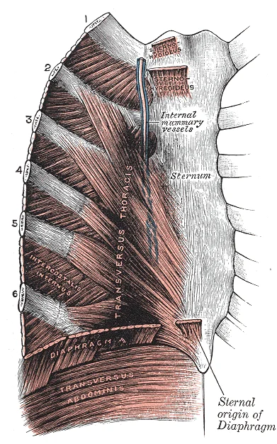
Figure 6Gray390
Long description
Posterior surface of sternum and costal cartilages, showing Transversus thoracis.

Figure 6: Posterior surface of sternum and costal cartilages, showing Transversus thoracis.
The central structure is the heart, enclosed within the pericardial sac. This sac consists of an outer fibrous capsule and an inner parietal pericardium lined with serous membrane. Between the sac and the heart lies the pericardial cavity, filled with lubricating serous fluid. When the lymphatic system is damaged in some way, such as by being blocked by cancer cells or destroyed by injury, protein-rich interstitial fluid accumulates (sometimes “backs up” from the lymph vessels) in the tissue spaces. This inappropriate accumulation of fluid referred to as lymphedema may lead to serious medical consequences. [1][2][3]
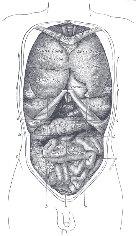
Figure 7Gray1034
Long description
Front view of the thoracic and abdominal viscera. a. Median plane. b b. Lateral planes. c c. Trans tubercular plane. d d. Subcostal plane. e e. Transpyloric plane.

Figure 7: Front view of the thoracic and abdominal viscera (a).
The left atrium lacks these ridges except in its auricle. The moderator band, a band of cardiac muscle covered by endocardium, reinforces the thin walls of the right ventricle and plays a crucial role in cardiac conduction. It arises from the inferior portion of the interventricular septum and crosses the interior space of the right ventricle to connect with the inferior papillary muscle. [3] [1]
The myocardium is the middle and thickest layer, made largely of cardiac muscle cells. It is built upon a framework of collagenous fibers, plus the blood vessels that supply the myocardium and the nerve fibers that help regulate the heart. The muscle pattern is elegant and complex, as the muscle cells swirl and spiral around the chambers of the heart. They form a figure 8 pattern around the atria and around the bases of the great vessels. Deeper ventricular muscles also form a figure 8 around the two ventricles and proceed toward the apex. [1]
The walls of the ventricle are lined with trabeculae carneae, ridges of cardiac muscle covered by endocardium. In addition to these muscular ridges, a band of cardiac muscle, also covered by endocardium, known as the moderator band (see ) reinforces the thin walls of the right ventricle and plays a crucial role in cardiac conduction. It arises from the inferior portion of the interventricular septum and crosses the interior space of the right ventricle to connect with the inferior papillary muscle. The muscular layer is much thicker in the left ventricle compared to the right. [1]
| Muscle | Origin | Insertion | Action | Nerve Supply |
|---|---|---|---|---|
| External Intercostal | Dorsal aspect of rib | Transverse process | Elevates ribs | Intercostal nerves |
| Pectinate Muscles | Right atrium wall | Right auricle | Contracts atrium | Autonomic |
| Papillary Muscles | Ventricular walls | Chordae tendineae | Tenses valves | Cardiac nerves |
| Moderator Band | Interventricular septum | Inferior papillary muscle | Conduction support | Purkinje fibers |
The right ventricle receives blood from the right atrium through the tricuspid valve. Each flap of the valve is attached to strong strands of connective tissue, the chordae tendineae, literally “tendinous cords,” or sometimes more poetically referred to as “heart strings”. There are several chordae tendineae associated with each of the flaps. They are composed of approximately 80 percent collagenous fibers with the remainder consisting of elastic fibers and endothelium. They connect each of the flaps to a papillary muscle that extends from the inferior ventricular surface. There are three papillary muscles in the right ventricle, called the anterior, posterior, and septal muscles, which correspond to the three sections of the valves. The mitral valve is connected to papillary muscles via chordae tendineae. There are two papillary muscles on the left—the anterior and posterior—as opposed to three on the right. [1]
Clinical pearlChordae Tendineae
These tendinous cords prevent the atrioventricular valves from prolapsing into the atria during ventricular systole.
When the ventricles begin to contract, pressure within the ventricles rises and blood flows toward the area of lowest pressure, which is initially in the atria. This backflow causes the cusps of the tricuspid and mitral (bicuspid) valves to close. These valves are tied down to the papillary muscles by chordae tendineae. During the relaxation phase of the cardiac cycle, the papillary muscles are also relaxed and the tension on the chordae tendineae is slight. However, as the myocardium of the ventricle contracts, so do the papillary muscles. This creates tension on the chordae tendineae, helping to hold the cusps of the atrioventricular valves in place and preventing them from being blown back into the atria. [1]
DefinitionMyocardium
The thick, muscular middle layer of the heart wall responsible for contraction.
The right coronary artery proceeds along the coronary sulcus and distributes blood to the right atrium, portions of both ventricles, and the heart conduction system. Normally, one or more marginal arteries arise from the right coronary artery inferior to the right atrium. The marginal arteries supply blood to the superficial portions of the right ventricle. On the posterior surface of the heart, the right coronary artery gives rise to the posterior interventricular artery, also known as the posterior descending artery. It runs along the posterior portion of the interventricular sulcus toward the apex of the heart, giving rise to branches that supply the interventricular septum and portions of both ventricles. [1]
NoteClinical Relevance
Understanding muscle attachments is vital for surgical approaches, such as rib resection for tumour removal.
The central structure is the pulmonary trunk, which arises from the right ventricle and bifurcates into the left and right pulmonary arteries. These vessels carry deoxygenated blood towards the lungs. The left and right coronary arteries originate from the aortic sinuses. The left coronary artery divides into the circumflex artery and the anterior interventricular artery, also known as the left anterior descending artery. The circumflex artery follows the coronary sulcus to the left. The anterior interventricular artery follows the anterior interventricular sulcus. These vessels supply the myocardium. Understanding this layout is essential for interpreting imaging and performing surgical procedures. [1]
The right ventricle pumps deoxygenated blood into the pulmonary trunk. This vessel leads toward the lungs and bifurcates into the left and right pulmonary arteries. These vessels branch many times before reaching the pulmonary capillaries. Gas exchange occurs here: carbon dioxide exits the blood and oxygen enters. The pulmonary trunk arteries and their branches are the only arteries in the post-natal body that carry relatively deoxygenated blood. Highly oxygenated blood returning from the pulmonary capillaries passes through vessels that join to form the pulmonary veins. These are the only post-natal veins carrying highly oxygenated blood. The pulmonary veins conduct blood into the left atrium. The left atrium pumps blood into the left ventricle. The left ventricle pumps oxygenated blood into the aorta. The aorta distributes blood to the systemic circuit. [1]
Coronary arteries supply blood to the myocardium and other components of the heart. The first portion of the aorta after it arises from the left ventricle gives rise to the coronary arteries. There are three dilations in the wall of the aorta just superior to the aortic semilunar valve. Coronary vessel branches that remain on the surface of the artery and follow the sulci are called epicardial coronary arteries. The left coronary artery distributes blood to the left side of the heart, the left atrium and ventricle, and the interventricular septum. The circumflex artery arises from the left coronary artery and follows the coronary sulcus to the left. Eventually, it will fuse with the small branches of the right coronary artery. The larger anterior interventricular artery, also known as the left anterior descending artery (LAD), is the second major branch arising from the left coronary artery. It follows the anterior interventricular sulcus around the pulmonary trunk. Along the way it gives rise to numerous smaller branches that interconnect with the branches of the posterior interventricular artery, forming anastomoses. An anastomosis is an area where vessels unite to form interconnections that normally allow blood to circulate to a region even if there may be partial blockage in another branch. [1]
The thoracic cavity contains a complex network of veins that return blood to the heart. Understanding this drainage is crucial for interpreting clinical signs such as superior vena cava obstruction.
The left brachiocephalic vein passes anterior to the great arteries of the aortic arch, while the right brachiocephalic vein descends vertically. The hemiazygos vein crosses the midline at the level of T9 to join the azygos vein. The internal thoracic veins drain into the brachiocephalic veins.
The superior vena cava is a large, short vessel that receives deoxygenated blood from the upper half of the body. It is formed by the union of the left and right brachiocephalic veins behind the first right costal cartilage. The SVC descends vertically to the right of the ascending aorta. It enters the right atrium at the level of the third right costal cartilage. The azygos vein drains into the posterior aspect of the SVC. The right superior intercostal vein also drains into the SVC. [1]
The inferior vena cava (IVC) returns blood from the lower half of the body. It passes through the caval opening of the diaphragm at the level of T8. The IVC enters the right atrium below the SVC. The hepatic veins drain directly into the IVC just before it enters the heart. [1]
The azygos system provides a crucial collateral pathway. The azygos vein arises from the right ascending lumbar vein. It ascends along the right side of the vertebral column. It arches over the root of the right lung to drain into the SVC. The hemiazygos vein drains the left lower intercostal spaces. It crosses the midline to join the azygos vein. The accessory hemiazygos vein drains the left upper intercostal spaces. It may drain into the azygos or hemiazygos vein.
Obstruction of the SVC leads to dilation of these veins. Blood flows backwards into the azygos system. This can lead to the development of collateral veins on the chest wall. The clinical significance of this anatomy is seen in conditions such as lung cancer or lymphoma.
DefinitionAzygos System
A network of veins on the right side of the thoracic vertebral column that drains into the superior vena cava.
| Vein | Drainage Area | Termination |
|---|---|---|
| Superior Vena Cava | Upper body | Right Atrium |
| Azygos Vein | Right thoracic wall | Superior Vena Cava |
| Hemiazygos Vein | Left lower thoracic wall | Azygos Vein |
| Internal Thoracic Vein | Anterior chest wall | Brachiocephalic Vein |
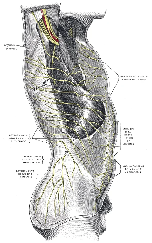
Figure 8Gray820
Long description
Cutaneous distribution of thoracic nerves. (Testut.)

Figure 8: Cutaneous distribution of thoracic nerves (Testut.).
Understanding this spatial arrangement is critical, as complications such as nerve root avulsion can occur during procedures requiring extrapleural dissection or excessive rib retraction.
The motor and cutaneous supply of the thoracic wall is primarily provided by the intercostal nerves. These are the anterior rami of the thoracic spinal nerves. Each intercostal nerve runs between the internal intercostal muscle and the innermost intercostal muscle.
| Structure | Innervation |
|---|---|
| Intercostal muscles | Anterior ramus of corresponding thoracic nerve |
| Skin of thoracic wall | Anterior ramus of corresponding thoracic nerve |
| Parietal pleura | Anterior ramus of corresponding thoracic nerve |
Lesions of these nerves can result from surgical complications. For instance, disarticulation of the costotransverse joint may lead to nerve root avulsion. This injury can cause significant pain and sensory loss in the corresponding dermatome. In severe cases, such as a spontaneous spinal fluid leak, the nerve root itself may be avulsed from the dural sac. Recognition of these anatomical relationships is essential for preventing iatrogenic injury during thoracic surgery. The proximity of the nerve roots to the vertebral column and ribs underscores the need for careful dissection in the costovertebral angle. [3]
The thoracic lymphatic system functions as a vital return pathway, removing interstitial fluid from tissues and returning it to the blood. This system also transports dietary lipids and immune cells, which originate from the bone marrow’s hematopoietic system. Primary lymphoid organs, specifically the bone marrow and thymus gland, are where adaptive immune lymphocytes proliferate and mature. Secondary lymphoid organs serve as sites where mature lymphocytes congregate to mount immune responses. [2]
DefinitionLymphedema
When the lymphatic system is damaged, such as by cancer blockage or injury, protein-rich interstitial fluid accumulates in tissue spaces, a condition known as lymphedema. [2]
The thoracic duct, the larger of the two, ascends through the posterior mediastinum. It receives lymph from the lower limbs, pelvis, abdomen, left thorax, left upper limb, and left side of the head and neck. It terminates by draining into the junction of the left internal jugular and left subclavian veins. The right lymphatic duct is shorter and drains the right thorax, right upper limb, and right side of the head and neck into the right venous angle. Lymph nodes act as major staging areas for immune response development. These small, bean-shaped organs are located throughout the system. [2]
Lymphatic capillaries form the open end of the system, allowing interstitial fluid entry via overlapping endothelial cells. When interstitial pressure is low, these endothelial flaps close to prevent backflow. As interstitial pressure increases, the spaces between cells open, permitting fluid entry. Collagen filaments anchor capillaries to surrounding structures; increased pressure pulls these filaments, opening the flaps further. In the small intestine, specialized capillaries called lacteals t [2]
Figure 9Aorta Anatomy
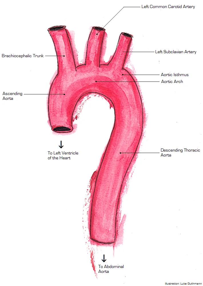
Long description
Aorta Anatomy. Labelled illustration from Wikimedia Commons, used under its open licence.

Figure 9: Aorta Anatomy.
The superior and inferior venae cavae are major systemic veins that return blood to the right atrium. These vessels lie in close relation to the mediastinal structures. The right atrium receives this blood and pumps it into the right ventricle. The left ventricle serves as the major pumping chamber for the systemic circuit. It ejects blood into the aorta through the aortic semilunar valve. This anatomical arrangement ensures efficient circulation. [1]
The thoracic cavity also contains lymphoid organs critical for immune function. The primary lymphoid organs are the bone marrow and thymus gland. These organs are where lymphocytes mature, proliferate, and are selected. This process enables the body to attack pathogens without harming its own cells. The thymus is located in the superior mediastinum. Tonsils are lymphoid nodules located along the inner surface of the pharynx. They are important in developing immunity to oral pathogens. The pharyngeal tonsil, or adenoid, is situated at the back of the throat. Histologically, tonsils do not contain a complete capsule. The epithelial layer invaginates deeply into the interior to form tonsillar crypts. These structures accumulate materials taken into the body through eating and breathing. [2]
DefinitionSubarachnoid-pleural fistula
An abnormal communication between the subarachnoid and pleural spaces that normally can arise from blunt or penetrating trauma or as a complication of chest operation. [3]
A rare complication of chest operation is the development of a fistula between the subarachnoid and pleural spaces. Common anatomic features involve the upper lobe of the lung with chest wall invasion into the region of the costovertebral angle. The paravertebral gutter region is particularly susceptible. Symptoms develop from the accumulation of pleural fluid, the reduced volume of cerebrospinal fluid, the presence of intracranial air, or the development of meningitis. Outcomes range from benign to catastrophic. [3]
| Structure | Location/Relation | Clinical Significance |
|---|---|---|
| Superior Vena Cava | Returns blood to right atrium | Major systemic vein |
| Left Ventricle | Ejects blood into aorta | Major pumping chamber |
| Thymus | Superior mediastinum | Lymphocyte maturation |
| Pharyngeal Tonsil | Back of throat | Immunity to oral pathogens |
| Subarachnoid-Pleural Space | Abnormal communication | Rare post-surgical complication |
Understanding the flow of blood through the pulmonary and systemic circuits is critical to all health professions. The differentiation and development of B and T cells is critical to the understanding of the adaptive immune response. Such swelling of the adenoid is an indication of an active immune response to infection. The necrotizing potential of preoperative or intraoperative radiotherapy can impede wound healing of these defects. This predisposes patients to fistula formation. [1] [2][3]
Understanding these relationships is fundamental for interpreting imaging studies accurately.
A systematic approach to the chest radiograph ensures no critical structure is overlooked. Begin by assessing the technical quality, including patient positioning and exposure. Then, evaluate the airways, lungs, and pleura. Next, examine the heart and mediastinum. Finally, inspect the bones and soft tissues. This methodical sequence reduces the risk of missing subtle abnormalities.
Computed tomography (CT) provides cross-sectional images with high spatial resolution. It is particularly useful for evaluating the mediastinum, lungs, and chest wall. CT can detect small nodules, lymphadenopathy, and vascular anomalies that may not be visible on plain radiographs. The ability to reconstruct images in different planes enhances the diagnostic yield.
Magnetic resonance imaging (MRI) offers superior soft tissue contrast. It is valuable for assessing the spinal cord, nerve roots, and mediastinal masses. MRI is particularly useful when CT is contraindicated or when detailed evaluation of soft tissue structures is required. It does not involve ionizing radiation, making it a safer option for repeated imaging.
Ultrasound is a dynamic, real-time imaging modality. It is primarily used for evaluating pleural effusions and guiding thoracentesis. Ultrasound can distinguish between free-flowing and loculated fluid. It is also useful for assessing chest wall masses and guiding biopsies. The portability of ultrasound machines makes it ideal for bedside evaluation.
NoteClinical Pearl
Always correlate imaging findings with clinical symptoms and physical examination findings for accurate diagnosis.
| Modality | Primary Use | Key Advantage |
|---|---|---|
| X-ray | Initial screening | Widely available, low cost |
| CT | Detailed anatomy | High spatial resolution |
| MRI | Soft tissue contrast | No ionizing radiation |
| Ultrasound | Pleural fluid | Dynamic, bedside capability |
At the base of this trunk lies the pulmonary semilunar valve, which prevents backflow.
Surface anatomy relies on palpable bony landmarks to locate deeper structures. The sternal angle marks the level of the second rib and the bifurcation of the trachea. Clinicians use these landmarks to estimate the position of the heart and great vessels. The apex beat is typically felt in the fifth intercostal space, mid-clavicular line, corresponding to the left ventricle. Palpation of the carotid pulse provides information on cardiac output. Auscultation points correspond to the valve areas: aortic, pulmonary, tricuspid, and mitral. These surface projections guide physical examination and procedural interventions.
Thoracic injuries can damage the intercostal nerves, leading to sensory deficits or motor weakness in the intercostal muscles. Phrenic nerve injury may occur during cardiac surgery, causing diaphragmatic paralysis. Vagus nerve damage can affect heart rate and gastrointestinal function. Surgical procedures, such as lung resection, carry risks of bleeding from the intercostal vessels. The internal thoracic artery is a common site for harvesting grafts in coronary artery bypass surgery. Knowledge of vascular anatomy prevents iatrogenic injury. [3]
Procedures such as thoracentesis require precise knowledge of the neurovascular bundle, which lies along the inferior border of each rib. Insertion must be superior to the rib to avoid nerve and vessel damage. Pericardiocentesis targets the subxiphoid approach to access the pericardial sac. The coronary sinus is a key structure in electrophysiology studies. The auricles are often used for thrombus assessment in atrial fibrillation. The sulci contain major coronary vessels, which are critical in ischaemic heart disease. Specific treatment of defects, such as fistulae, depends on size and symptoms. Larger fistulae may require surgical closure using omentum or muscle patches. [3]
| Structure | Location | Clinical Significance |
|---|---|---|
| Pulmonary Trunk | Base of right ventricle | Site of pulmonary semilunar valve |
| Auricles | Superior surface of atria | Thin-walled, fill with blood [1] |
| Coronary Sinus | Posterior atrioventricular sulcus | Drains into right atrium |
| Small Cardiac Vein | Parallel to right coronary artery | Drains posterior right heart |
The human heart comprises four chambers, with each side containing one atrium and one ventricle. The atria act as receiving chambers, contracting to push blood into the ventricles. The ventricles serve as the primary pumping chambers, propelling blood to the lungs or the rest of the body. [1]
The pulmonary circuit transports blood to the lungs for oxygenation and carbon dioxide delivery. The systemic circuit transports oxygenated blood to body tissues and returns deoxygenated blood to the heart. [1]
The endocardium lines the heart chambers and covers the valves. It consists of simple squamous epithelium called endothelium. This endothelium is continuous with the lining of blood vessels. [1]
The interventricular septum separates the two ventricles. It is normally intact after fetal development. This septum is substantially thicker than the interatrial septum. The increased thickness allows the ventricles to generate far greater pressure during contraction. [1]
During ventricular relaxation, the atrioventricular valves are open. The semilunar valves are closed at this stage. Atrial contraction pumps blood into the ventricles while both chambers are relaxed. [1]
During ventricular contraction, the atrioventricular valves close. The semilunar valves open to allow ejection. Blood is ejected into the pulmonary trunk and aorta. Closure of the atrioventricular valves prevents backflow into the atria. [1]
The right and left coronary arteries branch first from the aorta. They arise from sinuses near the base of the aorta. These arteries are generally located in the sulci. [1]
Cardiac veins parallel the small cardiac arteries. They generally drain into the coronary sinus. [1]
Exam trapSeptal Thickness
Do not confuse the interatrial and interventricular septa. The interventricular septum is substantially thicker because ventricles generate far greater pressure than atria.
DefinitionEndocardium
The endocardium is the innermost layer of the heart wall, made of endothelium, continuous with blood vessel linings.
| Phase | Atrioventricular Valves | Semilunar Valves |
|---|---|---|
| Ventricular Relaxation | Open | Closed |
| Ventricular Contraction | Closed | Open |
NoteCoronary Origin
Coronary arteries are the first branches of the aorta, arising from sinuses near its base.
Students frequently conflate physiological adaptation with pathological hypertrophy. It is a critical error to assume that an enlarged heart is always a result of exercise training. While athletic hearts pump blood more effectively at lower rates, pathological enlargement, such as in hypertrophic cardiomyopathy, has an unknown cause and is often undiagnosed. This condition can cause sudden death in apparently otherwise healthy young people, distinguishing it from benign athletic adaptation. [1]
Another frequent mistake involves the microscopic nature of serous membranes. In most organs, visceral serous membranes are microscopic. However, the epicardium is a macroscopic layer. It consists of a simple squamous epithelium called a mesothelium, reinforced with loose, irregular, or areolar connective tissue that attaches to the pericardium. This mesothelium secretes the lubricating serous fluid that fills the pericardial cavity and reduces friction as the heart contracts. [1]
Students also misinterpret the thickness of the ventricular walls. Although the ventricles on the right and left sides pump the same amount of blood per contraction, the muscle of the left ventricle is much thicker and better developed than that of the right ventricle. This is because the left ventricle must generate a great amount of pressure to overcome the high resistance required to pump blood into the long systemic circuit. The right ventricle does not need to generate as much pressure, since the pulmonary circuit is shorter and provides less resistance. [1]
A common anatomical error concerns the septa. The word septum is derived from the Latin for “something that encloses;” in this case, a septum (plural = septa) refers to a wall or partition that divides the heart into chambers. The septa are physical extensions of the myocardium lined with endocardium. Located between the two atria is the interatrial septum. Normally in an adult heart, the interatrial septum bears an oval-shaped depression known as the fossa ovalis, a remnant of an opening in the fetal heart known as the foramen ovale. The foramen ovale allowed blood in the fetal heart to pass directly from the right atrium to the left atrium, allowing some blood to bypass the pulmonary circuit. Within seconds after birth, a flap of tissue known as the septum primum that previously acted as a valve closes the foramen ovale and establishes the typical cardiac circulation pattern. [1]
Finally, students underestimate the vulnerability of coronary circulation. The anastomoses in the heart are very small. Therefore, this ability is somewhat restricted in the heart so a coronary artery blockage often results in death of the cells (myocardial infarction) supplied by the particular vessel. [1]
At the bedside, understanding the wall of the heart is essential. The wall of the heart is composed of three layers of unequal thickness. From superficial to deep, these are the epicardium, the myocardium, and the endocardium. The outermost layer of the wall of the heart is also the innermost layer of the pericardium, the epicardium, or the visceral pericardium discussed earlier. Recognising these layers helps in interpreting murmurs and pericardial friction rubs. [1]
In the operating theatre, knowledge of valve mechanics is vital. At the base of the aorta is the aortic semilunar valve, or the aortic valve, which prevents backflow from the aorta. It normally is composed of three flaps. When the ventricle relaxes and blood attempts to flow back into the ventricle from the aorta, blood will fill the cusps of the valve, causing it to close and producing an audible sound. Surgeons rely on this anatomical detail to repair or replace valves effectively. [1]
On imaging, the distinction between fetal and adult circulation remnants is key. The fossa ovalis is a remnant of the foramen ovale. Identifying this structure on echocardiography confirms normal postnatal closure. If the septum primum fails to close properly, it indicates a patent foramen ovale, which has clinical implications for stroke risk.
In examinations, students must recall the dual pattern of circulation. Recall that the heart’s contraction cycle follows a dual pattern of circulation—the pulmonary and systemic circuits—because of the pairs of chambers that pump blood into the circulation. In order to develop a more precise understanding of cardiac function, it is first necessary to explore the internal anatomical structures in more detail. This conceptual framework underpins all questions regarding cardiac output and pressure gradients. [1]
Cardiac Anatomy Review
- StartIdentify heart layers
- Question: Outer layer?
Yes
- Epicardium (Visceral Pericardium)
No
- Check next layer
- Middle layerMyocardium (Thickest in Left Ventricle)
- Question: Inner layer?
Yes
- Endocardium
No
- Error in identification
The epicardium is a macroscopic layer, not microscopic like most visceral serous membranes. Athletic heart enlargement differs from pathological hypertrophic cardiomyopathy. The left ventricle is thicker than the right due to higher systemic resistance. The septum divides the heart; septa are myocardial extensions lined with endocardium. The fossa ovalis is the adult remnant of the fetal foramen ovale. The aortic valve has three flaps and prevents backflow from the aorta. Coronary anastomoses are small, making myocardial infarction likely upon blockage. The heart wall consists of epicardium, myocardium, and endocardium from superficial to deep.
Answer each one in your head first, then open the answer.
What is the composition of the epicardium?
Show answerHide answer
Simple squamous epithelium called a mesothelium, reinforced with loose, irregular, or areolar connective tissue.Why is the left ventricular muscle thicker?
Show answerHide answer
To overcome the high resistance required to pump blood into the long systemic circuit.What structure closes the foramen ovale after birth?
Show answerHide answer
A flap of tissue known as the septum primum.How many flaps does the aortic valve normally have?
Show answerHide answer
Three.What is the consequence of small coronary anastomoses?
Show answerHide answer
A coronary artery blockage often results in death of the cells (myocardial infarction) supplied by the particular vessel.
- Betts JG, Young KA, Wise JA, et al. Heart Anatomy. OpenStax, Anatomy and Physiology 2e (CC BY-NC-SA 4.0). Accessed .
- Betts JG, Young KA, Wise JA, et al. Anatomy of the Lymphatic and Immune Systems. OpenStax, Anatomy and Physiology 2e (CC BY-NC-SA 4.0). Accessed .
- Shamji MF, Sundaresan S, Da Silva V, et al. Subarachnoid-pleural fistula: applied anatomy of the thoracic spinal nerve root. ISRN surgery 2011;2011:168959. doi:10.5402/2011/168959. Accessed .
Figure credits
- Figure 1: Henry Vandyke Carter, via Wikimedia Commons, Public domain, Public domain.
- Figure 2: OpenStax College, via Wikimedia Commons, CC BY 3.0, CC BY 3.0. resized.
- Figure 3: Henry Vandyke Carter, via Wikimedia Commons, Public domain, Public domain.
- Figure 4: Henry Vandyke Carter, via Wikimedia Commons, Public domain, Public domain.
- Figure 5: OpenStax College, via Wikimedia Commons, CC BY 3.0, CC BY 3.0.
- Figure 6: Henry Vandyke Carter, via Wikimedia Commons, Public domain, Public domain.
- Figure 7: Henry Vandyke Carter, via Wikimedia Commons, Public domain, Public domain.
- Figure 8: Henry Vandyke Carter, via Wikimedia Commons, Public domain, Public domain.
- Figure 9: Luke Guthmann, via Wikimedia Commons, CC BY-SA 3.0, CC BY-SA 3.0.
Educational content for medical students and doctors. It is not a substitute for clinical judgement or local guidelines.