Autonomic innervation of the thorax
Innervation of thoracic viscera by sympathetic and parasympathetic nerves.
Figure 1Gray846
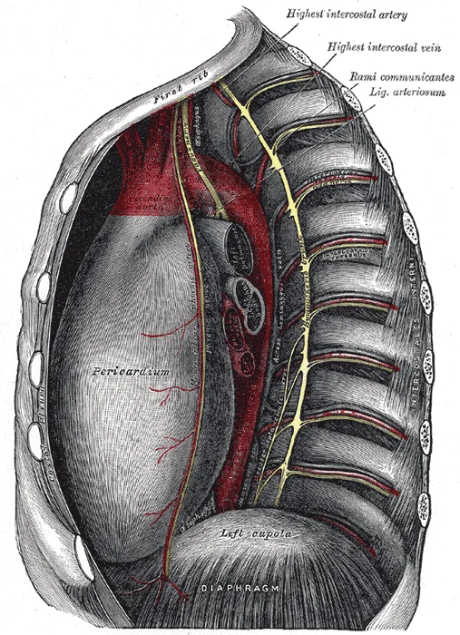
Long description
Thoracic portion of the sympathetic trunk.

Figure 1: Thoracic portion of the sympathetic trunk.
The autonomic nervous system regulates internal organs through a balance of two aspects, or divisions[1]. These are the sympathetic division and the parasympathetic division[1]. The sympathetic system is associated with the fight-or-flight response[1]. Parasympathetic activity is referred to by the epithet of rest and digest[1]. Homeostasis is the balance between the two systems[1]. At each target effector, dual innervation determines activity[1]. For example, the heart receives connections from both the sympathetic and parasympathetic divisions[1]. One causes heart rate to increase, whereas the other causes heart rate to decrease[1].
The autonomic system targets cardiac and smooth muscle, as well as glandular tissue. Somatic responses are solely based on skeletal muscle contraction. The autonomic nervous system regulates organ systems through circuits that resemble the reflexes described in the somatic nervous system. The role of visceral reflexes is to maintain a balance of function in the organ systems of the body. The two divisions of the autonomic system each play a role in effecting change, usually in competing directions. The sympathetic system increases heart rate, whereas the parasympathetic system decreases heart rate. The competing inputs can contribute to the resting tone of the organ system. Heart rate is normally under parasympathetic tone, whereas blood pressure is normally under sympathetic tone. [2]
DefinitionDual Innervation
The presence of both sympathetic and parasympathetic fibers on a single target organ, allowing for opposing regulatory effects.
The cardiac and pulmonary plexuses are networks of autonomic nerves located in the mediastinum. They receive contributions from both the sympathetic trunk and the vagus nerve. These plexuses distribute fibers to the heart, lungs, and esophagus. The sympathetic fibers originate from the upper thoracic spinal cord segments. They travel via the sympathetic trunk to reach the plexuses. The parasympathetic fibers are carried by the vagus nerve. They synapse in terminal ganglia near or within the walls of the target organs. [2]
| Component | Origin | Effect on Heart |
|---|---|---|
| Sympathetic | Thoracic sympathetic trunk | Increases heart rate and force |
| Parasympathetic | Vagus nerve | Decreases heart rate |
The pericardium is innervated by somatic and autonomic fibers. The fibrous pericardium receives sensory innervation from the phrenic nerves. These nerves provide pain sensation from the parietal layer. The visceral layer, or epicardium, receives autonomic innervation via the cardiac plexuses. This innervation regulates the coronary blood flow and myocardial contractility. Pain from the visceral pericardium is often referred to the neck or shoulder due to shared spinal segments.
Neurons in the dorsal nucleus of the vagus nerve and the nucleus ambiguus project through the vagus nerve (cranial nerve X) to the terminal ganglia of the thoracic and abdominal cavities[1]. Parasympathetic preganglionic fibers primarily influence the heart, bronchi, and esophagus in the thoracic cavity[1]. The postganglionic fibers from the ganglia activated by the vagus nerve are often incorporated into the structure of the organ[1]. Where an autonomic neuron connects with a target, there is a synapse[1]. The electrical signal of the action potential causes the release of a signaling molecule[1]. Synapses of the autonomic system are classified as either cholinergic, meaning that acetylcholine (ACh) is released[1]. The terms cholinergic and adrenergic refer not only to the signaling molecule that is released but also to the class of receptors that each binds[1]. That neuron, connected to the smooth muscle, is a postganglionic parasympathetic neuron that can be controlled by a fiber found in the vagus nerve. [2]
The autonomic innervation of the thorax arises from two distinct embryological and functional systems: the sympathetic division, originating from the thoracolumbar spinal cord, and the parasympathetic division, derived from the cranial vagus nerve. Understanding their formation requires tracing the path from the central nervous system to the thoracic viscera.
The sympathetic outflow begins in the lateral horn of the spinal cord segments T1 to T12. Preganglionic neurons in these regions project axons through the ventral spinal roots. These axons then enter the sympathetic chain via the white ramus communicans. The sympathetic chain itself is a network of ganglia running alongside the vertebral column. There are typically 23 ganglia on either side of the spinal column, with 12 located in the thoracic region. These ganglia appear as clusters of neurons linked by axonal bridges. [1]
DefinitionSympathetic Chain
A series of 23 ganglia on each side of the vertebral column, including 12 thoracic ganglia, linked by axonal bridges.
Not all sympathetic fibres synapse within this chain. Some branches pass through the chain ganglia to form splanchnic nerves. This divergence allows a single preganglionic neuron to influence up to 10–20 targets, facilitating a broad systemic response. [1]
The parasympathetic innervation of the thorax is primarily provided by the vagus nerve (cranial nerve X). The cranial component of the parasympathetic system is based in nuclei of the brain stem, specifically the dorsal motor nucleus of the vagus. The vagus nerve descends through the neck and enters the thorax. In the neck, it gives off the recurrent laryngeal nerves. The left recurrent laryngeal nerve hooks under the aortic arch, while the right hooks under the subclavian artery. These nerves provide motor and sensory innervation to the larynx. [2]
Within the thorax, the vagus nerves contribute to the cardiac and pulmonary plexuses. The postganglionic parasympathetic fibres are short and originate from terminal ganglia located near or within the target effector organs. These terminal ganglia are often referred to as intramural ganglia when found within the walls of the target organ. The postganglionic fibres release acetylcholine (ACh), which binds to muscarinic receptors on the target cells. [1]
The phrenic nerves are somatic motor nerves, not autonomic, but they are crucial anatomical landmarks in the thorax. They arise from the cervical spinal cord segments C3, C4, and C5. The phrenic nerves descend through the superior mediastinum, anterior to the root of the lung, to innervate the diaphragm. Their course is closely related to the pericardium and the fibrous pericardium. Damage to the phrenic nerve results in paralysis of the diaphragm on the affected side.
The thoracic sympathetic trunk runs vertically along the heads of the ribs. It receives white rami communicantes from T1 to T12. Grey rami communicantes return to all 12 thoracic spinal nerves. The sympathetic trunk also gives off cardiac branches that join the cardiac plexus. These branches increase heart rate and force of contraction by releasing norepinephrine, which binds to adrenergic receptors. The sympathetic tone of blood vessels is maintained by this system, as most systemic blood vessels lack parasympathetic input. [2] [1]
The intercostal nerves are the ventral rami of the thoracic spinal nerves T1 to T11. They run in the intercostal spaces between the internal and innermost intercostal muscles. While primarily somatic, they carry sympathetic fibres that have passed through the grey rami communicantes. These sympathetic fibres innervate the smooth muscle of the blood vessels and sweat glands in the thoracic wall. Sweat glands receive only sympathetic input, making them a unique example of single innervation. [2]
The thoracic sympathetic trunk is located posteriorly, adjacent to the vertebral column.
The vagus nerves are located within the pulmonary plexus. The phrenic nerve is anterior to the lung. The intercostal nerves are lateral, within the costal groove.
The autonomic innervation of the thorax involves a complex interplay between sympathetic and parasympathetic pathways, each with distinct origins, courses, and effector targets. Understanding these pathways requires tracing the anatomical route from the central nervous system to the thoracic viscera, including the heart, lungs, and great vessels.
The sympathetic supply to the thorax originates from the thoracic segments of the spinal cord. Preganglionic sympathetic neurons reside in the lateral horn of the spinal cord grey matter. Their axons exit via the ventral roots and enter the sympathetic trunk through white rami communicantes. These preganglionic fibers release acetylcholine (ACh), which acts on nicotinic receptors on the ganglionic neurons.
Within the sympathetic trunk, preganglionic fibers may synapse at the level of entry or ascend/descend to synapse at other thoracic levels. Postganglionic fibers then leave the sympathetic trunk via grey rami communicantes to join the spinal nerves, or they form specific splanchnic nerves. For thoracic organs, postganglionic fibers often arise from the cervical and upper thoracic sympathetic ganglia. These fibers travel along the blood vessels to reach the heart and lungs. The postganglionic sympathetic fibers release norepinephrine, which binds to adrenergic receptors on the target organs. For example, in the heart, norepinephrine binding causes depolarization and an increase in heart rate.
The parasympathetic supply to the thorax is provided by the vagus nerves (cranial nerve X). The vagus nerves descend through the neck and enter the thorax, contributing to the cardiac and pulmonary plexuses. Preganglionic parasympathetic neurons originate in the dorsal motor nucleus of the vagus in the medulla oblongata. Their axons travel within the vagus nerves to ganglia located near or within the target organs.
These ganglia are typically small and situated in the walls of the heart or lungs. The preganglionic fibers release ACh, which acts on nicotinic receptors on the postganglionic neurons. The postganglionic fibers are short and release ACh, which acts on muscarinic receptors on the effector cells. In the heart, parasympathetic stimulation leads to hyperpolarization and a decrease in heart rate. Without this parasympathetic input, the heart would work at a rate of approximately 100 beats per minute. The parasympathetic system slows it down to the resting heart rate of 60–80 bpm. [2]
The sympathetic and parasympathetic fibers converge to form the cardiac and pulmonary plexuses. The cardiac plexus is located near the base of the heart and provides innervation to the heart muscle and conduction system. The pulmonary plexus is located at the hilum of the lungs and innervates the bronchial tree and pulmonary blood vessels. These plexuses ensure that both branches of the autonomic nervous system can modulate thoracic organ function simultaneously.
The autonomic innervation of the thorax is primarily sympathetic, with parasympathetic contributions via the vagus nerves. Understanding the specific branches requires distinguishing between the sympathetic chain ganglia and the prevertebral ganglia, although the latter are predominantly abdominal.
DefinitionSympathetic Chain
A series of ganglia located along the vertebral column that receives preganglionic sympathetic input and distributes postganglionic fibers to the body wall and viscera.
The sympathetic supply to the thorax originates from the thoracic spinal cord segments. Preganglionic neurons exit via the ventral roots and enter the sympathetic trunk. At the level of T1, the preganglionic fibre synapses with the T1 chain ganglion. This direct connection allows for rapid innervation of thoracic structures. The fibres entering the chain ganglion are myelinated and are termed white rami communicantes. This myelination gives them their characteristic white appearance. After synapsing within the ganglion, the postganglionic fibres are unmyelinated. These unmyelinated axons are referred to as gray rami communicantes. They return to the spinal nerve to be distributed to somatic structures, but specific branches also arise directly from the ganglia to supply thoracic viscera. [1]
The trachea is situated centrally, anterior to the esophagus. The branch lies adjacent to the tracheal cartilages. This anatomical arrangement ensures that sympathetic signals can modulate tracheal smooth muscle tone directly.
| Structure | Type | Origin | Target |
|---|---|---|---|
| White Rami Communicantes | Motor (Preganglionic) | Thoracic Spinal Nerves | Sympathetic Chain Ganglia |
| Gray Rami Communicantes | Motor (Postganglionic) | Sympathetic Chain Ganglia | Spinal Nerves (Somatic) |
| Direct Ganglionic Branches | Motor (Postganglionic) | Sympathetic Chain Ganglia | Thoracic Viscera (e.g., Trachea) |
| Vagus Nerve Branches | Motor/Parasympathetic | Medulla Oblongata | Thoracic Viscera (Heart, Lungs) |
| Visceral Sensory Fibres | Sensory | Thoracic Viscera | Spinal Cord (via Spinal Nerves) |
However, these are associated with controlling organs in the abdominal cavity. They receive inputs from splanchnic nerves. They are not typically involved in the direct innervation of the thorax. Therefore, the thoracic sympathetic supply is largely mediated through the chain ganglia themselves. [2] [1]
Sensory innervation follows a different path. Visceral sensory fibres from the mediastinal region, where the heart is located, enter the spinal cord at the same level as the spinal nerves from the shoulder and arm. This convergence explains referred pain. Projections from the medial and inferior divisions of the cervical ganglia do enter the spinal cord at the middle to lower cervical levels. This is where the somatosensory fibres enter. Consequently, pain from the heart may be perceived in the left shoulder and arm. This is a classic example of visceral sensation being misinterpreted by the brain as somatic pain. [2] [1]
The parasympathetic system provides contrasting control. The vagus nerves supply the heart and lungs. However, the sympathetic system determines the tone of systemic blood vessels. These tissues have adrenergic receptors that respond to the release of norepinephrine from postganglionic sympathetic fibers. The parasympathetic system has no significant input to the systemic blood vessels. [2]
The autonomic innervation of the thorax involves a complex interplay between sympathetic and parasympathetic pathways, and lesions at specific levels produce distinct clinical deficits. Understanding these deficits requires mapping the anatomical course of the nerves to their functional outcomes.
DefinitionReferred Pain
Visceral sensations that rise to conscious perception are often felt in unexpected somatic areas, such as heart pain projecting to the left shoulder and arm, due to convergence of visceral and somatosensory fibers at the same spinal cord level. [2]
Lateral to the trunk, the white rami communicantes connect the spinal nerves to the sympathetic chain. Superiorly, the stellate ganglion is formed by the fusion of the inferior cervical and first thoracic ganglia.
| Level | Structure Affected | Resulting Deficit |
|---|---|---|
| T1-T4 | Cardiac nerves | Loss of sympathetic innervation to the heart, potentially leading to bradycardia or altered heart rate response. |
| T2-T6 | Pulmonary and oesophageal branches | Disruption of bronchoconstriction and oesophageal motility control. |
| T1 | Stellate Ganglion | Horner’s syndrome (ptosis, miosis, anhidrosis) due to interruption of sympathetic supply to the head and neck. |
| T5-T9 | Greater splanchnic nerve | Loss of sympathetic input to upper abdominal viscera, affecting gastric and hepatic function. |
| Branch | Origin | Termination |
|---|---|---|
| White Rami Communicantes | Spinal Nerves T1-L2 | Sympathetic Chain Ganglia |
| Grey Rami Communicantes | Sympathetic Chain Ganglia | Spinal Nerves (all levels) |
| Splanchnic Nerves | Sympathetic Chain | Prevertebral Ganglia |
When particularly strong visceral sensations rise to the level of conscious perception, the sensations are often felt in unexpected places. For example, strong visceral sensations of the heart will be felt as pain in the left shoulder and left arm. This irregular pattern of projection of conscious perception of visceral sensations is called referred pain. Depending on the organ system affected, the referred pain will project to different areas of the body. The location of referred pain is not random, but a definitive explanation of the mechanism has not been established. The most broadly accepted theory for this phenomenon is that the visceral sensory fibers enter into the same level of the spinal cord as the somatosensory fibers of the referred pain location. [2]
Lesions affecting the sympathetic chain at the thoracic level can therefore manifest not only through direct motor or secretory deficits but also through aberrant pain perception. A lesion at T1, for instance, interrupts the sympathetic outflow to the head, causing Horner’s syndrome, while simultaneously potentially altering cardiac sensation referral patterns. Conversely, lesions higher in the cervical region may spare the thoracic sympathetic outflow but affect other autonomic functions. The key is to recognize that the spinal cord level of entry for visceral afferents dictates the somatic location of referred pain, providing a crucial diagnostic clue in clinical practice.
Clinical examination of the nerve
Horner syndrome
Horner syndrome results from disruption of the sympathetic pathway to the eye. The sympathetic outflow originates in the lateral horn of the thoracolumbar spinal cord. Specifically, the pathway involves spinal nerve T1, which innervates the eye. This nerve tracks up through the sympathetic chain until it reaches the superior cervical ganglion. The synapse with the ganglionic neuron occurs at this superior location. These ganglia are part of the sympathetic chain and are also called paravertebral ganglia. The postganglionic fiber then projects to the iris. It releases norepinephrine onto the radial fibers of the iris. Contraction of these smooth muscle fibers dilates the pupil. Disruption at any point causes ptosis, miosis, and anhidrosis. [2] [1]
Recurrent laryngeal injury
The recurrent laryngeal nerve is a branch of the vagus nerve. It loops under the aortic arch on the left and the subclavian artery on the right. Injury leads to vocal cord paralysis. This is often iatrogenic during thyroid or cardiac surgery. The clinical sign is hoarseness or breathy voice.
Ortner syndrome
Ortner syndrome is cardiovocal syndrome. It involves hoarseness due to recurrent laryngeal nerve palsy. The cause is compression by cardiovascular structures. Common causes include left atrial enlargement. Pulmonary hypertension can also cause dilation of the pulmonary artery. This compresses the nerve against the ligamentum arteriosum. The result is unilateral vocal cord paralysis.
DefinitionSympathetic Pathway
The sympathetic supply to the head travels from T1 up the chain to the superior cervical ganglion, where postganglionic fibers innervate the eye.
| Structure | Origin | Course | Termination | Action |
|---|---|---|---|---|
| Preganglionic Fiber | Lateral horn T1 | Ventral root to spinal nerve | Superior cervical ganglion | Synapses via nicotinic receptor |
| Postganglionic Fiber | Superior cervical ganglion | Travels with carotid arteries | Iris radial fibers | Releases norepinephrine |
| Target Muscle | - | - | Radial fibers of iris | Pupil dilation |
The sympathetic system causes divergent responses. Preganglionic fibers release acetylcholine. This excites the ganglionic neuron. The postganglionic fiber releases norepinephrine. This binds to adrenergic receptors. In the eye, this dilates the pupil. The parasympathetic system opposes this. It acts via the Edinger–Westphal nucleus. The signal travels through the oculomotor nerve. It synapses in the ciliary ganglion. This constricts the pupil. Clinical examination assesses these pathways. Horner syndrome indicates sympathetic failure. Ortner syndrome indicates vascular compression. Recurrent laryngeal injury indicates surgical risk. Understanding the anatomy prevents errors. The T1 segment is critical. The superior cervical ganglion is key. The iris radial fibers are the effector. Norepinephrine is the transmitter. Acetylcholine is the preganglionic transmitter. Nicotinic receptors mediate the synapse. Adrenergic receptors mediate the effect. The lateral horn is the origin. The ventral root carries the fiber. The spinal nerve carries it to the chain. The chain ganglia are paravertebral. The synapse can be superior or inferior. T1 is the classic example. The eye is the target. Light levels influence the pathway. Low light triggers dilation. High light triggers constriction. The optic nerve carries afferent signals. The retinal ganglion cells are involved. The diencephalon processes the signal. The sympathetic system responds to low light. The parasympathetic system responds to high light. The ciliary ganglion is in the posterior orbit. The oculomotor nerve carries the parasympathetic fibers. The Edinger–Westphal nucleus is the source. The radial fibers are smooth muscle. This is the physiological basis. Clinical signs reflect anatomical disruption. Ptosis is due to Muller muscle. Miosis is due to radial muscle failure. Anhidrosis is due to facial sweat gland loss. Lesions can be central, pre-ganglionic or post-ganglionic. T1 root avulsion causes Horner syndrome. Carotid dissection causes Horner syndrome. Pancoast tumour causes Horner syndrome. These are common causes. The superior cervical ganglion is a common site. The synapse is at the ganglion. The postganglionic fiber is long. It travels to the eye. The norepinephrine release is key. The adrenergic receptor binding is key. The physiological change is dilation. This is the sympathetic response. The parasympathetic response is constriction. The ciliary ganglion mediates this. The oculomotor nerve carries the signal. The Edinger–Westphal nucleus is the origin. The retinal ganglion cells detect light. The optic nerve carries the signal. The diencephalon processes the input. The sympathetic system is activated by low light. The parasympathetic system is activated by high light. The iris radial fibers contract for dilation. The iris sphincter contracts for constriction. The ciliary ganglion innervates the sphincter. The superior cervical ganglion innervates the radial fibers. The T1 segment is the source. The lateral horn is the origin. The ventral root is the exit. The spinal nerve is the conduit. The sympathetic chain is the route. The paravertebral ganglia are the synapse sites. The superior cervical ganglion is the specific site for the eye. The postganglionic fiber is the final link. The norepinephrine is the chemical messenger. The adrenergic receptor is the target. The physiological effect is pupil dilation. This is the sympathetic function. The parasympathetic function is pupil constriction. This is the autonomic balance. Clinical examination tests this balance. Horner syndrome is a key sign. Ortner syndrome is a vascular sign. Recurrent laryngeal injury is a surgical sign. These are the clinical correlates. [1][2]
- Divergent Architecture: The sympathetic system coordinates the "fight or flight" response by diverging from a limited central nervous system region to a wide array of ganglia, activating respiratory, cardiovascular, and musculoskeletal systems simultaneously. [1]
- Adrenal Medulla Exception: Preganglionic sympathetic fibers that project to the adrenal medulla do not terminate in a ganglion; instead, they synapse directly on chromaffin cells, which release signaling molecules into the bloodstream. [1]
- Neural Crest Origin: Chromaffin cells in the adrenal medulla are neurosecretory cells that develop from the neural crest, reinforcing the functional view of the gland as a sympathetic ganglion. [1]
- Cholinergic Receptor Classes: The cholinergic system utilizes two distinct receptor classes: nicotinic receptors, which are ligand-gated cation channels, and muscarinic receptors, which are G protein–coupled receptors. [1]
- Receptor Specificity: Nicotinic and muscarinic receptors are differentiated by their specific ligands; nicotine binds only to nicotinic receptors, while muscarine binds only to muscarinic receptors, with no cross-reactivity. [1]
- Adrenergic Receptor Types: The adrenergic system features two main receptor types, alpha and beta, all of which are G protein–coupled receptors, unlike the mixed channel types of the cholinergic system. [1]
- Alpha Subtypes: There are two specific types of alpha-adrenergic receptors, termed alpha-1 and alpha-2, which mediate distinct physiological responses in target tissues. [1]
- Beta Subtypes: There are three specific types of beta-adrenergic receptors, termed beta-1, beta-2, and beta-3, allowing for varied tissue-specific signaling. [1]
- Epinephrine vs Norepinephrine: The chemical difference between norepinephrine and epinephrine is the addition of a methyl group (CH3) in epinephrine; the prefix "nor-" indicates this methyl group is missing. [1]
- Terminology Origin: The term "adrenergic" derives from the adrenal gland, where "ad-" means "on top of" and "renal" means "kidney," reflecting the gland's anatomical position. [1]
- Naming Conventions: In scientific usage, epinephrine is preferred in the United States, while adrenaline is preferred in Great Britain, a convention persisting from historical proprietary drug names. [1]
- Synonymous Molecules: Norepinephrine and noradrenaline are two names for the same molecule, just as epinephrine and adrenaline refer to the same chemical entity. [1]
- Ganglion Termination: Students often incorrectly assume all preganglionic sympathetic fibers terminate in ganglia; the exception is the adrenal medulla, where they synapse on chromaffin cells.
- Receptor Mechanism: Do not confuse the mechanism of cholinergic receptors; nicotinic receptors are ion channels, whereas muscarinic receptors are G protein–coupled, a distinction critical for pharmacological understanding.
- Adrenergic Classification: Adrenergic receptors are not classified by drug binding like cholinergic receptors; instead, they are all G protein–coupled receptors, a key structural difference.
- Methyl Group Detail: Remember that the "nor-" prefix specifically refers to the absence of the methyl group found in epinephrine, a detail frequently tested in biochemistry and pharmacology.
Students frequently confuse the anatomical and functional distinctions between the sympathetic and parasympathetic divisions, leading to errors in clinical reasoning and examination answers. The first major pitfall is assuming all postganglionic sympathetic fibres are adrenergic. While most release noradrenaline, there are critical exceptions. The postganglionic connections to sweat glands in the skin and blood vessels supplying skeletal muscle are, however, exceptions; those fibers release ACh onto muscarinic receptors. Students must remember that sympathetic cholinergic fibres exist specifically for thermoregulation and skeletal muscle vasodilation, not just general inhibition. [1] [2]
A second common error involves the length and myelination of autonomic pathways. Students often assume preganglionic sympathetic fibres are long because they originate in the spinal cord. In reality, because the sympathetic ganglia are adjacent to the vertebral column, preganglionic sympathetic fibers are relatively short, and they are myelinated. Conversely, postganglionic sympathetic fibres are long because of the relatively greater distance from the ganglion to the target effector. These fibres are unmyelinated. Understanding this structural difference explains why preganglionic fibres can synapse with multiple postganglionic neurons, facilitating the coordinated systemic change. [1]
The third pitfall is misidentifying the neurotransmitters at the ganglionic synapse. Both divisions use acetylcholine (ACh) at the ganglion. The parasympathetic preganglionic fibres release ACh onto nicotinic receptors to excite the ganglionic neurons. Similarly, sympathetic preganglionic fibres release ACh onto nicotinic receptors on the postganglionic cell body. Confusing nicotinic with muscarinic receptors at the ganglion is a frequent mistake. Muscarinic receptors are found on the target effector organs, not the ganglia.
A fourth error concerns the terminology of "neuron" versus "fiber". The term “postganglionic neuron” may be used to describe the projection from a ganglion to the target. However, the problem with that usage is that the cell body is in the ganglion, and only the fiber is postganglionic. Typically, the term neuron applies to the entire cell. Precision in language is required when describing the axon projecting from the ganglion.
The fifth pitfall relates to the adrenal medulla. Students often treat the adrenal medulla as a standard postganglionic synapse. It is not. The sympathetic system has a specialized preganglionic connection to the adrenal medulla that causes epinephrine and norepinephrine to be released into the bloodstream rather than exciting a neuron that contacts an organ directly. This hormonal component means that the sympathetic chemical signal can spread throughout the body very quickly and affect many organ systems at once. This is a unique endocrine function, not a direct neural innervation. [1]
Finally, students may confuse visceral sensation with conscious perception. Visceral senses are not primarily a part of conscious perception, those sensations sometimes make it to conscious awareness. If a visceral sense is strong enough, it will be perceived. However, the sensory homunculus—the representation of the body in the primary somatosensory cortex—only has a small region allotted for the perception of internal stimuli. Do not assume all autonomic changes are felt; only strong stimuli, such as a large bolus of food or cold air in the larynx, reach awareness. [2]
This knowledge is applied at the bedside, in the operating theatre, on imaging, and in examinations to diagnose and manage autonomic dysfunction. At the bedside, understanding the efferent branch of the visceral reflex arc begins with the projection from the central neuron along the preganglionic fiber. This fiber then makes a synapse on the ganglionic neuron that projects to the target effector. Disruption at any point alters clinical signs. For example, Horner’s syndrome results from interruption of sympathetic supply to the head, causing ptosis and miosis. [2]
In the operating theatre, anaesthetists must manage blood pressure and heart rate by targeting specific receptors. Knowledge of vascular tone is crucial. There are a limited number of blood vessels that respond to sympathetic input in a different fashion. Blood vessels in skeletal muscle, particularly those in the lower limbs, are more likely to dilate. It does not have an overall effect on blood pressure to alter the tone of the vessels, but rather allows for blood flow to increase for those skeletal muscles that will be active in the fight-or-flight response. This explains why systemic vasoconstriction does not always raise blood pressure proportionally during stress. [2]
On imaging, radiologists identify structures based on their autonomic relations. The sympathetic chain lies adjacent to the vertebral column, consistent with the short preganglionic fibres. Parasympathetic ganglia are terminal or intramural, located close to or within the wall of target effectors. This distinction helps localise lesions. For instance, a lesion in the brain stem or sacral spinal cord affects parasympathetic output, which is based in the brain stem and sacral spinal cord. [1]
In examinations, questions often test the specific actions of autonomic nerves on the eye. The postganglionic fiber then projects to the iris, where it releases ACh onto the circular fibers of the iris—another smooth muscle. When those fibers contract, the pupil constricts to limit the amount of light hitting the retina. This is a parasympathetic action. Sympathetic stimulation dilates the pupil via radial muscle contraction. Confusing these actions is a common error. [2]
The parasympathetic output is based in the brain stem and sacral spinal cord. Neurons from particular nuclei in the brain stem or from the lateral horn of the sacral spinal cord (preganglionic neurons) project to terminal (intramural) ganglia located close to or within the wall of target effectors. These preganglionic fibers also release ACh onto nicotinic receptors to excite the ganglionic neurons. The postganglionic fibers then contact the target tissues within the organ to release ACh, which binds to muscarinic receptors to induce rest-and-digest responses. This pathway is targeted in glaucoma treatment and urinary retention management. [2] [1]
Autonomic Pathway Identification
- Identify Target OrganThoracic structure
- Question: Is it a blood vessel in skeletal muscle?
Yes
- Sympathetic CholinergicReleases ACh on muscarinic receptors
No
- Question: Is it a sweat gland?
Yes
- Sympathetic CholinergicReleases ACh on muscarinic receptors
No
- Question: Is it cardiac/pulmonary?
Yes
- Sympathetic Adrenergic / Parasympathetic CholinergicCheck receptor type
No
- Standard SympatheticNoradrenaline on adrenergic receptors
- Preganglionic sympathetic fibres are short and myelinated because ganglia are near the vertebral column.
- Postganglionic sympathetic fibres are long and unmyelinated due to the distance to effectors.
- Sweat glands and skeletal muscle vessels receive sympathetic cholinergic innervation.
- The adrenal medulla acts as a modified sympathetic ganglion releasing hormones into blood.
- Parasympathetic preganglionic fibres release ACh onto nicotinic receptors at ganglia.
- Parasympathetic postganglionic fibres release ACh onto muscarinic receptors at effectors.
- Visceral sensation requires strong stimuli to reach conscious awareness.
- Parasympathetic preganglionic neurons originate in brain stem or sacral spinal cord.
Answer each one in your head first, then open the answer.
What is the myelination status of preganglionic sympathetic fibres?
Show answerHide answer
MyelinatedWhich receptors do postganglionic sympathetic fibres to sweat glands act on?
Show answerHide answer
MuscarinicWhere are parasympathetic preganglionic cell bodies located?
Show answerHide answer
Brain stem and sacral spinal cordWhat neurotransmitter is released by postganglionic parasympathetic fibres?
Show answerHide answer
AcetylcholineWhy are sympathetic postganglionic fibres unmyelinated?
Show answerHide answer
They are long due to distance from ganglion to effector
- Betts JG, Young KA, Wise JA, et al. Divisions of the Autonomic Nervous System. OpenStax, Anatomy and Physiology 2e (CC BY-NC-SA 4.0). Accessed .
- Betts JG, Young KA, Wise JA, et al. Autonomic Reflexes and Homeostasis. OpenStax, Anatomy and Physiology 2e (CC BY-NC-SA 4.0). Accessed .
Figure credits
Educational content for medical students and doctors. It is not a substitute for clinical judgement or local guidelines.