Thoracic duct
The body's largest lymphatic vessel, draining most lymph into left venous angle.
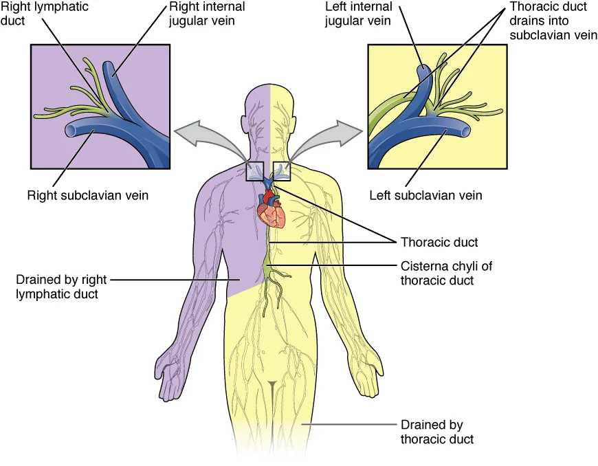
The thoracic duct is the largest lymphatic vessel in the human body. It is the main drainage channel for lymph from the lower body, left thorax, left upper limb, and the left side of the head and neck. Its function is to return filtered lymph, including chyle, to the bloodstream at the left venous angle, where the left internal jugular and left subclavian veins converge. The overall lymphatic drainage system is asymmetrical: the right lymphatic duct drains only the upper right side of the body, while the thoracic duct handles lymph from all remaining regions. [1]
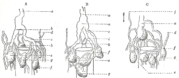
The thoracic duct begins just beneath the diaphragm in the cisterna chyli, a sac-like chamber that receives lymph from the lower abdomen, pelvis, and lower limbs via the left and right lumbar trunks and the intestinal trunk. The cisterna chyli is located at the level of the second lumbar vertebra, although this structure is not present in all individuals. From its origin, the duct ascends through the thorax to reach its termination.
The thoracic duct originates in the abdomen from the convergence of several lymphatic trunks, often beginning in the cisterna chyli. It then ascends through the aortic hiatus of the diaphragm into the posterior mediastinum. In the thorax, it lies anterior to the vertebral bodies, between the aorta and the azygos vein; the classical surgical landmark for ligation is the lympho-fatty tissue between these vessels at the level of the eighth–ninth thoracic vertebra. The duct continues upward to the root of the neck, where it arches laterally to empty into the left venous angle.
Superficial lymphatic vessels of the subcutaneous tissues follow the same routes as veins, whereas deep lymphatic vessels of the viscera generally follow the paths of arteries. This principle helps predict the course of the thoracic duct and its tributaries. [1]
The thoracic duct terminates by draining into the left venous angle, the junction of the left internal jugular and left subclavian veins. In most individuals, it terminates as a single duct; a pooled analysis of 1091 thoracic ducts found a single termination in 78.41% of cases (95% CI: 70.91–85.09%). A bifid or double duct termination was found in 13.76% (95% CI: 10.10–17.87%). These anatomical variations increase the risk of accidental injury during surgical procedures.
The thoracic duct carries chyle, which contains emulsified fats, proteins, sugars, lymphocytes, and immunoglobulins, contributing to fluid balance, fat absorption, and immune function. The distal part of the duct near its termination is also at risk during radical neck dissection (level IV), thyroidectomies, and lateral neck dissection. Operative treatments for chyle leak include video-assisted thoracoscopic duct ligation and percutaneous lymphangiography-guided thoracic duct cannulation and embolization.
Clinical pearlSurgical landmark
The thoracic duct is classically ligated between the aorta and azygos vein at the level of the eighth–ninth thoracic vertebra.
| Region | Draining vessel |
|---|---|
| Right upper quadrant (head, thorax, right upper limb) | Right lymphatic duct |
| Lower body, left thorax, left upper limb, left head and neck | Thoracic duct |
Figure 1Gray600
Long description
Modes of origin of thoracic duct. (Poirier and Charpy.) a. Thoracic duct. a’. Cisterna chyli. b, c’ Efferent trunks from lateral aortic glands. d. An efferent vessel which pierces the left crus of the diaphragm. e. f. Lateral aortic glands. h. Retroaortic glands. i. Intestinal trunk. j. Descending b

Figure 1: Modes of origin of thoracic duct (Poirier and Charpy.) a.
The thoracic duct is the largest lymphatic vessel in the body, responsible for returning most lymph and chyle to the venous circulation. It begins in the abdomen, ascends through the thorax, and terminates in the neck. Its anatomy is highly variable, which has important surgical implications.
The thoracic duct typically arises from the cisterna chyli, a dilated lymphatic sac located on the anterior surface of the body of the second lumbar vertebra. The cisterna chyli is formed by the confluence of the right and left lumbar lymphatic trunks and the intestinal trunk, which drain lymph from the lower limbs, pelvis, abdomen, and gastrointestinal tract. However, the cisterna chyli is present in only about 55% of individuals. When absent, the lumbar trunks drain directly into the thoracic [1]
It lies on the anterior surface of the vertebral bodies, between the azygos vein on the right and the descending thoracic aorta on the left. As it ascends, it crosses from the right side of the vertebral column to the left side, typically at the level of the T5–T6 vertebrae. Throughout its intrathoracic course, the duct receives tributaries that drain lymph from the posterior mediastinal nodes, intercostal spaces, and the left bronchomediastinal trunk. The duct then passes behind the aortic arch and the left subclavian artery, entering the root of the neck.
Figure 2Gray622
Long description
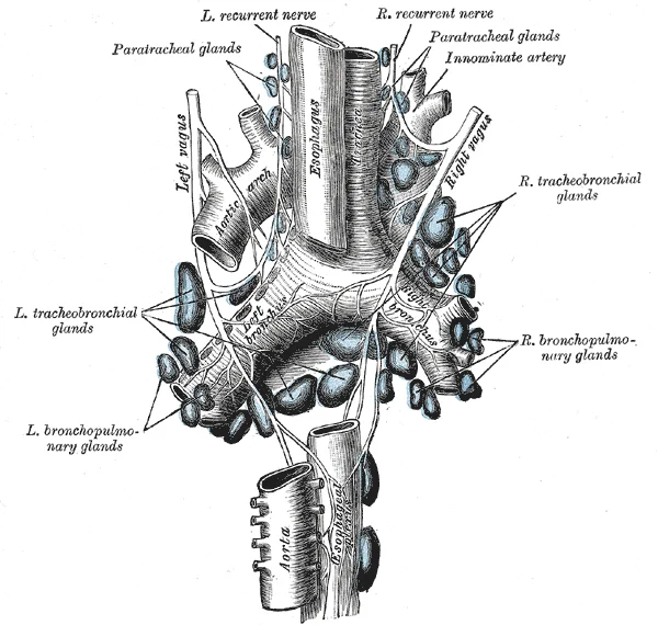
The tracheobronchial lymph glands. (From a figure designed by M. Hallé.)

Figure 2: The tracheobronchial lymph glands.
In the neck, the thoracic duct arches laterally and anteriorly, passing behind the carotid sheath and in front of the vertebral vessels, to terminate in the venous system. The most common termination site is the left venous angle, the junction of the left internal jugular and left subclavian veins, occurring in 45.29% of cases. Other termination sites include the left internal jugular vein (24.19%), the left subclavian vein (16.32%), the left external jugular vein (1.51%), and the left brachioce
Because of its long course and variable anatomy, the thoracic duct is vulnerable to injury during surgical procedures. During esophageal resections or thoracic surgeries, duct damage may lead to chylothorax, with a prevalence of up to 8.0%. Chylous leakage causes serious complications including malnutrition, immunosuppression, respiratory distress, fluid loss, and electrolyte imbalance. Prophylactic thoracic duct ligation during esophagectomy has been proposed to reduce this risk, though evidence for its benefit is debated; one systematic review reported significantly reduced odds of chylothorax (OR = 0.47), while another series found no significant difference (1.2% vs. 0.5%). [1]
Clinical pearlSurgical pearl
The thoracic duct crosses from right to left at T5–T6; therefore, injury in the lower thorax leaks on the right, while injury in the upper thorax or neck leaks on the left.
| Termination site | Prevalence |
|---|---|
| Left venous angle | 45.29% |
| Left internal jugular vein | 24.19% |
| Left subclavian vein | 16.32% |
| Left external jugular vein | 1.51% |
| Left brachiocephalic vein | 1.02% |
| Other rare sites (e.g., left vertebral vein, right venous angle) | <1% |
Figure 3Gray599
Long description
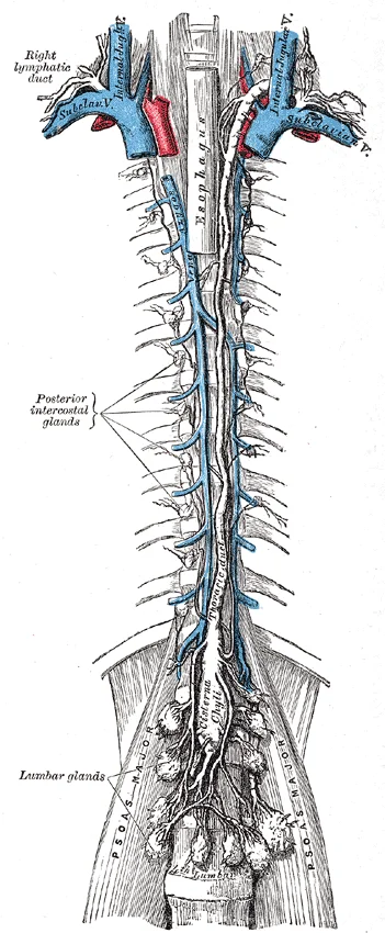
The thoracic and right lymphatic ducts. Labelled illustration from Wikimedia Commons, used under its open licence.

Figure 3: The thoracic and right lymphatic ducts.
The thoracic duct is the largest lymphatic vessel in the body, and its branching pattern is clinically significant because variations increase the risk of iatrogenic injury during head, neck, and thoracic surgeries. A meta-analysis of anatomical studies found that while a single terminal vessel is the most common arrangement, almost a quarter of cases exhibit branching of the duct. This means that in roughly 1 in 4 individuals, the thoracic duct divides into two or more branches, particularly near its termination.
DefinitionThoracic duct branching
The division of the thoracic duct into two or more separate channels, most commonly near its termination in the venous angle, occurring in almost a quarter of cases.
The branching most frequently occurs at the cranial end of the duct, where it approaches the junction of the left internal jugular and left subclavian veins (the left venous angle). The duct may divide into two, three, or occasionally more tributary-like branches before each enters the venous system separately. Less commonly, branching can occur along the course of the duct within the posterior mediastinum, where the duct may split and then reunite, forming a plexiform or ladder-like arrangement.
Near the level of the seventh cervical vertebra, the duct divides into two branches: a larger medial branch that arches forward to enter the left venous angle, and a smaller lateral branch that passes behind the internal jugular vein before joining the subclavian vein.
| Pattern | Frequency | Clinical relevance |
|---|---|---|
| Single vessel termination | Over three-quarters of cases | Standard surgical anatomy; lower risk of missing a duct during ligation |
| Two or more branches | Almost a quarter of cases | Higher risk of chylous leak if a branch is missed during lymphadenectomy |
| Plexiform network | Rare | May require careful dissection to identify all channels |
The clinical importance of this branching cannot be overstated. During a left-sided neck dissection or supraclavicular lymph node biopsy, a surgeon who identifies and ligates only one branch may leave another patent, resulting in a postoperative chylous fistula. Similarly, during thoracic duct ligation (TDL) performed as part of an esophagectomy for cancer, failure to recognise a branched duct may lead to persistent chylothorax. The meta-analysis emphasises that detailed anatomical knowledge of these variations is essential to minimise the risk of accidental injury.
The thoracic duct is the largest lymphatic vessel in the body and serves as the main conduit for returning lymph from most of the body to the venous circulation. It drains all lymph from the lower limbs, pelvis, abdomen, left half of the thorax, left upper limb, and the left side of the head and neck. In practical terms, it collects lymph from every region except the right upper limb, the right side of the head and neck, and the right hemithorax, which are drained by the smaller right lymphatic duct.
The duct is drawn as a single, slightly beaded tube ascending from the abdomen, through the thorax, to the root of the neck. Below the diaphragm, the duct receives the intestinal and lumbar lymphatic trunks, which carry lymph from the gastrointestinal tract, liver, and lower limbs. As it ascends through the thorax, it lies on the anterior surface of the vertebral bodies, between the azygos vein on its right and the descending aorta on its left. At the level of the fifth thoracic vertebra, it crosses from the right side of the vertebral column to the left side, passing behind the oesophagus. It then continues upward along the left border of the oesophagus to reach the thoracic inlet.
At the root of the neck, the duct arches laterally, forming the thoracic duct arch, which rises about 3 to 4 cm above the clavicle. It then turns downwards and forwards to empty into the junction of the left internal jugular and left subclavian veins, the left venous angle. Just before its termination, the duct receives the left jugular, left subclavian, and left bronchomediastinal trunks, which bring lymph from the left head and neck, left upper limb, and left thorax respectively. The duct therefore delivers a mixture of chyle from the gut and lymph from these other territories into the systemic venous blood.
The cisterna chyli, a dilated sac at the lower end of the thoracic duct, lies on the anterior surface of the second lumbar vertebra, behind the right crus of the diaphragm. It receives the right and left lumbar trunks and the intestinal trunk. From this sac, the duct passes upwards through the aortic hiatus of the diaphragm at the level of the twelfth thoracic vertebra, entering the posterior mediastinum. In the thorax, it is closely related to the oesophagus, the azygos vein, and the descending aorta, and it crosses from right to left at about the fifth thoracic vertebral level.
Because of its extensive course, the thoracic duct drains lymph from a remarkably large territory. Any obstruction or injury to the duct, for example during oesophagectomy, can lead to a chyle leak, a serious complication with a prevalence between 1.0% and 9.0%. The duct’s close anatomical relationship to the oesophagus places it at significant risk during such surgery. Untreated chyle leaks can cause hypovolaemia, electrolyte abnormalities, and immunosuppression from loss of immunoglobulins and T lymphocytes, with mortality rates up to 50%. The high rate of anatomical variation in the duct’s course, which arises from its complex embryonic development from paired vessels during the seventh and eighth gestational weeks, further increases the risk of iatrogenic injury.
The thoracic duct does not exist in isolation; it is part of a complex network of lymphatic channels that ensure drainage even if the main conduit is obstructed. Understanding these connections is vital for appreciating how lymph bypasses blockages and how infections or malignancies might spread to unexpected regions.
The thoracic duct typically begins at the cisterna chyli, a dilated sac located just beneath the diaphragm. It receives lymph from the paired lumbar trunks, which drain the lower limbs and pelvic organs, and the intestinal trunk, which drains the abdominal viscera. The lumbar trunks arise from the lumbar lymphatic plexuses surrounding the aorta. These plexuses collect lymph from the lower extremities, pelvic walls, and pelvic viscera. The intestinal trunk drains the stomach, i
While the thoracic duct drains the majority of the body, the right lymphatic duct handles drainage for the right upper quadrant. This short vessel collects lymph from the right side of the head and neck, the right upper limb, and the right thorax. It empties into the junction of the right internal jugular and right subclavian veins. These include transverse connections across the midline at the level of the neck and thorax. Such connections allow lymph to bypass an obstruction in one duct by flowing into the other. For example, if the thoracic duct is blocked, lymph from the left side may cross to the right side and drain via the right lymphatic duct.
Figure 42203 Lymphatic Trunks and Ducts System
Long description
The thoracic duct drains a much larger portion of the body than does the right lymphatic duct.

Figure 4: The thoracic duct drains a much larger portion of the body than does the right lymphatic duct.
Figure 5Gray601
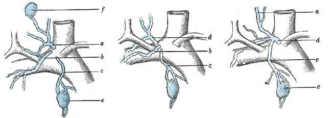
Long description
Terminal collecting trunks of right side. a. Jugular trunk. b. Subclavian trunk. c. Bronchomediastinal trunk. d. Right lymphatic trunk. e. Gland of internal mammary chain. f. Gland of deep cervical chain. (Poirier and Charpy.)

Figure 5: Terminal collecting trunks of right side (a).
Within the thorax, the thoracic duct has extensive anastomoses with intercostal lymphatic vessels. These connections link the posterior intercostal spaces with the duct itself. Additionally, mediastinal lymphatic trunks connect the anterior mediastinum to the thoracic duct. These pathways provide alternative routes for lymph flow from the chest wall and mediastinal organs. The anastomoses are particularly important in conditions such as lymphatic obstruction due to tumour compression or fibrosis. They ensure that lymph can still reach the venous system, albeit potentially with reduced efficiency.
The presence of these collaterals explains why lymphoedema may develop in unexpected patterns. If the thoracic duct is ligated or obstructed, lymph may reflux into the cisterna chyli or divert through intercostal channels. This can lead to swelling in the lower limbs or abdomen, even if the primary blockage is in the chest. Furthermore, malignant cells can travel through these anastomoses, leading to metastasis in regions distant from the primary tumour. For instance, cancer from the left breast might spread to the right axillary nodes via transverse anastomoses. Recognising these pathways helps clinicians predict the spread of disease and plan surgical interventions.
Clinical pearlKey Concept
Collateral lymphatic pathways ensure drainage continuity but can also facilitate the spread of pathology across body quadrants.
| Structure | Drainage Area | Termination |
|---|---|---|
| Lumbar Trunks | Lower limbs, pelvic walls, pelvic viscera | Cisterna chyli |
| Intestinal Trunk | Stomach, intestines, pancreas, liver, spleen | Cisterna chyli |
| Right Lymphatic Duct | Right head/neck, right upper limb, right thorax | Right venous angle |
| Thoracic Duct | Rest of body | Left venous angle |
The thoracic duct (TD) is the largest lymphatic vessel in the body, yet its anatomy is far from constant. Considerable variability exists in both its course and its termination, particularly in the cervical segment. For the medical student, understanding these variations is not merely academic; it is essential for safe surgical access to the neck and mediastinum, for interpreting lymphangiograms, and for planning procedures such as thoracic duct ligation or cannulation.
The cervical portion of the thoracic duct is widely regarded as the most variable part of the entire vessel. In its typical position, it lies at the base of the left side of the neck, where its relations are well defined: anteriorly by the carotid sheath, medially by the esophagus, laterally by the omohyoid muscle, and posteriorly by the prevertebral fascia. However, even within this region, the duct's behaviour is unpredictable. It usually extends 3 to 5 cm above the clavicle before arching anteriorly over the subclavian artery, but the exact height and angle of this arch vary between individuals.
The most clinically significant variations concern the termination of the thoracic duct. In the standard description, the duct ends at the left venous angle, the junction of the left internal jugular and left subclavian veins. Yet the literature documents numerous alternative termination sites, including the left internal jugular vein, the left subclavian vein, and even the left external jugular vein. Moreover, the duct does not always terminate as a single trunk; it may divide into multiple bra
Given this variability, no single "normal" pattern can be relied upon. For the surgeon, the key takeaway is that the duct's cervical course and termination must be individually identified in every patient, as the classic single-trunk termination at the left venous angle is only one of several possible arrangements. This variability explains why chyle leaks and iatrogenic injuries are more common when the duct is ligated blindly rather than dissected and visualised.
The thoracic duct is the largest lymphatic vessel in the body, responsible for returning lymph from most of the body to the venous system. It typically arises in the abdomen, passes through the thorax, and terminates in the neck. Understanding its exact course and relations is essential for interpreting chylothorax, planning neck dissections, and performing thoracic duct embolization.
Origin and formation. The thoracic duct usually begins in the abdomen at the upper lumbar level, where the intestinal trunk and the left and right lumbar trunks converge. This dilated origin is the cisterna chyli, which lies on the anterior surface of the body of the second lumbar vertebra (L2), just to the right of the abdominal aorta and behind the right crus of the diaphragm. The cisterna chyli is present in about 50% of individuals; when absent, the trunks join directly to form the duct.
Course in the abdomen and thorax. From the cisterna chyli, the thoracic duct passes upward through the aortic hiatus of the diaphragm at the level of the twelfth thoracic vertebra (T12), entering the posterior mediastinum. In the lower thorax, it lies on the anterior surface of the vertebral bodies, between the azygos vein on the right and the descending aorta on the left. As it ascends, it gradually crosses to the left side of the vertebral column, typically at the level of the fifth thoracic vertebra (T5), and then continues upward behind the aortic arch and the left subclavian artery. In the superior mediastinum, it lies to the left of the oesophagus and behind the left common carotid artery and vagus nerve.
Termination in the neck. The thoracic duct enters the root of the neck by passing behind the left common carotid artery and the left internal jugular vein. It then arches laterally and downwards, anterior to the vertebral artery and the thyrocervical trunk, before opening into the venous system at the junction of the left internal jugular and left subclavian veins, the left venous angle. These morphometric values are relevant for intravascular navigation during thoracic duct embolization.
Branches and tributaries. Along its course, the thoracic duct receives tributaries from the posterior mediastinal lymph nodes, the intercostal lymph trunks, and, in the neck, the left jugular, left subclavian, and left bronchomediastinal trunks. The duct may have a variable number of valves, which are more numerous near its termination)Skip.
Relations and clinical relevance. Because the thoracic duct crosses from right to left at about T5, injury to the duct in the lower thorax produces a right-sided chylothorax, whereas injury above T5 produces a left-sided chylothorax. The duct also passes behind the oesophagus, so oesophageal surgery may damage it.
Clinical pearlClinical pearl
A chylous effusion after thoracic surgery on the right side suggests injury below T5; on the left side, injury above T5.
| Feature | Detail |
|---|---|
| Origin | Cisterna chyli at L2, right of aorta |
| Diaphragmatic passage | Aortic hiatus at T12 |
| Crosses midline | Right to left at about T5 |
| Termination | Left venous angle (IJV–subclavian junction) |
| Termination diameter | Mean 3.79 mm |
| Cysterna chyli length | Mean 18.25 mm |
Thoracic duct course
- Cisterna chyli (L2)
- Aortic hiatus (T12)
- Posterior mediastinum, right of aorta
- Crosses to left at T5
- Behind aortic arch and left subclavian artery
- Arches in neck, opens at left venous angle
Clinical pearlOrigin and the Cisterna Chyli
The thoracic duct typically begins at the cisterna chyli, a sac-like chamber that receives lymph from the lower abdomen, pelvis, and lower limbs by way of the left and right lumbar trunks and the intestinal trunk.
Clinical pearlTermination Prevalence
The most frequent site of termination is the left internal jugular–subclavian joint, with a pooled prevalence of 45.29%. This junction represents the meeting point of the left internal jugular vein and left subclavian vein, forming the venous angle where the thoracic duct typically drains.
Exam trapTermination Variations
Do not assume the duct always ends at the joint; it terminates at the left internal jugular vein in 24.19% of cases and at the left subclavian vein in 16.32% of cases. Rare variations include termination in the right brachiocephalic vein or directly into the superior vena cava, which occurs in less than 5% of cases.
Clinical pearlVessel Structure
Lymphatic vessels, including the thoracic duct, possess a three-tunic structure similar to veins and contain one-way valves. These valves are positioned close together, creating a characteristic beaded appearance. The valves prevent backflow of lymph and ensure单向 flow toward the venous system.
Clinical pearlLymphatic Network
The system begins as blind-ending capillaries that feed into progressively larger vessels before emptying into the bloodstream via ducts. These capillaries are found in most tissues except the central nervous system, bone marrow, and avascular tissues like cartilage and epidermis. [1]
Clinical pearlNode Distribution
Lymph travels through lymph nodes, which are found in clusters near the neck, chest, abdomen, armpits, and groin. There are approximately 500–600 lymph nodes in the human body. The nodes serve as filtering stations where lymphocytes encounter antigens and mount immune responses. [1]
This reduction in survival is particularly significant in patients with malignancies, where the thoracic duct may be involved in tumor spread.
Clinical pearlMortality Risk
TDL is linked to a significantly higher hazard for mortality at 6 months (HR 1.83), 12 months (HR 1.54), 24 months (HR 1.21), and 28 months (HR 1.14) compared to no ligation.
Clinical pearlImmune Transport
The thoracic duct serves as a conduit for lymphocytes, including natural killer (NK) cells, which are first-line defenses against certain cancers and viruses. NK cells contain cytotoxic granules in their extensive cytoplasm and share their cell-killing mechanism with cytotoxic T cells.
NoteDevelopmental Considerations
In pediatric patients, the thoracic duct may have different anatomical variations compared to adults. Children have approximately the same number of lymph nodes (500-600) but they are relatively smaller in size. The immune system is still developing in children, making the lymphatic system particularly important for antigen exposure and immune education.
The thoracic duct typically originates from the cisterna chyli, a dilated lymphatic sac. The cisterna chyli is present in 55.49% of individuals, most commonly located at the L1–L2 vertebral levels (19.08% prevalence). The mean length of this origin sac is 18.25 mm. In the absence of a cisterna chyli, the thoracic duct may originate directly from the union of lumbar lymphatic trunks, which occurs in approximately 45% of cases. The cisterna chyli
The duct ascends from the abdomen into the thorax through the aortic hiatus of the diaphragm at the T12 level. It travels through the lymphatic system's hierarchy, which starts as blind-ending capillaries that merge into larger vessels. These vessels are characterized by a three-tunic structure and the presence of frequent one-way valves that give the duct a beaded appearance. As it ascends, the duct crosses anterior to the vertebral column, typically between T5 and T7 levels, and then crosses to the left side of the thorax at approximately the T4-T5 level. [1]
As it ascends, the duct serves as a transport route for lymphocytes moving from secondary lymphoid organs—such as the spleen (approximately 12 cm long) and various lymph nodes—back into the circulation. The thoracic duct receives tributaries from the left jugular, left subclavian, and left bronchomediastinal trunks, which typically converge near the venous angle. In the neck, the duct arches laterally and anteriorly to empty into the venous system. The duct is accompanied by the sympathetic chain and is in close proximity to the esophagus, thoracic aorta, and thoracic duct itself.
The duct terminates by emptying its contents into the venous system of the neck and upper thorax. The distribution of termination sites is as follows:
| Termination Site | Pooled Prevalence |
|---|---|
| Left internal jugular–subclavian joint | 45.29% |
| Left internal jugular vein | 24.19% |
| Left subclavian vein | 16.32% |
In approximately 14% of cases, the thoracic duct may have multiple termination sites or variations in its termination pattern. The termination point is clinically significant as it represents the site where lymph returns to the bloodstream, completing the lymphatic circulation. The left venous angle is the most common termination point, but variations can occur based on anatomical development and individual differences. In elderly patients, the termination point may be more variable due to age-related changes in vascular anatomy.
Damage to the thoracic duct or its ligation can lead to significant clinical outcomes. While postoperative chylothorax may occur, the more critical concern in surgical contexts is the impact on survival. The thoracic duct is particularly vulnerable during procedures involving the neck, chest, or abdomen, such as esophagectomy, lung resection, or neck dissection.
| Outcome | Finding | Reference |
|---|---|---|
| Overall Survival (OS) | Reduced by 3.5 months on average at 60-month follow-up | |
| 6-month Mortality Hazard | HR 1.83 | |
| 12-month Mortality Hazard | HR 1.54 | |
| 24-month Mortality Hazard | HR 1.21 | |
| 28-month Mortality Hazard | HR 1.14 |
Lymphatic Flow Path
- Blind-ending capillariesInitial collection of interstitial fluid
- Lymphatic vesselsThree-tunic structure with one-way valves
- Lymph nodesFiltering stations (500-600 total)
- Thoracic DuctMain collector ascending from cisterna chyli
- Venous JunctionTermination at left jugular-subclavian joint (45.29%)
Exam trapMisconception about lymph flow
Lymph is not actively pumped by the heart but is forced through vessels by body movements, skeletal muscle contractions, and breathing. One-way valves keep lymph moving toward the heart[1].
Exam trapOverlooking termination variability
The thoracic duct predominantly terminates in the region of the left internal jugular and left subclavian veins, with Types 1, 2, and 3 accounting for 85.80% of cases. However, students often assume it always terminates in the same location.
Exam trapAssuming single vessel termination
The thoracic duct typically terminates as a single vessel in the majority of cases (78.41%), but it divides into two vessels before termination in 13.76% of cases and rarely into three or more vessels (8.54%).
Exam trapConfusion about cisterna chyli
The cisterna chyli is said to be the origin of the thoracic duct, with a pooled prevalence of 55.49%. When absent, the thoracic duct is formed by a less distinct plexus of lymphatic channels.
Exam trapMisunderstanding lymph composition
Lymph is interstitial fluid that has entered the lymphatic system. Of the 20 liters of plasma released into interstitial space daily, 17 liters is reabsorbed directly by blood vessels, while the remaining 3 liters enters the lymphatic system[1].
Exam trapIgnoring clinical implications
Understanding thoracic duct anatomy is crucial to reduce the risk of inadvertent injury during surgical procedures involving the head, neck, and thorax. Students often fail to connect anatomical knowledge to clinical practice.
NoteClinical significance
Knowledge of thoracic duct anatomy is essential for surgeons operating in the head, neck, and thorax to avoid inadvertent injury, which could lead to chylothorax or other complications.
Clinical pearlImaging interpretation
Radiologists must recognize thoracic duct anatomy on imaging studies. Its termination patterns and variations are important when interpreting lymphangiograms, CT scans, and MRI studies of the neck and thorax.
Clinical pearlSurgical planning
In thoracic surgery, particularly esophagectomy and lung cancer resection, surgeons must be aware of thoracic duct location to prevent injury. The duct typically ascends through the posterior mediastinum, crossing from right to left at the T5-T6 level.
Clinical pearlManagement of chylothorax
When chylothorax occurs due to thoracic duct injury, knowledge of duct anatomy guides treatment options. Conservative management is first-line, but surgical intervention may be necessary, with options including ligation of the thoracic duct or pleurodesis.
NoteExamination relevance
Medical students are frequently tested on thoracic duct anatomy in examinations. Common questions include its origin, course, termination patterns, and clinical significance of injury.
Thoracic duct injury management
- Suspected thoracic duct injuryConsider chylothorax if milky fluid in chest drain
- Question: Is patient hemodynamically unstable?
Yes
- Immediate surgical intervention
No
- Conservative management: NPO, TPN, octreotide
- Question: Improvement within 5-7 days?
Yes
- Continue conservative management
No
- Consider surgical intervention: thoracic duct ligation
- The thoracic duct drains excess interstitial fluid (3 liters daily) back into the bloodstream[1]
- Lymph flow depends on body movements, muscle contractions, and breathing, not cardiac pumping[1]
- The thoracic duct predominantly terminates near the left internal jugular and subclavian veins (85.80% of cases)
- It typically terminates as a single vessel (78.41%), but may divide into two (13.76%) or more vessels (8.54%)
- The cisterna chyli is present in 55.49% of cases and is considered the origin of the thoracic duct
- Understanding thoracic duct anatomy reduces surgical complications in head, neck, and thoracic procedures
Answer each one in your head first, then open the answer.
How much interstitial fluid enters the lymphatic system daily?
Show answerHide answer
3 litersWhere does the thoracic duct predominantly terminate?
Show answerHide answer
Left internal jugular and left subclavian veinsWhat percentage of cases show the thoracic duct terminating as a single vessel?
Show answerHide answer
78.41%What is the prevalence of the cisterna chyli?
Show answerHide answer
55.49%What factors drive lymph flow through the thoracic duct?
Show answerHide answer
Body movements, skeletal muscle contractions, and breathing
- Betts JG, Young KA, Wise JA, et al. Anatomy of the Lymphatic and Immune Systems. OpenStax, Anatomy and Physiology 2e (CC BY-NC-SA 4.0). Accessed .
Figure credits
- Figure 1: Henry Vandyke Carter, via Wikimedia Commons, Public domain, Public domain.
- Figure 2: Henry Vandyke Carter, via Wikimedia Commons, Public domain, Public domain.
- Figure 3: Henry Vandyke Carter, via Wikimedia Commons, Public domain, Public domain.
- Figure 4: OpenStax College, via Wikimedia Commons, CC BY 3.0, CC BY 3.0.
- Figure 5: Henry Vandyke Carter, via Wikimedia Commons, Public domain, Public domain.
Educational content for medical students and doctors. It is not a substitute for clinical judgement or local guidelines.