Oesophagus
Anatomical relations of the thoracic oesophagus including vascular, lymphatic and neural structures.
The oesophagus is a muscular tube connecting the pharynx to the stomach, traversing the posterior mediastinum. Its anatomical relationships are critical for surgical safety and diagnostic imaging. The structure lies anterior to the vertebral column, posterior to the trachea and heart, and lateral to the aorta and azygos system.
The azygos vein ascends on the right side of the vertebral column, arching over the right main bronchus to drain into the superior vena cava. These veins provide collateral circulation and are at risk during oesophagectomy.
Histological examination is fundamental for diagnosing pathological changes. In conditions affecting the oesophagus, such as Barrett's oesophagus, the normal squamous epithelium is replaced by intestinal-type columnar epithelium. This metaplastic change is a response to chronic acid exposure. Diagnosis relies on identifying this change in biopsy specimens. Sometimes, therefore, instead of one, the material for microscopic examination is taken from two or even three places. The clipping from [2]
A penetrating aortic ulcer into the thoracic esophagus can cause aortoesophageal fistula[1]. [4]
Oesophageal varices are dilated submucosal veins resulting from portal hypertension. They are located in the lower third of the oesophagus. Rupture leads to life-threatening haematemesis. The triad of mid thoracic pain, sentinel arterial hemorrhage, and final exsanguination after a symptom free interval has been termed as Chiari's triad[1]. [4]
Clinical pearlSurgical Pearl
Knowledge of the complete anatomy of the TD is of utmost importance to both reduce the risk of accidental damage to the vessel and also to aid in the surgical treatment of the complication that already occurred.
| Feature | Prevalence |
|---|---|
| Single vessel termination | 78.41% |
| Bifid or double duct | 13.76% |
| Left venous angle termination | 45.29% |
The view is anterior, showing the duct's course relative to surrounding structures. It terminates within 1 cm of the left venous angle. This anatomical relationship is critical for surgeons to avoid injury during head and neck procedures. [1]
The oesophagus itself is a muscular tube connecting the pharynx to the stomach. In the cervical region, it lies immediately posterior to the trachea and anterior to the prevertebral fascia. The thoracic duct, a major lymphatic vessel, runs alongside the oesophagus in this segment. Variations in the termination of the thoracic duct are common.
Understanding these variations is essential for surgical safety. The cervical segment of the thoracic duct may be the most variable portion of this major lymphatic vessel. Numerous termination patterns have been described in the literature and major anatomical textbooks, including the left venous angle, the left internal jugular vein, the left subclavian vein, and even the left external jugular vein, amongst others. Surgeons must be aware of these anatomical nuances to reduce the risk of potential injury to the thoracic duct during various head and neck, as well as thoracic, surgeries. The oesophagus serves as a key landmark in this region, providing a consistent reference point amidst the variability of the lymphatic system. [1]
The oesophagus is a muscular tube connecting the pharynx to the stomach, traversing the neck, thorax, and abdomen. Its anatomical course is defined by its relations to surrounding structures, which are critical for understanding clinical presentations and surgical approaches. The organ is divided into cervical, thoracic, and abdominal segments, each with distinct relationships.
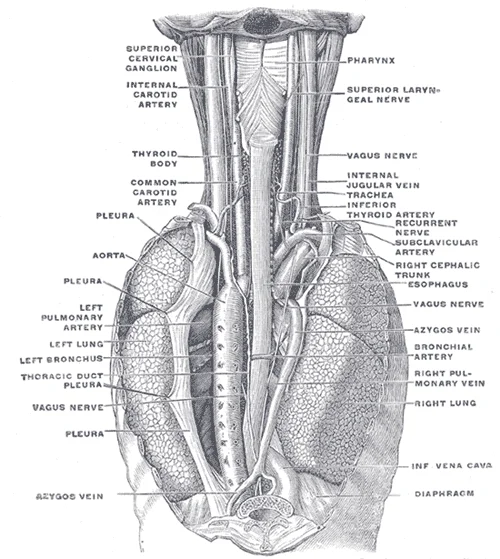
Figure 1Gray1032
Long description
The position and relation of the esophagus in the cervical region and in the posterior mediastinum. Seen from behind. (Poirier and Charpy.)

Figure 1: The position and relation of the esophagus in the cervical region and in the posterior mediastinum (seen from behind).
The thoracic oesophagus lies within the posterior mediastinum. It is closely related to the trachea anteriorly, the vertebral column posteriorly, and the azygos vein on the right. The azygos system of veins arches over the right main bronchus to drain into the superior vena cava. This proximity is clinically significant because varices in the azygos system can compress the oesophagus or indicate portal hypertension. The descending thoracic aorta is a key landmark.
Constrictions of the oesophagus are important sites for pathology and instrumentation. The first constriction is at the pharyngoesophageal junction, where the oesophagus begins. The second constriction occurs where the oesophagus is crossed by the aortic arch and the left main bronchus. The third constriction is at the oesophageal hiatus of the diaphragm. These constrictions are natural points of narrowing.
The cervical oesophagus lies anterior to the prevertebral fascia and is related to the trachea anteriorly. The recurrent laryngeal nerves run in the tracheoesophageal grooves. The thyroid gland lies anterior and lateral to the upper part of the cervical oesophagus. The carotid sheaths, containing the common carotid arteries and internal jugular veins, lie lateral to the oesophagus.
It enters the stomach at the cardia. The left vagus nerve forms the anterior vagal trunk, and the right vagus nerve forms the posterior vagal trunk. These nerves are crucial for gastric secretion and motility. The left gastric artery and vein lie near the abdominal oesophagus, making them important in surgical procedures such as oesophagectomy.
Clinical pearlClinical Pearl
Surgeons need to be aware of the prevalence and topography of the CC as iatrogenic damage to this lymphatic structure during abdominal dissection may lead to chylous ascites or chylothorax. [1]
| Structure | Relation |
|---|---|
| Trachea | Anterior (upper part) |
| Aortic Arch | Anterior (middle part) |
| Left Main Bronchus | Anterior (middle part) |
| Azygos Vein | Right lateral |
| Thoracic Aorta | Left lateral |
| Vertebral Column | Posterior |
| Diaphragm | Inferior |
The close anatomical relationship of the TD to the esophagus, the lymphatic vessel is at significant risk of injury during esophagectomies. This highlights the importance of careful dissection in the thoracic cavity. The oesophagus is a vital conduit, and its relations dictate both its function and its vulnerability to disease and iatrogenic injury. Understanding these relationships is fundamental for medical students and clinicians alike. [1]
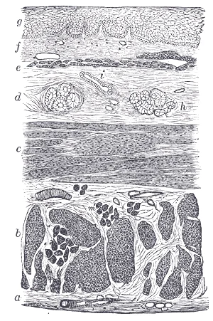
The oesophagus is a muscular tube connecting the pharynx to the stomach, serving as the conduit for food and liquid. Its interior is lined by mucosa, submucosa, and muscular layers, each with distinct structural features that facilitate peristalsis and protection. The mucosa consists of non-keratinised stratified squamous epithelium, which provides durability against abrasion from swallowed solids. This epithelium rests on a basement membrane and is supported by the lamina propria, a layer of loose connective tissue containing blood vessels and lymphatics. The muscularis mucosae, a thin layer of smooth muscle, separates the mucosa from the submucosa.
Figure 2Gray1033
Long description
Section of the human esophagus. (From a drawing by V. Horsley.) Moderately magnified. The section is transverse and from near the middle of the gullet. a. Fibrous covering. b. Divided fibers of longitudinal muscular coat. c. Transverse muscular fibers. d. Submucous or areolar layer. e. Muscularis mu

Figure 2: Section of the human esophagus (From a drawing by V.
DefinitionMucosal Layers
The oesophageal mucosa comprises stratified squamous epithelium, lamina propria, and muscularis mucosae, designed for protection and limited movement.
The submucosa is a dense connective tissue layer containing larger blood vessels, lymphatics, and the submucosal (Meissner’s) plexus, which regulates glandular secretion. The oesophageal glands proper, located in the submucosa, secrete mucus to lubricate the lumen. The myenteric (Auerbach’s) plexus, situated between these two muscle layers, controls motility.
The lumen is central, lined by the stratified squamous epithelium. Beneath the epithelium lies the lamina propria, appearing as a lighter pink connective tissue zone. The submucosa is a thick, pale layer containing glandular structures and blood vessels.
| Layer | Composition | Nerve Supply | Action |
|---|---|---|---|
| Inner Circular | Smooth muscle (upper 2/3), Skeletal (lower 1/3) | Autonomic | Constricts lumen |
| Outer Longitudinal | Smooth muscle (upper 2/3), Skeletal (lower 1/3) | Autonomic | Shortens tube |
This transition is crucial for understanding motility disorders. The vascular supply includes branches from the inferior thyroid artery superiorly and the left gastric artery inferiorly, with venous drainage following the arterial pattern. Lymphatic drainage flows to cervical, thoracic, and abdominal nodes, relevant in metastatic spread.
NoteMuscle Transition
The muscularis externa transitions from skeletal to smooth muscle, affecting control mechanisms from voluntary to autonomic.
This anatomical feature facilitates the spread of malignancy into adjacent tissues. The internal structure is thus optimised for both protection and propulsion, with layered organisation supporting its physiological role.
The oesophagus receives its blood supply from a segmental arrangement of arteries, a feature that is critical for surgical planning and understanding the consequences of tissue injury. Unlike many other tubular structures, the oesophageal vasculature does not form a single continuous longitudinal trunk but rather consists of distinct arterial branches that enter the organ at various levels. This segmental nature means that each part of the oesophagus is supplied by specific vessels arising from adjacent anatomical regions.
Understanding this layout is essential because excessive dissection can disrupt these segmental supplies. [3]
NoteClinical Relevance
The esophageal blood supply is predominantly segmental in nature and any excessive dissection around the esophagus may get further devitalized and the surgeon gets committed to esophagectomy lest the danger of disruption and sepsis. [4]
This segmental pattern has profound implications for surgical interventions. If a surgeon performs extensive dissection around the oesophagus, they risk devitalizing the tissue by cutting off these segmental blood sources. Such devitalization significantly increases the risk of anastomotic leak or disruption, particularly after procedures involving oesophageal closure or reconstruction. Consequently, preserving the arterial supply is paramount. In cases where dense adhesions are present, careful surgical technique that avoids unnecessary dissection can help preserve this vital blood flow.
| Segment | Primary Arterial Source |
|---|---|
| Cervical | Inferior thyroid artery |
| Thoracic | Oesophageal branches from thoracic aorta |
| Abdominal | Left gastric artery and left inferior phrenic artery |
The preservation of this blood supply is crucial for healing. When the oesophagus is injured, such as in cases of perforation, the integrity of the arterial supply determines the potential for natural healing. Conservative treatment strategies often rely on the assumption that the segmental blood supply remains intact. For instance, in a case where the oesophagus was not dissected due to dense adhesions, the preserved arterial blood supply enabled the oesophagus to heal naturally, aided by the small size of the rent. This outcome underscores the importance of respecting the segmental vascular anatomy during any surgical or diagnostic procedure involving the oesophagus. [4]
The venous drainage of the oesophagus is characterised by a rich submucosal plexus that forms longitudinal channels. These channels do not drain into a single uniform vein but instead communicate with the systemic venous systems of the neck, thorax, and abdomen. This arrangement creates potential portosystemic anastomoses, which are clinically significant in portal hypertension. The drainage pattern varies according to the segment of the oesophagus being considered, reflecting its embryological origin and anatomical relations.
These veins are small, short vessels that accompany the inferior thyroid artery. They drain into the left brachiocephalic vein. This drainage pathway connects the oesophageal plexus to the superior vena cava system. This dual supply means that pathology in either structure can affect the other. The veins are valveless, allowing for bidirectional flow under certain pathological conditions. [1]
The azygos vein runs along the right side of the vertebral column. It ascends through the posterior mediastinum. The hemiazygos vein runs on the left side. It crosses to the right to join the azygos vein. These veins provide a direct route for blood to reach the superior vena cava. The azygos system also receives intercostal veins. This arrangement ensures efficient drainage from the thoracic wall and mediastinal structures. The oesophageal branches join the azygos vein at various levels.
This vein is a branch of the portal vein. It runs along the lesser curvature of the stomach. It receives oesophageal branches from the lower oesophagus. This connection creates an anastomosis between the portal and systemic venous systems. In portal hypertension, blood can flow retrograde from the portal system into the oesophageal veins. This leads to the formation of oesophageal varices. These varices are thin-walled and prone to rupture. Rupture causes life-threatening haemorrhage. The left gastric vein is therefore a critical structure in understanding upper gastrointestinal bleeding.
Clinical pearlClinical Pearl
Oesophageal varices arise from portosystemic anastomoses, primarily involving the left gastric vein and oesophageal branches of the azygos system.
| Segment | Primary Veins | Destination |
|---|---|---|
| Upper | Inferior thyroid veins | Left brachiocephalic vein |
| Middle | Azygos and hemiazygos veins | Superior vena cava |
| Lower | Left gastric vein | Portal vein |
The lymphatic drainage of the oesophagus is segmental, following the vessel supply, which creates distinct pathways for metastatic spread. Understanding these routes is essential for surgical planning and staging. The upper third drains primarily into the deep cervical and superior mediastinal nodes. The middle third drains into the posterior mediastinal nodes and the tracheobronchial nodes. The lower third drains into the left gastric nodes and the coeliac nodes. This segmental nature means that a tumour in one region may not necessarily spread to the next, but advanced disease can bypass these barriers. [2] [3][4]
The posterior mediastinal nodes are a critical group for oesophageal cancer staging. They receive lymph from the middle and lower thirds of the oesophagus. These nodes are located along the lower thoracic aorta and the oesophageal wall itself. Metastasis to these nodes upstages the disease significantly. The potential for the thoracic duct to divide into two or more branches in almost a quarter of cases increases the risk of injury to these critical lymphatic structures during dissection. This anatomical variation requires careful identification of the duct to prevent chylothorax. [1]
Clinical pearlClinical Pearl
Always consider the thoracic duct's variability when dissecting the posterior mediastinum to avoid iatrogenic injury.
The TNM staging system relies heavily on the extent of spread to these lymph nodes. The 'N' category defines the number and location of involved nodes. Involvement of posterior mediastinal nodes is classified differently from distant nodal groups. Accurate assessment of these nodes guides treatment decisions, including the choice between surgery and neoadjuvant therapy. The integration of imaging and surgical staging is vital for determining resectability.
| Segment | Primary Drainage Nodes | Secondary/Distant Nodes |
|---|---|---|
| Upper Third | Deep Cervical, Superior Mediastinal | Paratracheal |
| Middle Third | Posterior Mediastinal, Tracheobronchial | Hilar |
| Lower Third | Left Gastric, Coeliac | Supraclavicular (rare) |
This indicates advanced metastatic spread. The presence of such nodes generally precludes curative resection. Therefore, precise mapping of the posterior mediastinal nodes is a cornerstone of preoperative evaluation. [2]
The oesophagus receives a complex innervation that integrates autonomic control with somatic sensation, a duality that explains both its physiological function and its characteristic patterns of referred pain. Understanding this supply requires tracing the pathways from the brainstem down to the muscular layers, noting how the nerve fibres change their nature as they descend.
The anterior vagal trunk, derived primarily from the left vagus, lies on the anterior surface of the oesophagus. The posterior vagal trunk, derived primarily from the right vagus, lies on the posterior surface. Both trunks descend along the oesophagus, giving off oesophageal branches.
DefinitionVagal Supply
The parasympathetic supply to the oesophagus is provided by the vagus nerves, which form anterior and posterior trunks that descend along the muscular wall.
The vagus nerves provide the primary parasympathetic innervation. In the neck, the right and left vagus nerves descend in the carotid sheaths. As they enter the thorax, they contribute to the oesophageal plexus. The anterior vagal trunk is formed mainly by fibres from the left vagus nerve and lies on the anterior aspect of the oesophagus. The posterior vagal trunk is formed mainly by fibres from the right vagus nerve and lies on the posterior aspect. These trunks descend along the oesophagus, pr [3]
Exam trapReferred Pain
Pain from the oesophagus is often referred to the epigastrium or between the shoulder blades due to shared spinal segments with somatic structures.
Somatic sensation is carried by different pathways. The upper third of the oesophagus, being striated muscle, receives somatic motor innervation from the recurrent laryngeal branches of the vagus nerves and sensory fibres from the cervical spinal nerves. The lower two-thirds, composed of smooth muscle, receives autonomic innervation. Pain fibres from the oesophagus travel with sympathetic fibres to the upper thoracic spinal segments (T1–T5). Consequently, oesophageal pain is often felt in the ch [3]
The sympathetic supply arises from the sympathetic trunk. Preganglionic fibres originate from the upper thoracic spinal cord (T1–T5) and synapse in the cervical and upper thoracic sympathetic ganglia. Postganglionic fibres then travel along the blood vessels to reach the oesophageal plexus. These fibres mediate vasoconstriction and modulate peristalsis. The interplay between the parasympathetic vagal stimulation, which promotes peristalsis and secretion, and sympathetic inhibition, which relaxes [3]
The oesophagus is a muscular tube connecting the pharynx to the stomach, serving as the conduit for food and liquid. Its wall structure is consistent with the general plan of the gastrointestinal tract but features distinct adaptations for its function. The mucosa is lined by non-keratinised stratified squamous epithelium, which provides robust protection against mechanical abrasion during swallowing. This epithelial layer rests on a lamina propria and a muscularis mucosae. The submucosa contains oesophageal glands proper, which secrete mucus to lubricate the bolus. The muscularis externa varies along the length of the organ; the upper third consists of skeletal muscle, the middle third contains a mixture of skeletal and smooth muscle, and the lower third is composed entirely of smooth muscle. This transition reflects the voluntary initiation of swallowing followed by involuntary peristalsis. The adventitia, rather than a serosa, covers the intrathoracic portion, allowing for mobility within the mediastinum.
DefinitionEpithelial Transition
The squamocolumnar junction marks the point where the oesophageal squamous epithelium meets the gastric columnar epithelium, a critical landmark in endoscopy and pathology.
Histological examination is fundamental for diagnosing pathological changes. In conditions affecting the oesophagus, such as Barrett's oesophagus, the normal squamous epithelium is replaced by intestinal-type columnar epithelium. This metaplastic change is a response to chronic acid exposure. Diagnosis relies on identifying this change in biopsy specimens. The clipping from
Exam trapBiopsy Sampling
Sampling must be targeted. Relying on a single random biopsy may miss focal lesions. Multiple samples from suspicious areas increase diagnostic yield.
The submucosa houses the oesophageal glands, which are mucous-secreting. These glands open into the lumen to facilitate the passage of the food bolus. The muscular layers are innervated by the autonomic nervous system and somatic motor neurons, coordinating the complex process of peristalsis. Lymphatic drainage follows the vascular supply to regional nodes. Understanding these histological and anatomical details is essential for interpreting clinical findings and planning interventions.
The oesophagus is a muscular conduit connecting the pharynx to the stomach, serving as a critical component of the digestive tract. Its development involves complex embryological processes that establish its anatomical position and functional integrity.
DefinitionEmbryological Origin
The oesophagus develops from the foregut, undergoing significant elongation and differentiation to form the muscular tube seen in adults.
During early gestation, the foregut gives rise to the respiratory and digestive systems. The oesophagus separates from the trachea through the formation of the tracheoesophageal septum. This separation is crucial for establishing distinct pathways for air and food. Failure of this process can lead to congenital anomalies such as oesophageal atresia or tracheoesophageal fistula.
The muscular wall of the oesophus develops from the splanchnic mesoderm. The upper third consists primarily of skeletal muscle, allowing for voluntary initiation of swallowing. The middle third contains a mixture of skeletal and smooth muscle, facilitating the transition to involuntary peristalsis. The lower third is composed entirely of smooth muscle, controlled by the autonomic nervous system to ensure efficient transport of the bolus into the stomach.
The mucosal lining is derived from the endoderm. It is initially lined by simple columnar epithelium but undergoes metaplasia to become stratified squamous non-keratinised epithelium. This adaptation provides resistance to mechanical abrasion from food passage. The submucosa contains glands that secrete mucus to lubricate the lumen, reducing friction during deglutition.
Neural development involves the migration of neural crest cells to form the myenteric (Auerbach’s) and submucosal (Meissner’s) plexuses. These plexuses coordinate peristaltic movements and glandular secretion. The vagus nerves provide parasympathetic innervation, enhancing motility and secretion, while sympathetic innervation modulates blood flow and inhibits motility.
Vascular development establishes the arterial supply from branches of the aorta and venous drainage into the azygos system and portal circulation. This dual drainage is clinically significant in portal hypertension, where collateral circulation can develop, leading to oesophageal varices.
Lymphatic development creates a rich network that drains into cervical, thoracic, and abdominal lymph nodes. This extensive drainage is relevant in the spread of oesophageal carcinoma, which often metastasises early due to the lack of a serosal layer in the oesophagus.
Understanding these embryological foundations is essential for recognising congenital defects and understanding the anatomical relationships that define the oesophagus in the mediastinum. The precise alignment of muscular layers and neural controls ensures the oesophagus functions as an efficient, low-resistance tube for nutrient transport.
The oesophagus is situated posterior to the trachea and the left main bronchus, lying anterior to the vertebral column. Understanding these spatial relationships is critical for interpreting cross-sectional imaging and planning surgical interventions.
The anatomical characteristics of the thoracic duct (TD) and cisterna chyli (CC) demonstrate considerable variability[1]. While previous reviews exist, the description is often obscured in literature and not reported as specific types[1]. Studies attempting to categorize variations have been inconsistent[1]. Therefore, a systematic search and meta-analysis were performed to clarify variations in anatomy and dimensions[1]. Our meta-analysis reveals significant variability in the anatomy of the TD and CC, particularly regarding TD termination patterns[1]. Despite the predominance of single-vessel terminations, almost a quarter of cases exhibit branching, highlighting the complexity of the anatomy of the TD. These findings demonstrate the importance of detailed anatomical knowledge for surgeons to minimize the risk of accidental injury during head and neck, as well as thoracic surgeries.
Understanding these anatomical characteristics is crucial because accidental injury to the TD can lead to complications that significantly decrease the patient’s quality of life[1]. During esophageal resections, reconstructions, or thoracic surgeries, TD damage may lead to chylothorax, with a prevalence of up to 8.0%[1]. Chylous fistula or chylothorax can cause serious systemic complications, including malnutrition, immunosuppression, and respiratory distress[1]. Therefore, surgeons performing procedures that risk TD injury must have a thorough understanding of TD anatomy to minimize accidental damage[1].
Clinical pearlClinical Relevance
Accidental injury to the thoracic duct during oesophageal surgery can result in chylothorax, a serious complication with a prevalence of up to 8.0% in thoracic procedures.
In agreement with the Boolean technique, the following search terms were employed: ((thoracic duct) OR (Hoorne’s canal)) AND (anatomy)[1]. The search terms were individually adapted to each database to minimize potential bias[1]. An additional search was conducted through the references of the identified studies at the end of the search stage to ensure the accuracy of the process[1].
| Procedure | Complication | Prevalence/Rate |
|---|---|---|
| Radical neck dissection | Chylous leakage | 1.0% to 3.0% |
| Esophageal resection | Chylothorax | Up to 8.0% |
The oesophagus is a muscular tube connecting the pharynx to the stomach, traversing the neck, thorax, and abdomen. It begins at the lower border of the cricoid cartilage, corresponding to the sixth cervical vertebra (C6), and terminates at the cardiac orifice of the stomach at the eleventh thoracic vertebra (T11). This length of approximately 25 centimetres is crossed by three physiological constrictions of clinical significance. The first constriction is cervical, located at the pharyngoesophageal junction where the cricopharyngeus muscle forms the upper oesophageal sphincter. The second is thoracic, where the aortic arch and the left main bronchus cross the anterior surface of the tube. These narrowings are common sites for the impaction of foreign bodies and the development of strictures. [2] [1]
The thoracic constriction is marked where the aortic arch crosses from the left, and the left main bronchus passes anteriorly. Understanding these relations is vital for interpreting barium swallows and endoscopic findings. [2]
The blood supply arises from multiple sources. The thoracic portion receives branches from the oesophageal branches of the thoracic aorta and the bronchial arteries. Venous drainage follows the arterial supply, with the lower third draining into the left gastric vein, which is part of the portal system. This portosystemic anastomosis is clinically important because oesophageal varices can develop in portal hypertension. [3]
Lymphatic drainage is segmental. The upper third drains to the deep cervical nodes, the middle third to the posterior mediastinal and tracheobronchial nodes, and the lower third to the cardiac and left gastric nodes. This pattern explains the metastatic spread of oesophageal carcinoma. The nerve supply includes the vagus nerves for parasympathetic innervation and sympathetic fibres from the sympathetic trunk. The intrinsic innervation is provided by the myenteric and submucosal plexuses. [4]
| Structure | Origin | Termination | Clinical Significance |
|---|---|---|---|
| Upper Constriction | Cricoid Cartilage | Pharynx | Foreign body impaction |
| Middle Constriction | Aortic Arch | Left Main Bronchus | Compression by enlarged nodes |
| Lower Constriction | Diaphragm | Stomach | Hiatus hernia site |
The oesophagus is a muscular tube connecting the pharynx to the stomach, passing through the neck, thorax, and diaphragm. It is not a single uniform structure but varies in diameter and relation to surrounding organs. Understanding its anatomy requires knowing its precise boundaries, vascular supply, and lymphatic drainage, which are critical for surgical planning and endoscopic interpretation.
The oesophagus begins at the lower border of the cricoid cartilage, corresponding to the C6 vertebra. It descends through the superior mediastinum, lying posterior to the trachea. In the thorax, it is closely related to the left recurrent laryngeal nerve, which hooks under the aortic arch. The vagus nerves form anterior and posterior oesophageal plexuses before reconstituting into anterior and posterior trunks. These trunks pass through the diaphragm with the oesophagus at the oesophageal hiatus, level T10.
The abdominal part of the oesophagus is short, measuring approximately 1–2 cm. It ends at the cardiac orifice of the stomach, located at T11. The lower oesophageal sphincter is a physiological sphincter formed by the tonic contraction of the circular muscle fibres. It is not a distinct anatomical ring but a zone of high pressure. These nerves provide parasympathetic innervation to the stomach and oesophagus.
The cervical oesophagus is supplied by the inferior thyroid artery. The abdominal oesophagus is supplied by the left gastric artery and left inferior phrenic artery. Venous drainage follows the arterial supply, with the lower oesophagus draining into the left gastric vein, which is part of the portal system. This connection is clinically significant in portal hypertension, leading to oesophageal varices. Lymph from the upper oesophagus drains to deep cervical nodes. The middle and lower oesophagus drain to posterior mediastinal and left gastric nodes.
Congenital anomalies are rare but include oesophageal atresia and tracheo-oesophageal fistula. The embryological development involves the fusion of the ventral foregut with the dorsal oesophagus. Failure of this process results in fistulas. The oesophagus has three natural constrictions: the cervical constriction at the cricopharyngeus, the thoracic constriction where the aortic arch and left main bronchus cross, and the diaphragmatic constriction at the hiatus. These sites are common locations for foreign body impaction and strictures.
DefinitionLower Oesophageal Sphincter
A functional sphincter, not an anatomical one, maintained by tonic muscle contraction and supported by the diaphragmatic crura.
NoteBlood Supply
The oesophagus has a rich collateral blood supply, which is why ischaemia is rare unless multiple sources are compromised.
Clinical pearlLymphatic Drainage
Upper third drains to cervical nodes; middle third to mediastinal nodes; lower third to gastric nodes. This dictates metastasis patterns.
Exam trapAortic Relation
The thoracic aorta is to the left of the oesophagus in the lower thorax, not the right.
DefinitionHiatus
The oesophageal hiatus is at T10, transmitting the oesophagus, vagus nerves, and gastric vessels.
NoteVarices
Oesophageal varices arise from portosystemic anastomoses between the left gastric vein and azygos system.
Clinical pearlRecurrent Laryngeal Nerve
The left recurrent laryngeal nerve loops under the aortic arch and ascends in the tracheo-oesophageal groove.
Exam trapLength
The total length is about 25 cm, but the abdominal segment is only 1–2 cm long.
Students frequently confuse the anatomical origin of the thoracic duct with that of the cisterna chyli, leading to errors in lymphatic mapping. A common misconception is that the thoracic duct begins at the level of the diaphragm or the heart. In reality, the cisterna chyli is said to be the origin of the thoracic duct. This distinction is vital for understanding lymphatic drainage patterns in the abdomen and thorax. The cisterna chyli receives lymph from the intestinal lymphatic trunk, the left lumbar trunk, and, more rarely, the right lumbar trunk. It also receives smaller contributions from the periaortic nodes and intercostal lymphatics. Students often overlook these specific tributaries, assuming a single uniform source. [1]
Another significant pitfall involves the prevalence of the cisterna chyli. Many students assume it is a constant anatomical structure present in every individual. However, its overall prevalence is controversial, with some studies reporting that the CC is present in the majority of the subjects, while others state that it is prevalent in less than half. When the CC is absent, the TD is formed by the merging of a less distinct plexus of lymphatic channels. This anatomical variation means that imaging or surgical dissection may not always reveal a distinct cistern, which can be mistaken for pathology or technical error. [1]
Histological examination is fundamental for diagnosing pathological changes. In conditions affecting the oesophagus, such as Barrett's oesophagus, the normal squamous epithelium is replaced by intestinal-type columnar epithelium. This metaplastic change is a response to chronic acid exposure. Diagnosis relies on identifying this change in biopsy specimens. The clipping from
Exam trapAnatomical Variation
Do not assume the cisterna chyli is always visible. Its pooled prevalence is 55.49%. If absent, the thoracic duct forms from a plexus. [1]
Knowledge of oesophageal and thoracic duct anatomy is critical in the operating theatre, particularly during oesophagectomy or mediastinal surgery. The thoracic duct ascends through the posterior mediastinum, lying anterior to the vertebral column and between the azygos vein and the aorta. Injury to this structure can lead to chylothorax, a serious complication. Surgeons must identify the cisterna chyli and the duct's course to avoid damage. The duct's origin from the cisterna chyli, which receives lymph from the intestinal and lumbar trunks, dictates the risk of chyle leakage from abdominal sources.
On imaging, such as CT or MRI, the thoracic duct may be visualized as a tubular structure in the posterior mediastinum. Radiologists must distinguish it from vascular structures and lymph nodes. The variable prevalence of the cisterna chyli means that a missing cistern on imaging does not necessarily indicate pathology. Instead, it may represent normal anatomical variation where the duct arises from a plexus. Recognizing this prevents unnecessary further investigation or misdiagnosis.
In examinations, students are often tested on the relations and tributaries of the thoracic duct. Questions may ask for the specific lymphatic trunks that feed into the cisterna chyli. The correct answer includes the intestinal lymphatic trunk and the left lumbar trunk. The right lumbar trunk contributes more rarely. This detailed knowledge is essential for answering questions on lymphatic drainage and surgical anatomy.
DefinitionThoracic Duct Origin
The cisterna chyli is the origin of the thoracic duct. It collects lymph from the intestinal and left lumbar trunks.
| Structure | Relation/Detail |
|---|---|
| Origin | Cisterna Chyli |
| Tributaries | Intestinal trunk, Left lumbar trunk |
| Prevalence of CC | 55.49% pooled |
| Absent CC | Plexus of channels |
Lymphatic Drainage Assessment
- Identify Cisterna ChyliCheck for presence (55.49% prevalence)
- Question: Is CC present?
Yes
- Trace to Thoracic DuctOrigin confirmed
No
- Identify PlexusFormed by merging channels
Clinical pearlClinical Relevance
Injury to the thoracic duct causes chylothorax. Know its course in the posterior mediastinum.
The cisterna chyli is the origin of the thoracic duct. It receives lymph from the intestinal and left lumbar trunks. The right lumbar trunk contributes more rarely. Pooled prevalence of the cisterna chyli is 55.49%. When absent, the thoracic duct forms from a plexus. Early mucosal lesions have no risk of nodal transmission. [2] Preinvasive carcinoma allows for conservative surgery. [2] EBUS-TBNA is first-line for mediastinal staging. [3]
Answer each one in your head first, then open the answer.
What is the origin of the thoracic duct?
Show answerHide answer
Cisterna chyliWhich trunks drain into the cisterna chyli?
Show answerHide answer
Intestinal and left lumbar trunksWhat is the pooled prevalence of the cisterna chyli?
Show answerHide answer
55.49%How is the thoracic duct formed if the cisterna chyli is absent?
Show answerHide answer
Merging of a less distinct plexusWhat is the first-line modality for mediastinal staging?
Show answerHide answer
EBUS-TBNA
- Plutecki D, Bonczar M, Wilk J, et al. The Anatomy of the Thoracic Duct and Cisterna Chyli: A Meta-Analysis with Surgical Implications. Journal of clinical medicine 2024;13(15):4285. doi:10.3390/jcm13154285. Accessed .
- Šarenac T, Mikov M. Cervical Cancer, Different Treatments and Importance of Bile Acids as Therapeutic Agents in This Disease. Frontiers in pharmacology 2019. Accessed .
- Matache SR, Afetelor AA, Voinea AM, Cosoveanu GC, Dumitru SM, Alexe M, Orghidan M, Smaranda AM, Dobrea VC, Șerbănoiu A, Mahler B, Savu CF. Balancing Accuracy, Safety, and Cost in Mediastinal Diagnostics: A Systematic Review of EBUS and Mediastinoscopy in NSCLC. Healthcare (Basel, Switzerland) 2025. Accessed .
- Chandrashekar G, Kumar VM, Kumar AK. Repair of aortoesophageal fistula due to a penetrating atherosclerotic ulcer of the descending thoracic aorta and literature review. Journal of cardiothoracic surgery 2007. Accessed .
Figure credits
Educational content for medical students and doctors. It is not a substitute for clinical judgement or local guidelines.