Great vessels
Anatomy of the great vessels connecting the heart to the lungs and systemic circulation.
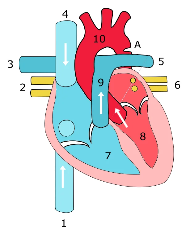
Figure 1Coarctation
Long description
Schetch showing heart with coarctation of the aorta. Image drawn by myself, inspired by illustration by Wapcaplet. Legend: A: Coarctation (narrowing) of the aorta. 1:inferior caval vein, 2:right pulmonary veins, 3: right

Figure 1: Schetch showing heart with coarctation of the aorta.
The single vessel exiting the right ventricle is the pulmonary trunk. At the base of the pulmonary trunk is the pulmonary semilunar valve, which prevents backflow of blood into the right ventricle during ventricular diastole. As the pulmonary trunk reaches the superior surface of the heart, it curves posteriorly and rapidly bifurcates (divides) into two branches, a left and a right pulmonary artery.
The femoral circumflex vein drains blood from the areas in proximity to the head and neck of the femur. [1]
DefinitionSystemic Venous Return
The superior and inferior venae cavae are the two major systemic veins returning blood to the right atrium. [2]
The superior vena cava is formed by the union of the left and right brachiocephalic veins. These veins drain the upper limbs, head, and neck. The right brachiocephalic vein is shorter and more vertical than the left. The left brachiocephalic vein crosses the midline anterior to the great arteries.
The ligamentum arteriosum is a fibrous remnant of the fetal ductus arteriosus. It connects the left pulmonary artery to the aortic arch. This structure anchors the pulmonary artery to the aorta.
| Structure | Origin/Termination | Key Feature |
|---|---|---|
| Pulmonary Trunk | Right Ventricle | Bifurcates into left/right pulmonary arteries |
| Superior Vena Cava | Brachiocephalic Veins | Drains head, neck, upper limbs |
| Inferior Vena Cava | Iliac Veins | Drains lower limbs, abdominopelvic region |
| Ligamentum Arteriosum | Left Pulmonary Artery | Connects to aortic arch |
The aorta originates from the left ventricle, arches posteriorly and to the left, then descends. The pulmonary trunk arises from the right ventricle, bifurcating into left and right pulmonary arteries. The superior vena cava forms from the union of brachiocephalic veins, draining into the right atrium. The inferior vena cava ascends through the diaphragm to enter the right atrium. [2]
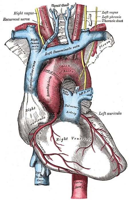
The aorta is the main arterial trunk of the systemic circuit. It originates from the left ventricle. The ascending aorta rises anteriorly. The aortic arch curves posteriorly and to the left. It gives off three major branches: the brachiocephalic trunk, the left common carotid artery, and the left subclavian artery. The arch continues as the descending aorta. The thoracic aorta runs alongside the vertebral column. It gives off visceral and parietal branches. The abdominal aorta begins at the aort [1]
The pulmonary trunk arises from the right ventricle. It is short and wide. It bifurcates into the left and right pulmonary arteries. These vessels carry deoxygenated blood to the lungs. This system is referred to as the pulmonary circuit[1]. The pulmonary arteries enter the lungs at the hilum. They branch extensively within the lung parenchyma. They accompany the bronchi. They terminate in the capillary beds surrounding the alveoli. Gas exchange occurs here. Oxygenated blood returns via the pulmonary veins. [2]
The superior vena cava drains blood from the upper body. It is formed by the union of the left and right brachiocephalic veins. It descends vertically in the superior mediastinum. It enters the right atrium. The inferior vena cava drains blood from the lower body. It ascends through the posterior abdominal wall. It passes through the caval opening of the diaphragm. It enters the right atrium. Blood returning from the systemic circuit enters the right atrium via the superior and inferior venae cavae [1][2]
Figure 2Gray556
Long description
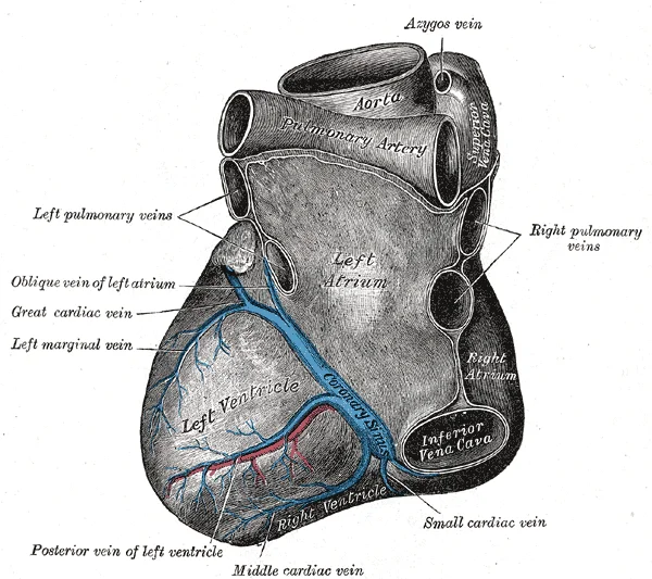
Base and diaphragmatic surface of heart.

Figure 2: Base and diaphragmatic surface of heart.
The great saphenous vein is located on the medial surface of the leg and thigh[1]. It collects blood from the superficial portions of these areas[1]. It drains into the femoral vein. The femoral vein receives the deep femoral vein and the femoral circumflex vein[1]. The deep femoral vein drains blood from the deeper portions of the thigh[1].
Figure 3Gray489
Long description
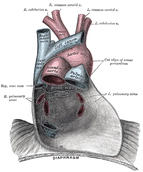
Posterior wall of the pericardial sac, showing the lines of reflection of the serous pericardium on the great vessels.

Figure 3: Posterior wall of the pericardial sac, showing the lines of reflection of the serous pericardium on the great vessels.
The aorta is the largest artery in the body. It arises from the left ventricle and eventually descends to the abdominal region, where it bifurcates at the level of the fourth lumbar vertebra into the two common iliac arteries. The aorta consists of the ascending aorta, the aortic arch, and the descending aorta. At the base of the aorta is the aortic semilunar valve that prevents backflow of blood into the left ventricle while the heart is relaxing. After exiting the heart, the ascending aorta moves in a superior direction for approximately 5 cm and ends at the sternal angle. Following this ascent, it reverses direction, forming a graceful arc to the left, called the aortic arch. The aortic arch descends toward the inferior portions of the body and ends at the level of the intervertebral disk between the fourth and fifth thoracic vertebrae. [1]
Emerging from the right ventricle at the base of the pulmonary trunk is the pulmonary semilunar valve, or the pulmonary valve. The pulmonary valve is comprised of three small flaps of endothelium reinforced with connective tissue. Unlike the atrioventricular valves, there are no papillary muscles or chordae tendineae associated with the pulmonary valve. When the ventricle relaxes, the pressure differential causes blood to flow back into the ventricle from the pulmonary trunk. This flow of blood fills the pocket-like flaps of the pulmonary valve, causing the valve to close and producing an audible sound.
Blood relatively high in oxygen concentration is returned from the pulmonary circuit to the left atrium via the four pulmonary veins. From the left atrium, blood moves into the left ventricle, which pumps blood into the aorta. The aorta and its branches—the systemic arteries—send blood to virtually every organ of the body. The venae cavae return deoxygenated blood to the right atrium, completing the circuit before pulmonary oxygenation. [1] [2]
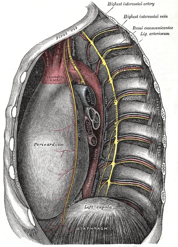
Beyond the arch, the descending aorta continues close to the bodies of the vertebrae and passes through an opening in the diaphragm known as the aortic hiatus. Superior to the diaphragm, the aorta is called the thoracic aorta. The thoracic aorta begins at the level of vertebra T5 and continues through to the diaphragm at the level of T12, initially traveling within the mediastinum to the left of the vertebral column. As it passes through the thoracic region, the thoracic aorta gives rise to several branches, which are collectively referred to as visceral branches and parietal branches. Those branches that supply blood primarily to visceral organs are known as the visceral branches and include the bronchial arteries, pericardial arteries, esophageal arteries, and the mediastinal arteries. Each bronchial artery (typically two on the left and one on the right) supplies systemic blood to the lungs and visceral pleura. The remaining thoracic aorta branches are collectively referred to as parietal branches or somatic branches, and include the intercostal and superior phrenic arteries. Each intercostal artery provides blood to the muscles of the thoracic cavity and vertebral column. The superior phrenic artery provides blood to the superior surface of the diaphragm. [1]
Figure 4Gray530
Long description
The thoracic aorta, viewed from the left side.

Figure 4: The thoracic aorta, viewed from the left side.
Figure 5Gray505
Long description
The arch of the aorta, and its branches.

Figure 5: The arch of the aorta, and its branches.
| Category | Branches | Supply |
|---|---|---|
| Visceral | Bronchial, Pericardial, Esophageal, Mediastinal | Lungs, Pericardium, Esophagus, Mediastinum |
| Parietal | Intercostal, Superior Phrenic | Thoracic Wall Muscles, Diaphragm |
DefinitionCoarctation
A congenital narrowing of the aorta, typically near the ductus arteriosus, which can lead to hypertension in the upper extremities and reduced perfusion in the lower body.
DefinitionAneurysm
A localized, abnormal dilation of a blood vessel wall, which may occur in the aorta due to weakening of the vessel layers.
The great vessels form the primary conduits for systemic circulation, originating from the heart and distributing blood to the body’s tissues. Understanding their territories requires tracing the path from the aortic arch through to the peripheral capillary beds.
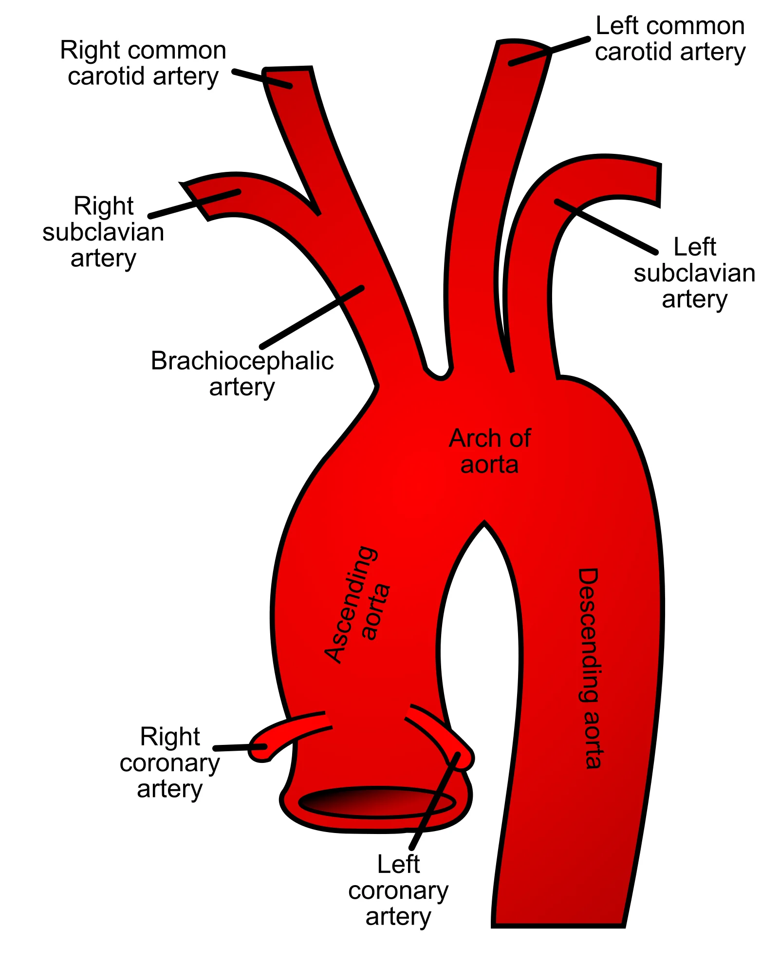
The aortic arch gives rise to three major branches that supply the head, neck, and upper limbs. These vessels are classified as elastic arteries due to their proximity to the heart and their ability to withstand high pressure. [1]
- Brachiocephalic Artery: This is the first and largest branch. It ascends obliquely and divides into the right common carotid artery and the right subclavian artery. [1]
- Left Common Carotid Artery: The second branch, it ascends directly into the neck to supply the head and brain.
- Left Subclavian Artery: The third branch, named for its position under the clavicle, supplies the left upper limb and parts of the brain and spinal cord. [1]
The brachiocephalic artery arises from the superior aspect of the arch, lying to the right of the midline. The left common carotid artery originates immediately to the left of the brachiocephalic trunk. The left subclavian artery is the final branch, emerging just before the arch descends into the thoracic aorta. The vertebral arteries, which supply the posterior brain, branch off the subclavian arteries. The internal thoracic arteries also arise from the subclavian arteries, running inferiorly along the sternum. [1]
Figure 6Gray506
Long description
Plan of the branches. Labelled illustration from Wikimedia Commons, used under its open licence.

Figure 6: Plan of the branches.
Blood flows from these elastic arteries into muscular arteries, then arterioles, and finally into systemic capillaries. The arterioles are the resistance vessels, offering the greatest resistance in the vascular network due to their small lumen. This resistance is crucial for regulating blood pressure. Vasodilation and vasoconstriction of arterioles play a more significant role in blood pressure regulation than changes in other vessel types. [3]
At the capillary level, exchange occurs between blood and tissue fluid. Oxygen and nutrients exit the capillaries for cellular metabolism, while carbon dioxide and waste products enter the blood. [2]
Systemic veins return deoxygenated blood to the right atrium. This blood has already passed through systemic capillaries and is relatively low in oxygen. In many regions, veins parallel the arteries that supplied the same area, creating a complementary pattern. However, venous anatomy shows greater variability than arterial anatomy. [1]
The heart itself is supplied by the coronary arteries. Anastomoses in the heart are very small, meaning collateral circulation is limited. Consequently, a blockage in a coronary artery often leads to myocardial infarction and cell death. [2]
Coronary veins drain the heart and generally parallel the surface arteries. The great cardiac vein follows the anterior interventricular sulcus, paralleling the anterior interventricular artery. It eventually flows into the coronary sinus on the posterior surface. Other veins, such as the middle cardiac vein, parallel the posterior interventricular artery. [1] [2]
Blood from the gastrointestinal tract is carried to the liver via the hepatic portal system for processing before entering the general systemic circulation. This ensures that nutrients and toxins are filtered by the liver before reaching the rest of the body. [1]
Blood pressure is the force exerted by blood upon vessel walls. It is typically measured in the brachial artery in clinical practice. Mean arterial pressure drives blood into tissues. Variables affecting flow include cardiac output, blood volume, viscosity, and vessel diameter. As blood volume increases, pressure and flow increase; conversely, decreased volume leads to decreased pressure and flow. [3]
Anastomoses are connections between blood vessels that allow blood to circulate to a region even if there may be partial blockage in another branch[1]. This concept is fundamental to understanding vascular resilience. Like a street that changes name as it passes through an intersection, an artery or vein can change names as it passes an anatomical landmark. For example, the left subclavian artery becomes the axillary artery as it passes through the body wall and into the axillary region, and then becomes the brachial artery as it flows from the axillary region into the upper arm. You will also find examples of anastomoses where two blood vessels that previously branched reconnect. Anastomoses are especially common in veins, where they help maintain blood flow even when one vessel is blocked or narrowed, although there are some important ones in the arteries supplying the brain.
The internal jugular vein receives blood from the brain and the superficial facial vein. The external jugular vein receives blood from the more superficial portions of the head, scalp, and cranial regions, including the temporal vein and maxillary vein. Although the external and internal jugular veins are separate vessels, there are anastomoses between them close to the thoracic region. Blood from the external jugular vein empties into the subclavian vein.
Intracranial circulation involves complex venous drainage. Many smaller veins of the brain stem and the superficial veins of the cerebrum lead to larger vessels referred to as intracranial sinuses. These include the superior and inferior sagittal sinuses, straight sinus, cavernous sinuses, left and right sinuses, the petrosal sinuses, and the occipital sinuses. Other cerebral veins and those from the eye socket flow into the cavernous sinus, which flows into the petrosal sinus and then into the internal jugular vein. [1]
NoteClinical Relevance
Anastomoses are especially common in veins, where they help maintain blood flow even when one vessel is blocked or narrowed.
This simple approach has proven effective for many students in mastering these major circulatory patterns. Another approach that works well for many students is to create simple line drawings similar to the ones provided, labeling each of the major vessels. It is beyond the scope of this text to name every vessel in the body. However, we will attempt to discuss the major pathways for blood and acquaint you with the major named arteries and veins in the body.
Individual variations in circulation patterns are not uncommon. While the standard anatomical arrangement of the great vessels is consistent, minor deviations can occur that are clinically significant. Understanding these variations requires a firm grasp of the surrounding structures, particularly the pericardium, which anchors and defines the space in which these vessels operate. [1]
The pericardium is the membrane that directly surrounds the heart and defines the pericardial cavity. It also surrounds the “roots” of the major vessels, or the areas of closest proximity to the heart. This structure consists of two distinct sublayers: the sturdy outer fibrous pericardium and the inner serous pericardium. The fibrous pericardium is made of tough, dense connective tissue that protects the heart and maintains its position in the thorax. The more delicate serous pericardium consists of two layers: the parietal pericardium, which is fused to the fibrous pericardium, and an inner visceral pericardium, or epicardium, which is fused to the heart and is part of the heart wall. The pericardial cavity, filled with lubricating serous fluid, lies between the epicardium and the pericardium. [2]
Inside the pericardium, the surface features of the heart are visible, including the four chambers. There is a superficial leaf-like extension of the atria near the superior surface of the heart, one on each side, called an auricle—a name that means “ear like”—because its shape resembles the external ear of a human. Auricles are relatively thin-walled structures that can fill with blood and empty into the atria or upper chambers of the heart. You may also hear them referred to as atrial appendages. Also prominent is a series of fat-filled grooves, each of which is known as a sulcus (plural = sulci), along the superior surfaces of the heart. Major coronary blood vessels are located in these sulci. The deep coronary sulcus is located between the atria and ventricles. Located between the left and right ventricles are two additional sulci that are not as deep as the coronary sulcus. [2] [1]
The heart resides within the pericardial sac and is located in the mediastinal space within the thoracic cavity. The pericardial sac consists of two fused layers: an outer fibrous capsule and an inner parietal pericardium lined with a serous membrane. Between the pericardial sac and the heart is the pericardial cavity, which is filled with lubricating serous fluid. The walls of the heart are composed of an outer epicardium, a thick myocardium, and an inner lining layer of endocardium. The human heart consists of a pair of atria, which receive blood and pump it into a pair of ventricles, which pump blood into the vessels. The right atrium receives systemic blood relatively low in oxygen and pumps it into the right ventricle, which pumps it into the pulmonary circuit. [1] [2]
In contrast to length, the diameter of blood vessels changes throughout the body, according to the type of vessel, as we discussed earlier. The diameter of any given vessel may also change frequently throughout the day in response to neural and chemical signals that trigger vasodilation and vasoconstriction. The vascular tone of the vessel is the contractile state of the smooth muscle and the primary determinant of diameter, and thus of resistance and flow. A decreased diameter means more of the blood contacts the vessel wall, and resistance increases, subsequently decreasing flow. [3]
The clinical assessment of the great vessels relies on understanding their anatomical course, palpable landmarks, and the haemodynamic consequences of structural pathology. A fundamental concept is pulse pressure, defined as the difference between systolic and diastolic pressure. For instance, a reading of 120 mm Hg systolic and 80 mm Hg diastolic yields a pulse pressure of 40 mmHg. Clinically, a pulse pressure should be at least 25 percent of the systolic pressure. Values below this threshold are termed narrow and may indicate low stroke volume, as seen in aortic stenosis or significant blood loss. [3]
DefinitionPulse Pressure
The numerical difference between systolic and diastolic blood pressure, reflecting stroke volume and arterial compliance.
Palpation of the pulse provides immediate clinical data on heart rate and ventricular strength. The radial artery in the wrist and the common carotid artery in the neck are standard sites for manual palpation. Other accessible sites include the temporal, facial, brachial, femoral, popliteal, posterior tibial, and dorsalis pedis arteries. A strong pulse indicates high systolic pressure and robust cardiac output, whereas a weak pulse suggests fallen systolic pressure and potential need for intervention. [3]
The brachiocephalic trunk is the first branch, arising from the right side of the arch. The left common carotid artery follows, supplying the head and neck. The left subclavian artery is the final major branch, supplying the upper limb. The descending thoracic aorta continues inferiorly. In the context of coarctation, a narrowing typically occurs just distal to the left subclavian artery origin. This location creates a pressure gradient, leading to hypertension in the upper body and hypotension in the lower body. The aneurysm may form at the site of the coarctation due to post-stenotic dilation. Dissection involves a tear in the intima, allowing blood to enter the media, potentially compromising branch vessel flow. Understanding these relations is critical for interpreting imaging and planning surgical access. [1]
Aneurysms can develop at sites of high stress or pre-existing weakness. Access points for vascular procedures often utilise the femoral or radial arteries due to their superficial location. Ligation of specific vessels may be necessary in trauma or tumour management, requiring precise knowledge of collateral circulation. For example, the internal carotid artery enters the cranial cavity through the carotid foramen. It gives rise to the anterior cerebral artery, supplying the frontal lobe, and the middle cerebral artery, which supplies the temporal and parietal lobes. The ophthalmic artery is another major branch, providing blood to the eyes. [1]
Exam trapHypovolemia Masking
An individual may remain asymptomatic until 10–20 percent of blood volume is lost due to effective regulatory mechanisms. [3]
Coarctation of the aorta is a congenital narrowing that often presents with differential blood pressures. The associated aneurysm forms due to turbulent flow and wall stress at the narrowing. Dissection in this region can extend proximally to the aortic root or distally into the abdominal aorta. Recognition of these anatomical variants is essential for accurate diagnosis and management. Mean arterial pressure (MAP) is calculated as diastolic BP plus one-third of the pulse pressure. This formula highlights the importance of diastolic pressure in maintaining organ perfusion. [3]
The celiac trunk divides into the left gastric artery to supply blood to the stomach and esophagus. [1]
The aorta is the main arterial trunk of the systemic circuit. It originates from the left ventricle. The ascending aorta rises anteriorly. The aortic arch curves posteriorly and to the left. It gives off three major branches: the brachiocephalic trunk, the left common carotid artery, and the left subclavian artery. The arch continues as the descending aorta. The thoracic aorta runs alongside the vertebral column. It gives off visceral and parietal branches. The abdominal aorta begins at the aort
| Structure | Supply Source |
|---|---|
| Urinary bladder | Internal iliac artery |
| Pelvic walls | Internal iliac artery |
| External genitalia | Internal iliac artery |
| Medial femoral region | Internal iliac artery |
| Uterus and vagina (female) | Internal iliac artery |
The external iliac vein is a large vein that drains blood from the leg to the common iliac vein. The pelvic organs and integument drain into the internal iliac vein. This vein forms from several smaller veins, including the umbilical veins that run on either side of the bladder. The external and internal iliac veins combine near the inferior portion of the sacroiliac joint to form the common iliac vein. The middle sacral vein drains the sacral region into the common iliac vein. The common iliac veins come together at the level of L5 to form the inferior vena cava. [1]
A typical heart is approximately the size of your fist: 12 cm (5 in) in length, 8 cm (3.5 in) wide, and 6 cm (2.5 in) in thickness. The weight of a female heart is approximately 250–300 grams (9 to 11 ounces). The weight of a male heart is approximately 300–350 grams (11 to 12 ounces). The human heart consists of four chambers: two atria and two ventricles. The ventricles serve as the primary pumping chambers of the heart. Located at the opening between the left atrium and left ventricle is the mitral valve. This valve consists of two cusps. The two cusps of the mitral valve are attached by chordae tendineae to two papillary muscles. [2]
DefinitionMitral Valve
Also called the bicuspid valve, it has two cusps attached to papillary muscles via chordae tendineae.
The pumping action of the heart propels the blood into the arteries, from an area of higher pressure toward an area of lower pressure. If blood is to flow from the veins back into the heart, the pressure in the veins must be greater than the pressure in the atria. The pressure in the atria during diastole is very low, often approaching zero when the atria are relaxed. Only one factor, the radius, can be changed rapidly by vasoconstriction and vasodilation. Small changes in the radius will greatly affect flow, since it is raised to the fourth power in the equation. [3]
Clinical pearlVascular Resistance
Radius is the only variable changed rapidly; small changes dramatically impact flow due to the fourth power relationship.
Students frequently confuse the primary arterial suppliers of the brain, leading to errors in localising neurological deficits. The internal carotid arteries and the vertebral arteries are the two primary suppliers of blood to the human brain. A common error is assuming the external carotid system contributes significantly to cerebral perfusion; it does not. Another pitfall involves the timing of ischaemic damage. Blood flow to the brain is remarkably constant, with approximately 20 percent of blood flow directed to this organ at any given time. Students often underestimate the speed of injury. When blood flow is interrupted, even for just a few seconds, a transient ischemic attack (TIA), or mini-stroke, may occur, resulting in loss of consciousness or temporary loss of neurological function. Furthermore, damage becomes irreversible quickly. Loss of blood flow for longer periods, typically between 3 and 4 minutes, will likely produce irreversible brain damage or a stroke, also called a cerebrovascular accident (CVA). [1]
A second major confusion relates to venous drainage patterns. Students often assume every vein has a corresponding artery. In both the neck and limb regions, there are often both superficial and deeper levels of veins. The deeper veins generally correspond to the complementary arteries. However, the superficial veins do not normally have direct arterial counterparts. This distinction is clinically vital for understanding thermoregulation. When the ambient temperature is warm, more blood is diverted to the superficial veins where heat can be more easily dissipated to the environment. In colder weather, there is more constriction of the superficial veins and blood is diverted deeper where the body can retain more of the heat. [1]
Thirdly, students misidentify the entry points of systemic venous return. The right atrium receives all of the systemic venous return. Most of the blood flows into either the superior vena cava or inferior vena cava. A frequent mistake is ignoring the coronary sinus. Most venous blood flow from the coronary veins flows directly into the coronary sinus and from there directly into the right atrium. The boundary for systemic drainage is anatomical. If you draw an imaginary line at the level of the diaphragm, systemic venous circulation from above that line will generally flow into the superior vena cava; this includes blood from the head, neck, chest, shoulders, and upper limbs. Beneath the diaphragm, systemic venous flow enters the inferior vava cava, that is, blood from the abdominal and pelvic regions and the lower limbs. [1] [2]
Finally, learners misunderstand the relationship between pressure and flow in different vascular beds. Blood moves from higher pressure to lower pressure. It is pumped from the heart into the arteries at high pressure. In the arterial system, if you increase pressure in the arteries (afterload), and cardiac function does not compensate, blood flow will actually decrease. In the venous system, the opposite relationship is true. Increased pressure in the veins does not decrease flow as it does in arteries, but actually increases flow. Since pressure in the veins is normally relatively low, for blood to flow back into the heart, the pressure in the atria during atrial diastole must be even lower. It normally approaches zero, except when the atria contract. [3]
This anatomical and physiological knowledge is applied at the bedside to interpret haemodynamic status. Clinicians measure systemic arterial blood pressure as a ratio of two numbers (e.g., 120/80 is a normal adult blood pressure), expressed as systolic pressure over diastolic pressure. The systolic pressure is the higher value (typically around 120 mm Hg) and reflects the arterial pressure resulting from the ejection of blood during ventricular contraction, or systole. The diastolic pressure is the lower value (usually about 80 mm Hg) and represents the arterial pressure of blood during ventricular relaxation, or diastole. Understanding this allows doctors to assess cardiac output and vascular resistance. The left ventricle is the major pumping chamber for the systemic circuit; it ejects blood into the aorta through the aortic semilunar valve. [3] [2]
The superior vena cava drains blood from the upper body. It is formed by the union of the left and right brachiocephalic veins. It descends vertically in the superior mediastinum. It enters the right atrium. The inferior vena cava drains blood from the lower body. It ascends through the posterior abdominal wall. It passes through the caval opening of the diaphragm. It enters the right atrium. Blood returning from the systemic circuit enters the right atrium via the superior and inferior venae ca
On imaging, radiologists use compliance principles to evaluate vascular health. Compliance is the ability of any compartment to expand to accommodate increased content. A metal pipe, for example, is not compliant, whereas a balloon is. The greater the compliance of an artery, the more effectively it is able to expand to accommodate surges in blood flow without increased resistance or blood pressure. Veins are more compliant than arteries and can expand to hold more blood. When vascular disease causes stiffening of arteries, compliance is reduced and resistance to blood flow is increased. The result is more turbulence, higher pressure within the vessel, and reduced blood flow. This increases the work of the heart.
In examinations, students must apply the physics of resistance to clinical scenarios. Blood flow refers to the movement of blood through a vessel, tissue, or organ, and is usually expressed in terms of volume of blood per unit of time. It is initiated by the contraction of the ventricles of the heart. Ventricular contraction ejects blood into the major arteries, resulting in flow from regions of higher pressure to regions of lower pressure, as blood encounters smaller arteries and arterioles, then capillaries, then the venules and veins of the venous system. The influence of lumen diameter on resistance is dramatic: A slight increase or decrease in diameter causes a huge decrease or increase in resistance. This is because resistance is inversely proportional to the radius of the blood vessel (one-half of the vessel’s diameter) raised to the fourth power (R = 1/r4). This means, for example, that if an artery or arteriole constricts to one-half of its original radius, the resistance to flow will increase 16 times. And if an artery or arteriole dilates to twice its initial radius, then resistance in the vessel will decrease to 1/16 of its original value and flow will increase 16 times. [3]
Haemodynamic Assessment
- Measure Blood PressureRecord systolic/diastolic ratio
- Question: Is systolic > 120 mm Hg?
Yes
- Check Arterial ComplianceReduced compliance increases resistance
No
- Assess Cardiac OutputLeft ventricle ejection status
- Evaluate Venous ReturnCheck atrial diastolic pressure
- Question: Is atrial pressure near zero?
Yes
- Normal Venous FlowFlow increases with venous pressure
No
- Check VenoconstrictionIncreases preload and contraction
The internal carotid and vertebral arteries are the two primary suppliers of blood to the human brain. Blood flow to the brain is remarkably constant, with approximately 20 percent of blood flow directed to this organ at any given time. Loss of blood flow for longer periods, typically between 3 and 4 minutes, will likely produce irreversible brain damage or a stroke. The deeper veins generally correspond to the complementary arteries. If you draw an imaginary line at the level of the diaphragm, systemic venous circulation from above that line will generally flow into the superior vena cava. The left ventricle is the major pumping chamber for the systemic circuit; it ejects blood into the aorta through the aortic semilunar valve. Coronary circulation is not continuous; rather, it cycles, reaching a peak when the heart muscle is relaxed and nearly ceasing while it is contracting. [2] Resistance is inversely proportional to the radius of the blood vessel (one-half of the vessel’s diameter) raised to the fourth power (R = 1/r4).
Answer each one in your head first, then open the answer.
What are the two primary suppliers of blood to the human brain?
Show answerHide answer
The internal carotid arteries and the vertebral arteriesHow much blood flow is directed to the brain at any given time?
Show answerHide answer
Approximately 20 percent of blood flowWhat happens if blood flow is interrupted for 3 to 4 minutes?
Show answerHide answer
It will likely produce irreversible brain damage or a strokeWhich veins generally correspond to the complementary arteries?
Show answerHide answer
The deeper veinsWhere does systemic venous circulation from above the diaphragm flow?
Show answerHide answer
Into the superior vena cava
- Betts JG, Young KA, Wise JA, et al. Circulatory Pathways. OpenStax, Anatomy and Physiology 2e (CC BY-NC-SA 4.0). Accessed .
- Betts JG, Young KA, Wise JA, et al. Heart Anatomy. OpenStax, Anatomy and Physiology 2e (CC BY-NC-SA 4.0). Accessed .
- Betts JG, Young KA, Wise JA, et al. Blood Flow, Blood Pressure, and Resistance. OpenStax, Anatomy and Physiology 2e (CC BY-NC-SA 4.0). Accessed .
Figure credits
- Figure 1: No machine-readable author provided. Ekko assumed (based on copyright claims)., via Wikimedia Commons, Public domain, Public domain.
- Figure 2: Henry Vandyke Carter, via Wikimedia Commons, Public domain, Public domain.
- Figure 3: Henry Vandyke Carter, via Wikimedia Commons, Public domain, Public domain.
- Figure 4: Henry Vandyke Carter, via Wikimedia Commons, Public domain, Public domain.
- Figure 5: Henry Vandyke Carter, via Wikimedia Commons, Public domain, Public domain.
- Figure 6: Henry Vandyke Carter, via Wikimedia Commons, Public domain, Public domain.
Educational content for medical students and doctors. It is not a substitute for clinical judgement or local guidelines.