Coronary circulation
Anatomical overview of coronary circulation, dominance, and blood flow dynamics.
The concept of coronary dominance is defined by the vessel that supplies the posterior interventricular artery. In a typical heart, the right coronary artery proceeds along the coronary sulcus and gives rise to the posterior interventricular artery, also known as the posterior descending artery. This vessel runs along the posterior portion of the interventricular sulcus toward the apex of the heart. It gives rise to branches that supply the interventricular septum and portions of both ventricles. When the right coronary artery provides this supply, the circulation is termed right-dominant. This arrangement is the most common anatomical variant. [1]
All vessels emptied into the left coronary vein. [3]
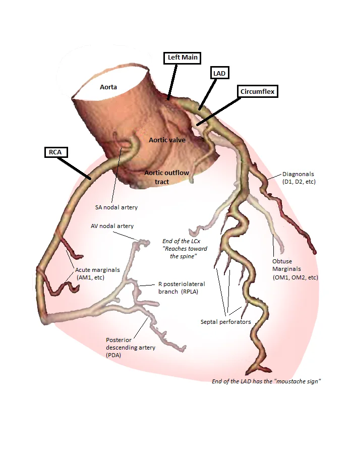
Figure 1Anatomy of coronary arteries
Long description
Anatomy of coronary arteries. Labelled illustration from Wikimedia Commons, used under its open licence.

Figure 1: Anatomy of coronary arteries.
The systemic circuit transports oxygenated blood to virtually all of the tissues of the body and returns relatively deoxygenated blood and carbon dioxide to the heart to be sent back to the pulmonary circulation. The right ventricle pumps deoxygenated blood into the pulmonary trunk, which leads toward the lungs and bifurcates into the left and right pulmonary arteries. The pulmonary trunk arteries and their branches are the only arteries in the post-natal body that carry relatively deoxygenated blood. The pulmonary veins conduct blood into the left atrium, which pumps the blood into the left ventricle, which in turn pumps oxygenated blood into the aorta. [1][2]
The right coronary artery distributes blood to the right atrium, portions of both ventricles, and the heart conduction system. Normally, one or more marginal arteries arise from the right coronary artery inferior to the right atrium. The marginal arteries supply blood to the superficial portions of the right ventricle. The blood exiting the systemic capillaries is lower in oxygen concentration than when it entered. The capillaries will ultimately unite to form venules, joining to form ever-larger veins, eventually flowing into the two major systemic veins. [1][3]
Most of the blood flows into either the superior vena cava or inferior vena cava. If you draw an imaginary line at the level of the diaphragm, systemic venous circulation from above that line will generally flow into the superior vena cava. The exception to this is that most venous blood flow from the coronary veins flows directly into the coronary sinus and from there directly into the right atrium. The remainder of the blood supply from the thorax drains into the azygos vein. Each intercostal vein drains muscles of the thoracic wall, each esophageal vein delivers blood from the inferior portions of the esophagus, each bronchial vein drains the systemic circulation from the lungs. [2]
DefinitionCoronary Dominance
Dominance is determined by the origin of the posterior descending artery, usually from the right coronary artery.
| Artery | Origin | Primary Territory |
|---|---|---|
| Right Coronary Artery | Aortic Root | Right Atrium, Right Ventricle |
| Left Coronary Artery | Aortic Root | Left Atrium, Left Ventricle |
| Posterior Interventricular | Right Coronary Artery | Interventricular Septum, Apex |
Coronary Flow Pathway
- Oxygenated BloodFrom Left Ventricle into Aorta
- Question: Enter Coronary Ostia?
Yes
- Right or Left Coronary Artery
No
- Systemic Circulation
The heart requires a dedicated blood supply because cardiomyocytes are active throughout life, necessitating a complex coronary circulation. This circulation is not continuous; it cycles, peaking when the heart muscle is relaxed and nearly ceasing during contraction. The coronary arteries arise from the first portion of the aorta after it leaves the left ventricle. Specifically, there are three dilations in the aortic wall just superior to the aortic semilunar valve. The third sinus, the right posterior aortic sinus, typically does not give rise to a vessel. Vessel branches that remain on the surface of the artery and follow the sulci are called epicardial coronary arteries. [1] [2]
The left coronary artery runs along the left side of the heart, while the right coronary artery descends along the right side. The left coronary artery quickly divides into two major branches. The circumflex artery arises from the left coronary artery and follows the coronary sulcus to the left. It eventually fuses with small branches of the right coronary artery. The larger anterior interventricular artery, also known as the left anterior descending artery (LAD), is the second major branch arising from the left coronary artery. It follows the anterior interventricular sulcus around the pulmonary trunk. Along its course, it gives rise to numerous smaller branches that interconnect with branches of the posterior interventricular artery, forming anastomoses. An anastomosis is an area where vessels unite to form interconnections that normally allow blood to circulate to a region even if there may be partial blockage in another branch. [1]
The venous drainage of the heart involves several distinct pathways. The coronary sinus is a large, thin-walled vein on the posterior surface of the heart lying within the atrioventricular sulcus and emptying directly into the right atrium. Vieussens identified communicating veins named ducti carnosi [1][3]
The coronary circulation is the specialized vascular network that supplies the myocardium with oxygenated blood. Understanding its anatomy requires tracing the origin, course, and branching patterns of the arteries that emerge from the aorta. The fundamental principle is that the heart must supply itself before it supplies the rest of the body.
The left coronary artery arises from the left side and quickly divides.
The first vessels that branch from the ascending aorta are the paired coronary arteries, which arise from two of the three sinuses in the ascending aorta just superior to the aortic semilunar valve. [2]
| Artery | Origin | Initial Course |
|---|---|---|
| Left Coronary Artery | Left posterior aortic sinus | Travels between the pulmonary trunk and left atrial appendage |
| Right Coronary Artery | Anterior aortic sinus | Travels in the right atrioventricular groove |
The right and left coronary arteries are the first to branch off the aorta and arise from two of the three sinuses located near the base of the aorta and are generally located in the sulci. The left coronary artery typically divides into two major branches: the anterior interventricular artery (also known as the left anterior descending artery) and the circumflex artery. The anterior interventricular artery runs in the anterior interventricular sulcus, supplying the anterior walls of both ventricles and the anterior two-thirds of the interventricular septum. The circumflex artery runs in the left atrioventricular groove, supplying the left atrium and the lateral and posterior walls of the left ventricle. [1]
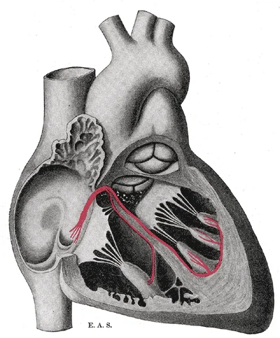
Figure 2Gray501
Long description
Schematic representation of the atrioventricular bundle of His. The bundle, represented in red originates near the orifice of the coronary sinus, undergoes slight enlargement to form a node, passes forward to the ventricular septum, and divides into two limbs. The ultimate distribution cannot be com

Figure 2: Schematic representation of the atrioventricular bundle of His.
The right coronary artery runs in the right atrioventricular groove. It gives off the marginal branch, which supplies the right ventricle. In most individuals, the right coronary artery continues to the posterior interventricular sulcus, where it becomes the posterior interventricular artery (also known as the posterior descending artery). This artery supplies the posterior one-third of the interventricular septum and the posterior walls of the ventricles.
Cardiac veins parallel the small cardiac arteries and generally drain into the coronary sinus.
DefinitionCoronary Dominance
Coronary dominance is determined by which artery gives rise to the posterior interventricular artery. Right dominance is the most common pattern.
Their course is closely related to the surface anatomy of the heart. The anterior interventricular artery is located in the anterior interventricular sulcus, which is a visible groove on the anterior surface of the heart. The circumflex artery is located in the left atrioventricular groove, which is less visible on the anterior surface but can be seen by retracting the left atrial appendage. The right coronary artery is located in the right atrioventricular groove, which is clearly visible on the anterior surface.
The branches of the coronary arteries supply specific regions of the myocardium. The anterior interventricular artery supplies the anterior wall of the right ventricle, the anterior wall of the left ventricle, and the anterior two-thirds of the interventricular septum. The circumflex artery supplies the lateral wall of the left ventricle and the left atrium. The right coronary artery supplies the right atrium, the right ventricle, and the posterior wall of the left ventricle. The posterior interventricular artery supplies the posterior one-third of the interventricular septum and the posterior wall of the left ventricle.
Exam trapCommon Misconception
Do not confuse the coronary arteries with the bronchial arteries. The bronchial arteries supply the lung tissue itself, not the heart muscle.
The coronary circulation is essential for maintaining cardiac function. Any obstruction in these arteries can lead to myocardial ischaemia or infarction. The specific region affected depends on the artery involved. For example, occlusion of the anterior interventricular artery can lead to an anterior wall myocardial infarction, which is often severe due to the large area of myocardium supplied by this vessel. Understanding the anatomy of the coronary arteries is therefore critical for diagnosing and treating coronary artery disease.
The coronary arteries encircle the heart, forming a ring-like structure that divides into the next level of branches that supplies blood to the heart tissues. This arterial network is defined by its dominance, which determines the blood supply to the apex and the posterior interventricular septum. In a right-dominant circulation, which is the most common pattern, the posterior interventricular artery arises from the right coronary artery. In left-dominant or co-dominant systems, it arises from the circumflex artery. The anterior interventricular artery, a branch of the left coronary artery, supplies the anterior walls of both ventricles and the anterior two-thirds of the interventricular septum. The marginal branches of the circumflex artery supply the lateral wall of the left ventricle. [2]
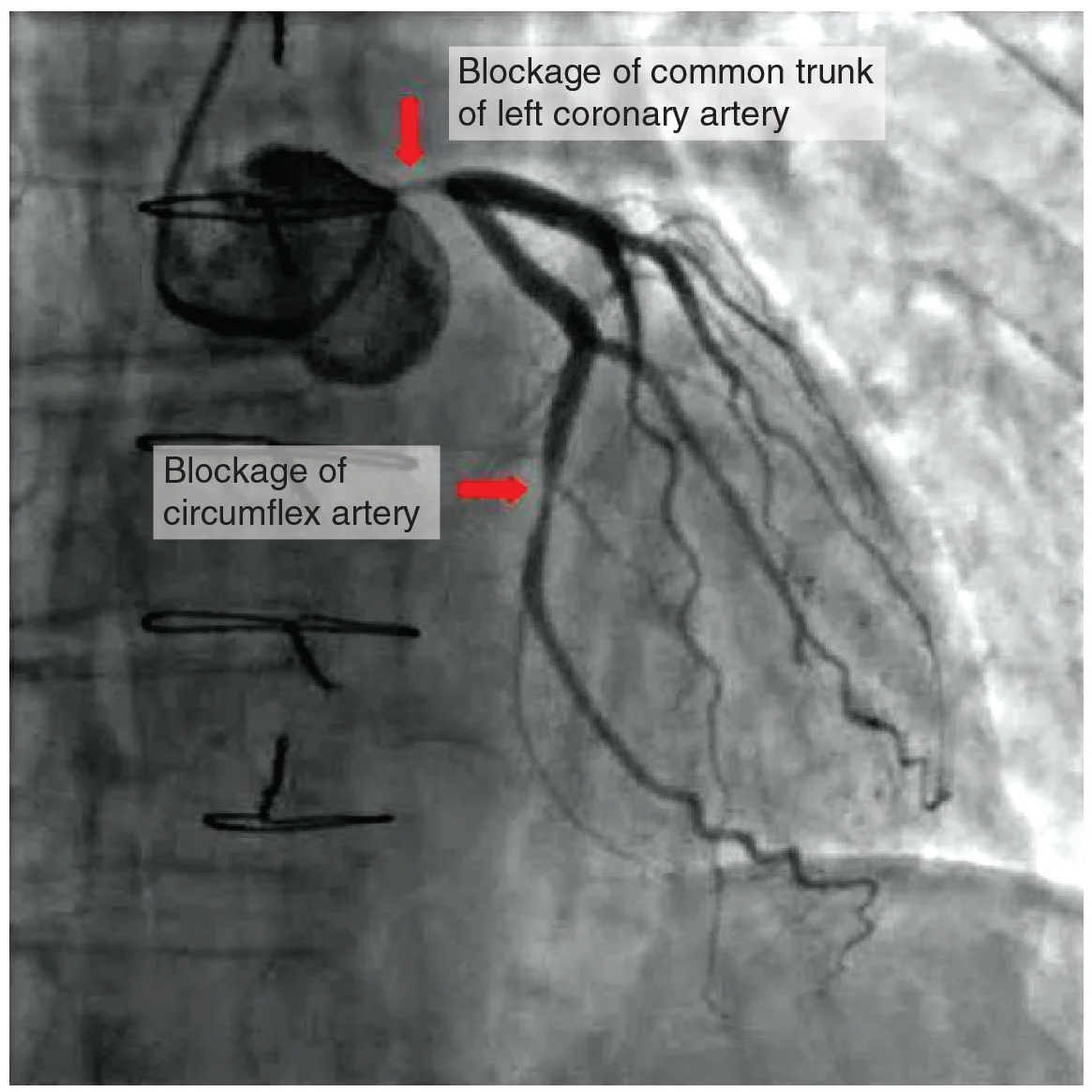
Figure 32016 Occluded Coronay Arteries
Long description
In this coronary angiogram (X-ray), the dye makes visible two occluded coronary arteries. Such blockages can lead to decreased blood flow (ischemia) and insufficient oxygen (hypoxia) delivered to the cardiac tissues. If uncorrected, this can lead to cardiac muscle death (myocardial infarction).

Figure 3: In this coronary angiogram (X-ray), the dye makes visible two occluded coronary arteries.
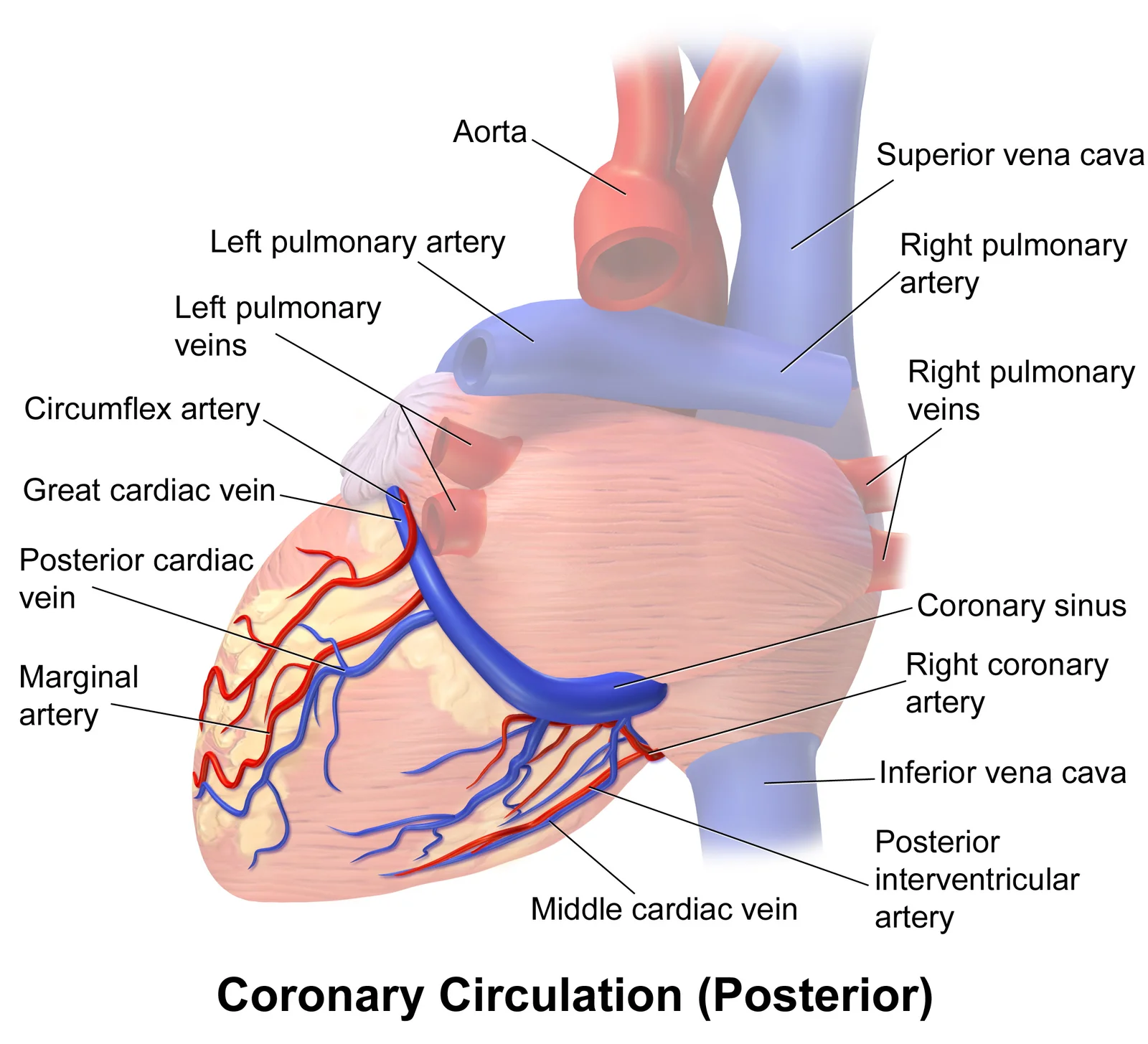
The left coronary artery bifurcates into the anterior interventricular artery and the circumflex artery. The anterior interventricular artery runs down the anterior interventricular sulcus towards the apex. The circumflex artery follows the left atrioventricular groove. The great cardiac vein parallels the anterior interventricular artery. The middle cardiac vein parallels the posterior interventricular artery. The coronary sinus is located in the posterior atrioventricular groove.
DefinitionCoronary Dominance
Coronary dominance is determined by which artery gives rise to the posterior interventricular artery, which supplies the posterior third of the interventricular septum and the inferior wall of the left ventricle.
Infarct territories correspond directly to these arterial distributions. An occlusion of the anterior interventricular artery causes an anterior wall myocardial infarction, affecting the anterior left ventricle and the anterior septum. Occlusion of the right coronary artery typically causes an inferior wall myocardial infarction, affecting the inferior left ventricle and the right ventricle. Occlusion of the circumflex artery causes a lateral wall myocardial infarction.
The great cardiac vein initially parallels the anterior interventricular artery and drains the areas supplied by this vessel. It receives several major branches, including the posterior cardiac vein, th [1]
| Vein | Parallel Artery | Territory Drained | Termination |
|---|---|---|---|
| Great cardiac vein | Anterior interventricular artery | Anterior walls of ventricles | Coronary sinus |
| Middle cardiac vein | Posterior interventricular artery | Inferior walls of ventricles | Coronary sinus |
| Small cardiac vein | Marginal artery (RCA) | Right ventricle | Coronary sinus |
| Posterior cardiac vein | Marginal artery (LCx) | Posterior left ventricle | Coronary sinus |
This venous system ensures that deoxygenated blood from the myocardium is returned to the right side of the heart for pulmonary circulation.
The coronary circulation is designed to supply the myocardium with oxygenated blood, but it also possesses a network of venous drainage and potential collateral pathways that are critical for understanding cardiac physiology and pathology. While arterial anastomoses are often considered insufficient to prevent infarction in acute occlusion, the venous system provides a crucial route for deoxygenated blood return.
The venous drainage of the heart is primarily collected by the coronary sinus, a large, thin-walled vessel located in the posterior atrioventricular groove. The coronary sinus opens into the right atrium. This vessel drains most of the coronary veins that return systemic blood from the heart. [1] [2]
Additionally, the oblique vein of the left atrium and the posterior vein of the left ventricle may drain directly into the coronary sinus or its tributaries.
The anatomy of these veins can be complex. Both of these veins open into the coronary sinus or the great cardiac vein.
This system helps describe with more precision the epicardial course of the cardiac veins. [3]
The middle cardiac vein is located in the posterior interventricular groove.
The deeper veins generally correspond to the complementary arteries. [2]
| Vein | Course | Termination |
|---|---|---|
| Great Cardiac Vein | Runs with anterior interventricular artery | Coronary Sinus |
| Middle Cardiac Vein | Runs with posterior interventricular artery | Coronary Sinus |
| Small Cardiac Vein | Runs with right marginal artery | Coronary Sinus |
| Oblique Vein of LA | Runs on posterior surface of LA | Coronary Sinus |
Understanding the anatomy of the coronary sinus and its tributaries is essential for procedures such as cardiac resynchronization therapy. This is distinct from the coronary venous system, which drains directly into the right atrium via the coronary sinus.
The coronary circulation is defined by the specific anatomical pathways of the arteries and veins that supply and drain the myocardium. While the standard pattern involves distinct left and right coronary arteries, significant anatomical variations exist that are critical for clinical procedures such as ablation and pacing. Understanding these variations requires a precise grasp of the coronary sinus and its tributaries, as well as the anastomotic networks within the heart.
The coronary venous system is crucial for draining deoxygenated blood from the cardiac structures. It serves as an access path for various electrophysiological diagnostic and treatment procedures. The coronary sinus (CS) ostium diameter varies between 5–15 mm. This dimension is influenced by gender and underlying heart disease. [3]
The great cardiac vein (GCV) and anterior interventricular vein (AIV) junction is a key landmark. Tavares et al. analyzed this junction in 52 coronary computed tomographic angiographies.
In case a CS ablation procedure is not possible because of the small caliber of the coronary veins, CS mapping with thin catheters could guide the endocardial approach. Like any treatment involving vascular access, these procedures pose risks even though they offer the opportunity of treating the arrhythmia. Potential complications include coronary artery injury, vein dissection or perforation. [3]
Clinical pearlClinical Pearl
Understanding the coronary sinus and its tributary veins distribution, dimensions and relationship with nearby structures could help the development of new ablation tools and strategies, with higher success rates. [3]
The anastomoses in the heart are very small. Therefore, this ability is somewhat restricted in the heart so a coronary artery blockage often results in death of the cells (myocardial infarction) supplied by the particular vessel. This contrasts with other vascular beds where extensive collateral circulation provides redundancy. For instance, the lingual artery has an extensive collateral circulation and a dense network of anastomoses, contributing to the tongue’s rich blood supply and its tendency to bleed profusely from injuries. It also provides some redundancy: if one artery is compromised, others can partially compensate. However, such extensive redundancy is not a feature of the coronary circulation itself. [1] [4]
| Feature | Description |
|---|---|
| Primary Function | Draining deoxygenated blood from cardiac structures |
| Clinical Utility | Access path for electrophysiological diagnostic and treatment procedures |
| Ostium Diameter | Varies between 5–15 mm |
| GCV-AIV Angle | Obtuse angle (133 ± 14º) in 79% of cases from lateral view |
| Anastomotic Size | Very small, restricting collateral flow |
Figure 4Blausen 0261 CoronaryVessels Posterior
Long description
Coronary Circulation (Posterior). Labelled illustration from Wikimedia Commons, used under its open licence.

Figure 4: Coronary Circulation (Posterior).
The coronary circulation is the primary determinant of myocardial viability, and its anatomical variations dictate clinical management in acute and chronic settings. Understanding the precise location of lesions, access points for intervention, and the consequences of occlusion is fundamental to cardiology.
The LAD runs in the anterior interventricular groove, supplying the anterior wall of the left ventricle and the anterior two-thirds of the interventricular septum. Occlusion here typically causes anterior wall myocardial infarction. The LCx courses in the left atrioventricular groove, supplying the lateral and posterior walls of the left ventricle. The RCA travels in the right atrioventricular groove, supplying the right ventricle, the inferior wall of the left ventricle, and the sinoatrial and atrioventricular nodes in most individuals. Bypass grafting sites are often marked on the distal branches of these arteries, such as the diagonal branches of the LAD or the obtuse marginal branches of the LCx, where flow restoration is critical.
DefinitionCoronary Artery Bypass Grafting
Surgical revascularisation using saphenous vein or radial artery grafts to bypass obstructed coronary segments, restoring blood flow to ischaemic myocardium.
Aneurysms of the coronary arteries are rare but serious complications, often associated with Kawasaki disease or atherosclerosis. In patients who have undergone coronary artery bypass grafting (CABG), the coronary sinus (CS) may exhibit significant changes in diameter, including stenosis or aneurysm formation, which complicates subsequent interventions. The CS itself is a major venous structure; in severe mitral regurgitation or post-CABG states, it tends to be displaced posteriorly and comes out of the atrio-ventricular plane, altering the strategy for catheter ablation. [3]
Access points for coronary interventions typically involve the femoral or radial arteries, leading to the aortic root where the coronary ostia are located. Pulse points are not directly on the coronary arteries but are assessed via peripheral pulses to ensure systemic perfusion. Ligation of coronary arteries is generally avoided due to the risk of infarction, except in specific experimental or palliative contexts.
Sites of infarction correspond directly to the territory supplied by the occluded vessel. Anterior infarctions result from LAD occlusion, inferior infarctions from RCA occlusion, and lateral infarctions from LCx occlusion. The venous drainage of the heart is primarily via the coronary sinus, which drains most of the myocardium. Other veins open directly into the right atrium or into any chamber via Thebesian vessels. [3]
| Artery | Course | Territory Supplied | Typical Infarction Site |
|---|---|---|---|
| LAD | Anterior interventricular groove | Anterior LV, anterior septum | Anterior wall |
| LCx | Left atrioventricular groove | Lateral LV, posterior LV | Lateral/Posterior wall |
| RCA | Right atrioventricular groove | RV, inferior LV, SA/AV nodes | Inferior wall |
The left ventricular septum (LVS) is a triangular area at the superior, epicardial region of the left ventricular outflow tract, bounded by the LAD, LCx, and an arched line from the left main bifurcation to the first septal perforator. Understanding this geometry is crucial for interpreting septal perforator branches and their role in septal perfusion. [3]
Assuming an average rate of contraction of 75 contractions per minute, a human heart would contract approximately 108,000 times in one day. [1]
DefinitionCardiac Dimensions
A typical heart is approximately the size of your fist: 12 cm (5 in) in length, 8 cm (3.5 in) wide, and 6 cm (2.5 in) in thickness. [1]
The shape of the heart is similar to a pinecone, rather broad at the superior surface and tapering to the apex. The weight of a female heart is approximately 250–300 grams (9 to 11 ounces). The weight of a male heart is approximately 300–350 grams (11 to 12 ounces). Exercise results in the addition of protein myofilaments that increase the size of the individual cells without increasing their numbers, a concept called hypertrophy. [1]
Exam trapValve Nomenclature
In a clinical setting, the valve is referred to as the mitral valve, rather than the bicuspid valve. [1]
Located at the opening between the left atrium and left ventricle is the mitral valve, also called the bicuspid valve or the left atrioventricular valve. Structurally, this valve consists of two cusps, compared to the three cusps of the tricuspid valve. The two cusps of the mitral valve are attached by chordae tendineae to two papillary muscles that project from the wall of the ventricle. [1]
Closure of the two atrioventricular valves prevents blood from being forced back into the atria. This occurs when the ventricles contract to eject blood into the pulmonary trunk and aorta. [1]
Systemic veins return blood to the right atrium. Since the blood has already passed through the systemic capillaries, it will be relatively low in oxygen concentration. In many cases, there will be veins draining organs and regions of the body with the same name as the arteries that supplied these regions and the two often parallel one another. [2]
This is rarer still, with a pooled prevalence of 0.09%. [4]
Students frequently confuse the anatomical boundaries and functional roles of the cardiac chambers and their associated valves. A primary error is misidentifying the primary pumping chamber for the systemic circulation. It is a common mistake to attribute this role to the right ventricle due to its anterior position. However, the left ventricle is the major pumping chamber for the systemic circuit; it ejects blood into the aorta through the aortic semilunar valve. The left ventricular wall is significantly thicker than the right, reflecting the higher pressure required for systemic distribution. [1]
Another frequent pitfall involves the description of the aorta's course and termination. Students often overlook the specific vertebral level at which the aorta bifurcates. The aorta is the largest artery in the body. It arises from the left ventricle and eventually descends to the abdominal region, where it bifurcates at the level of the fourth lumbar vertebra into the two common iliac arteries. This landmark is crucial for distinguishing the thoracic from the abdominal aorta. After crossing through the diaphragm at the aortic hiatus, the thoracic aorta is called the abdominal aorta. This vessel remains to the left of the vertebral column and is embedded in adipose tissue behind the peritoneal cavity. It formally ends at approximately the level of vertebra L4, where it bifurcates to form the common iliac arteries. [2]
Students also struggle with the complex muscle architecture of the heart. They often assume a simple linear contraction pattern. The middle and thickest layer is the myocardium, made largely of cardiac muscle cells. It is built upon a framework of collagenous fibers, plus the blood vessels that supply the myocardium and the nerve fibers that help regulate the heart. It is the contraction of the myocardium that pumps blood through the heart and into the major arteries. The muscle pattern is elegant and complex, as the muscle cells swirl and spiral around the chambers of the heart. They form a figure 8 pattern around the atria and around the bases of the great vessels. Deeper ventricular muscles also form a figure 8 around the two ventricles and proceed toward the apex. More superficial layers of ventricular muscle wrap around both ventricles. This complex swirling pattern allows the heart to pump blood more effectively than a simple linear pattern would. [1]
A further confusion arises regarding the venous drainage of the heart, specifically the coronary sinus and its tributaries. Students often fail to distinguish the great cardiac vein from the anterior interventricular vein. The AIV, described in some works as a component of the GCV, originates at the apex, and runs into the anterior interventricular sulcus, where it is accompanied by the left anterior descending coronary artery. The junction between the GCV and the AIV usually has an ‘S-shape’ that passes through the superior, epicardial portion of the left ventricle, known as the left ventricular summit (LVS) region. The AIV receives some septal tributaries draining the anterior portion of the interventricular septum. This anatomical relationship is critical for understanding coronary circulation patterns. [3]
Finally, students may overlook the significance of embryonic remnants in adult anatomy. The left atrial oblique vein, also known as the vein of Marshall, is an embryonic remnant of the left superior vena cava which is enclosed within the ligament of Marshall. During embryogenesis, the anterior cardinal vein obliterates, resulting in a fold that is the ligament of Marshall. A detailed description of these structures dates back to 1850 when John Marshall, a cardiac surgeon, described the left atrial oblique vein as “remarkably constant structure” that frequently “measures from half an inch to an inch in length, and sometimes is as large as a crow-quill, but more commonly it is smaller, and will admit only the head of a pin”. These anatomic structures that have been extensively described, in view of their importance in the pathogenesis of atrial fibrillation. [3]
Knowledge of coronary circulation anatomy is essential at the bedside for interpreting haemodynamic changes. When assessing a patient with suspected myocardial infarction, clinicians must understand which vessel supplies which region. The AIV is currently use as an alternative pacing site in patients with tricuspid valve valvopathies or prosthetic tricuspid valve. It is also used for mapping and ablation of idiopathic ventricular arrythmias originating from the LVS. This clinical application highlights the importance of knowing the precise course of the anterior interventricular vein.
In the operating theatre, surgeons rely on detailed anatomical landmarks to avoid injury during procedures. The valve of Vieussens marks the transition from the CS to GCV and it could cause difficulties when advancing the catheter and positioning the LV lead in CRT procedures. It is a structure present in about 60% of cases with variable morphology, with one to three leaflets that are concave/ flat, and are being inserted at the anterior (43.3%), antero-superior (16.7%), superior (15.6%), inferior (8.9%), posteroinferior or anteroinferior wall of the CS. Recognising this structure prevents procedural complications during cardiac resynchronisation therapy.
On imaging, radiologists identify key structures to diagnose pathology. The septum between the atria and ventricles is known as the atrioventricular septum. It is marked by the presence of four openings that allow blood to move from the atria into the ventricles and from the ventricles into the pulmonary trunk and aorta. Located in each of these openings between the atria and ventricles is a valve, a specialized structure that ensures one-way flow of blood. The valves between the atria and ventricles are known generically as atrioventricular valves. The valves at the openings that lead to the pulmonary trunk and aorta are known generically as semilunar valves. The interventricular septum is visible in . In this figure, the atrioventricular septum has been removed to better show the bicuspid and tricuspid valves; the interatrial septum is not visible, since its location is covered by the aorta and pulmonary trunk. This description aids in interpreting echocardiographic views. [1]
In examinations, students must accurately describe the aorta's course. The aorta consists of the ascending aorta, the aortic arch, and the descending aorta, which passes through the diaphragm and a landmark that divides into the superior thoracic and inferior abdominal components. Arteries originating from the aorta ultimately distribute blood to virtually all tissues of the body. At the base of the aorta is the aortic semilunar valve that prevents backflow of blood into the left ventricle while the heart is relaxing. After exiting the heart, the ascending aorta moves in a superior direction for approximately 5 cm and ends at the sternal angle. This precise measurement and anatomical detail are frequently tested.
The MAAV is one of the veins that travels close to the LVS. This vein originates from the GCV and courses towards mitro-aortic continuity. After identifying it using retrograde venography, Komatsu et al. have canulated the ‘communicating vein’, as it was named, using a 2F microcatheter. The term ‘communicating vein’ is not defining the mitro-aortic annular vein, as it was also used for other branches that originate more distally, at the level of the GCV-AIV junction. Understanding these variations is crucial for advanced interventional cardiology. [3]
The left ventricle is the major pumping chamber for the systemic circuit; it ejects blood into the aorta through the aortic semilunar valve. The aorta is the largest artery in the body. It arises from the left ventricle and eventually descends to the abdominal region, where it bifurcates at the level of the fourth lumbar vertebra into the two common iliac arteries. After crossing through the diaphragm at the aortic hiatus, the thoracic aorta is called the abdominal aorta. This vessel remains to the left of the vertebral column and is embedded in adipose tissue behind the peritoneal cavity. It formally ends at approximately the level of vertebra L4, where it bifurcates to form the common iliac arteries. The middle and thickest layer is the myocardium, made largely of cardiac muscle cells. It is built upon a framework of collagenous fibers, plus the blood vessels that supply the myocardium and the nerve fibers that help regulate the heart. It is the contraction of the myocardium that pumps blood through the heart and into the major arteries. The muscle pattern is elegant and complex, as the muscle cells swirl and spiral around the chambers of the heart. They form a figure 8 pattern around the atria and around the bases of the great vessels. Deeper ventricular muscles also form a figure 8 around the two ventricles and proceed toward the apex. More superficial layers of ventricular muscle wrap around both ventricles. This complex swirling pattern allows the heart to pump blood more effectively than a simple linear pattern would. The septum between the atria and ventricles is known as the atrioventricular septum. It is marked by the presence of four openings that allow blood to move from the atria into the ventricles and from the ventricles into the pulmonary trunk and aorta. Located in each of these openings between the atria and ventricles is a valve, a specialized structure that ensures one-way flow of blood. The valves between the atria and ventricles are known generically as atrioventricular valves. The valves at the openings that lead to the pulmonary trunk and aorta are known generically as semilunar valves. The interventricular septum is visible in . In this figure, the atrioventricular septum has been removed to better show the bicuspid and tricuspid valves; the interatrial septum is not visible, since its location is covered by the aorta and pulmonary trunk. The AIV, described in some works as a component of the GCV, originates at the apex, and runs into the anterior interventricular sulcus, where it is accompanied by the left anterior descending coronary artery. The junction between the GCV and the AIV usually has an ‘S-shape’ that passes through the superior, epicardial portion of the left ventricle, known as the left ventricular summit (LVS) region. The AIV receives some septal tributaries draining the anterior portion of the interventricular sep The left atrial oblique vein, also known as the vein of Marshall, is an embryonic remnant of the left superior vena cava which is enclosed within the ligament of Marshall. During embryogenesis, the anterior cardinal vein obliterates, resulting in a fold that is the ligament of Marshall. A detailed description of these structures dates back to 1850 when John Marshall, a cardiac surgeon, described the left atrial oblique vein as “remarkably constant structure” that frequently “measures from half an inch to an inch in length, and sometimes is as large as a crow-quill, but more commonly it is smaller, and will admit only the head of a pin”. These anatomic structures that have been extensively described, in view of their importance in the pathogenesis of atrial fibrillation. The MAAV is one of the veins that travels close to the LVS. This vein originates from the GCV and courses towards mitro-aortic continuity. After identifying it using retrograde venography, Komatsu et al. have canulated the ‘communicating vein’, as it was named, using a 2F microcatheter. The term ‘communicating vein’ is not defining the mitro-aortic annular vein, as it was also used for other branches that originate more distally, at the level of the GCV-AIV junction.
Answer each one in your head first, then open the answer.
Which chamber is the major pumping chamber for the systemic circuit?
Show answerHide answer
The left ventricleAt what vertebral level does the aorta bifurcate?
Show answerHide answer
The fourth lumbar vertebraWhat is the name of the embryonic remnant of the left superior vena cava?
Show answerHide answer
The left atrial oblique vein or vein of MarshallWhere does the anterior interventricular vein originate?
Show answerHide answer
At the apexWhat structure marks the transition from the CS to GCV?
Show answerHide answer
The valve of Vieussens
- Betts JG, Young KA, Wise JA, et al. Heart Anatomy. OpenStax, Anatomy and Physiology 2e (CC BY-NC-SA 4.0). Accessed .
- Betts JG, Young KA, Wise JA, et al. Circulatory Pathways. OpenStax, Anatomy and Physiology 2e (CC BY-NC-SA 4.0). Accessed .
- Zehan IG, Eötvös CA, Moldovan MP, et al. Angiographic Anatomy of the Left Coronary Veins: Beyond Conventional Cardiac Resynchronization Therapy. Current cardiology reports 2025;27(1):58. doi:10.1007/s11886-025-02204-z. Accessed .
- Na J, Kakazu A, Muthana A, et al. Lingual artery: Angiographic anatomy and variations review for neurosurgeons. Surgical neurology international 2025;16:156. doi:10.25259/sni_282_2025. Accessed .
Figure credits
- Figure 1: Npatchett, via Wikimedia Commons, CC BY-SA 4.0, CC BY-SA 4.0.
- Figure 2: Henry Vandyke Carter, via Wikimedia Commons, Public domain, Public domain.
- Figure 3: OpenStax College, via Wikimedia Commons, CC BY 3.0, CC BY 3.0.
- Figure 4: BruceBlaus. When using this image in external sources it can be cited as: Blausen.com staff (2014). "Medical gallery of Blausen Medical 2014". WikiJournal of Medicine 1 (2). DOI:10.15347/wjm/2014.010. ISSN 2002-4436., via Wikimedia Commons, CC BY 3.0, CC BY 3.0.
Educational content for medical students and doctors. It is not a substitute for clinical judgement or local guidelines.