Heart anatomy
Comprehensive guide to human heart anatomy, covering location, valves, conduction system, and surface landmarks.
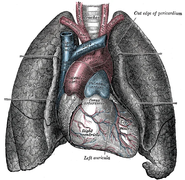
The human heart is located within the thoracic cavity, medially between the lungs in the space known as the mediastinum[1]. Within the mediastinum, the heart is separated from the other mediastinal structures by a tough membrane known as the pericardium, or pericardial sac, and sits in its own space called the pericardial cavity[1]. The dorsal surface of the heart lies near the bodies of the vertebrae, and its anterior surface sits deep to the sternum and costal cartilages[1]. The great veins, the superior and inferior venae cavae, and the great arteries, the aorta and pulmonary trunk, are attached to the superior surface of the heart, called the base[1].
Figure 1Gray490
Long description
Front view of heart and lungs. Labelled illustration from Wikimedia Commons, used under its open licence.

Figure 1: Front view of heart and lungs.
DefinitionPericardium
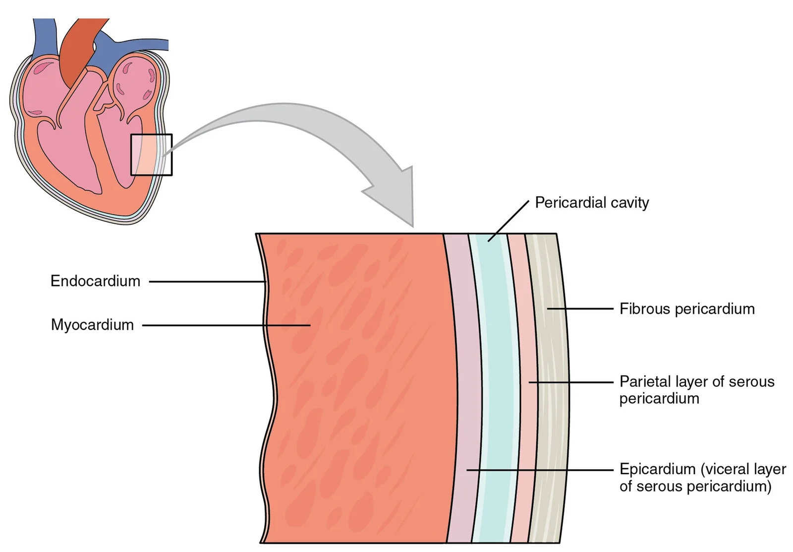
The membrane that directly surrounds the heart and defines the pericardial cavity is called the pericardium or pericardial sac[1].
Figure 22004 Heart Wall
Long description
The pericardial membrane that surrounds the heart consists of three layers and the pericardial cavity. The heart wall also consists of three layers. The pericardial membrane and the heart wall share the epicardium.

Figure 2: The pericardial membrane that surrounds the heart consists of three layers and the pericardial cavity.
The membrane that directly surrounds the heart and defines the pericardial cavity is called the pericardium or pericardial sac[1]. It also surrounds the “roots” of the major vessels, or the areas of closest proximity to the heart[1]. The pericardium, which literally translates as “around the heart,” consists of two distinct sublayers: the sturdy outer fibrous pericardium and the inner serous pericardium[1]. The fibrous pericardium is made of tough, dense connective tissue that protects the heart and maintains its position in the thorax[1]. The more delicate serous pericardium consists of two layers: the parietal pericardium, which is fused to the fibrous pericardium, and an inner visceral pericardium, or epicardium, which is fused to the heart and is part of the heart wall[1]. The pericardial cavity, filled with lubricating serous fluid, lies between the epicardium and the pericardium[1].
DefinitionCardiac Skeleton
Since these openings and valves structurally weaken the atrioventricular septum, the remaining tissue is heavily reinforced with dense connective tissue called the cardiac skeleton, or skeleton of the heart[1].
Figure 32008 Internal Anatomy of the HeartN
Long description
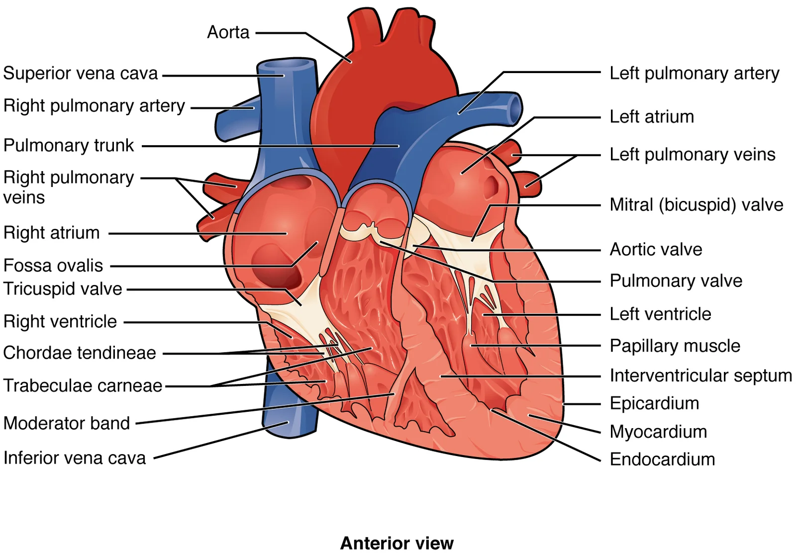
This anterior view of the heart shows the four chambers, the major vessels and their early branches, as well as the valves. The presence of the pulmonary trunk and aorta covers the interatrial septum, and the atrioventricular septum is cut away to show the atrioventricular valves.

Figure 3: This anterior view of the heart shows the four chambers, the major vessels and their early branches, as well as the valves.
Since these openings and valves structurally weaken the atrioventricular septum, the remaining tissue is heavily reinforced with dense connective tissue called the cardiac skeleton, or skeleton of the heart[1]. It includes four rings that surround the openings between the atria and ventricles, and the openings to the pulmonary trunk and aorta, and serve as the point of attachment for the heart valves[1]. The cardiac skeleton also provides an important boundary in the heart el
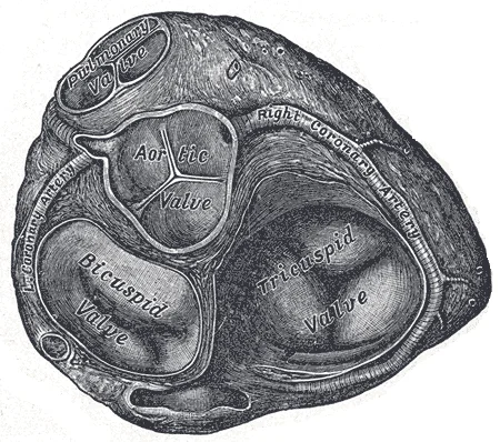
A transverse section through the heart slightly above the level of the atrioventricular septum reveals all four heart valves along the same plane[1]. The valves ensure unidirectional blood flow through the heart[1]. Between the right atrium and the right ventricle is the right atriove
Figure 4Gray494
Long description
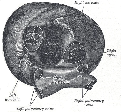
Heart seen from above. Labelled illustration from Wikimedia Commons, used under its open licence.

Figure 4: Heart seen from above.
| Vein | Parallel Artery | Drainage Area |
|---|---|---|
| Great cardiac vein | Anterior interventricular artery | Areas supplied by anterior interventricular artery |
| Posterior cardiac vein | Marginal artery (circumflex) | Areas supplied by marginal artery branch |
| Middle cardiac vein | Posterior interventricular artery | Areas supplied by posterior interventricular artery |
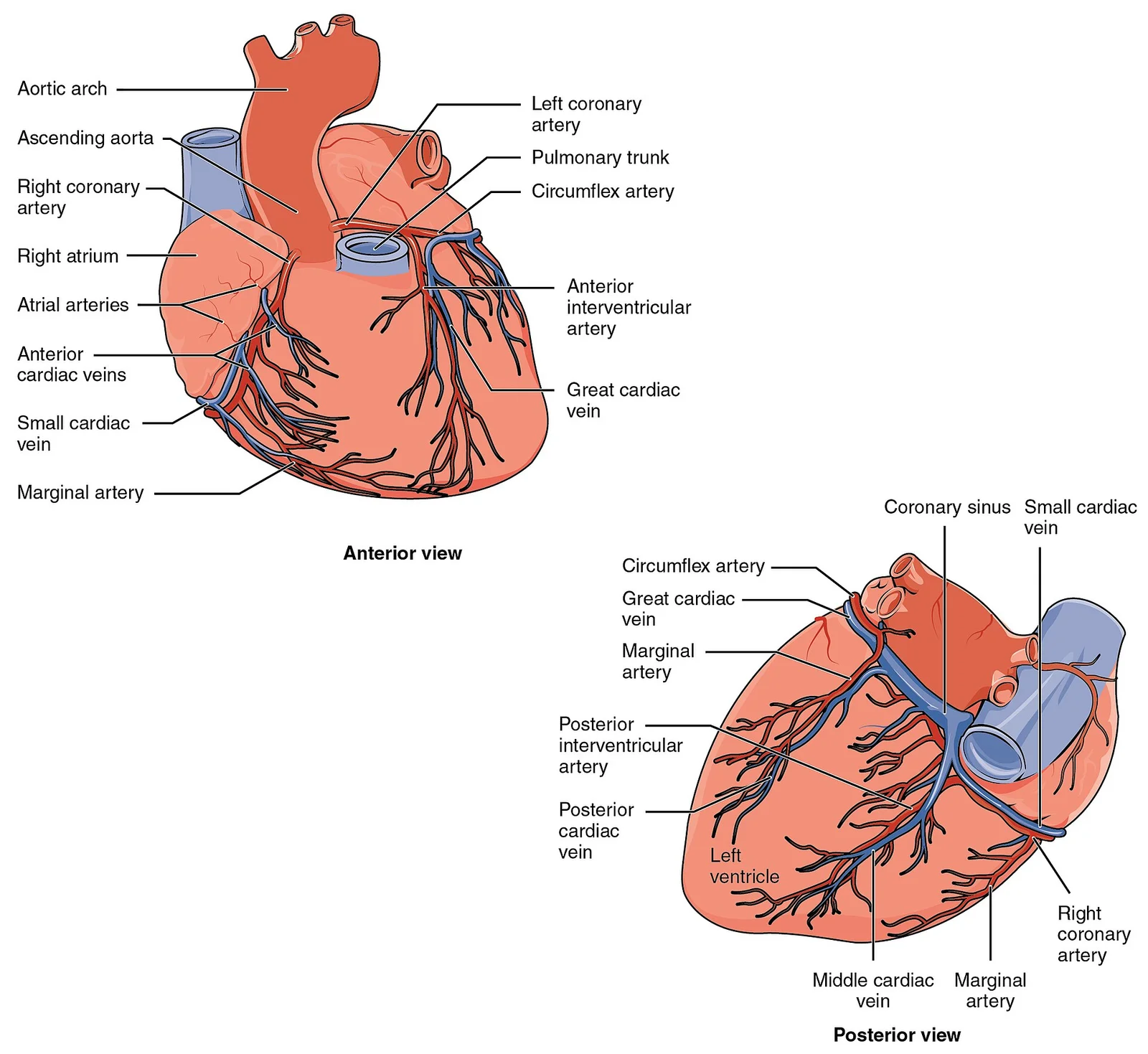
Coronary veins drain the heart and generally parallel the large surface arteries[1]. The great cardiac vein can be seen initially on the surface of the heart following the interventricular sulcus, but it eventually flows along the coronary sulcus into the coronary sinus on the posterior surface[1]. The great cardiac vein initially parallels the anterior interventricular artery and drains the areas supplied by this vessel[1]. The posterior cardiac vein parallels and drains the areas supplied by the marginal artery branch of the circumflex artery[1]. The middle cardiac vein parallels and drains the areas supplied by the posterior interventricular artery[1].
Figure 52014ab Coronary Blood Vessels
Long description
The anterior view of the heart shows the prominent coronary surface vessels. The posterior view of the heart shows the prominent coronary surface vessels.

Figure 5: The anterior view of the heart shows the prominent coronary surface vessels.
NoteConducting Zone
The cardiac skeleton also provides an important boundary in the heart electrical conduction system[1].
The conducting zone relies on the electrical insulation provided by the cardiac skeleton, which separates the atrial and ventricular myocardium electrically, ensuring that impulses travel through the atrioventricular node before reaching the ventricles. This structural arrangement is critical for coordinated contraction.
Figure 6Gray492
Long description
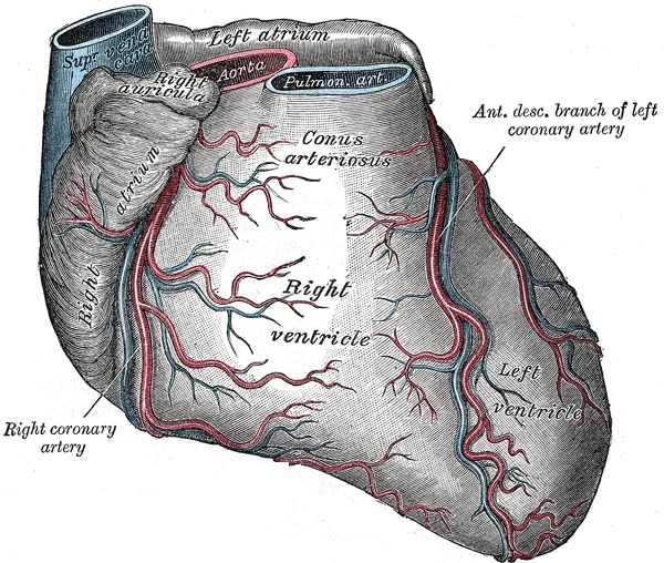
coronary sulcus or human heart skeleton. Labelled illustration from Wikimedia Commons, used under its open licence.

Figure 6: Coronary sulcus or human heart skeleton.
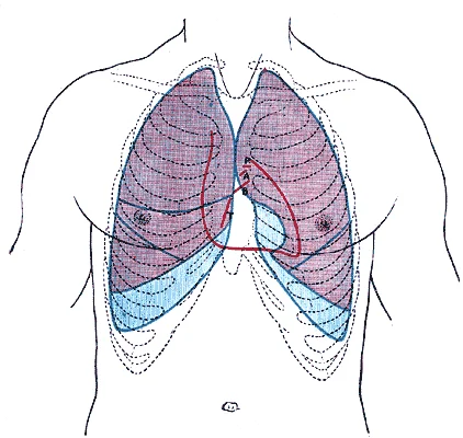
The heart is a muscular organ positioned in the thoracic cavity with its apex pointing toward the left side of the chest. The inferior tip of the heart, the apex, lies just to the left of the sternum between the junction of the fourth and fifth ribs near their articulation with the costal cartilages. The right side of the heart is deflected anteriorly, while the left side is deflected posteriorly. This orientation creates a cardiac notch in the medial surface of the superior lobe of the left lung. [1]
The heart consists of four chambers: two atria (upper chambers) and two ventricles (lower chambers). The right atrium and left atrium act as receiving chambers, contracting to push blood into the right and left ventricles respectively. The ventricles serve as the primary pumping chambers, with the right ventricle pumping blood to the lungs and the left ventricle pumping blood to the rest of the body. [1] [2]
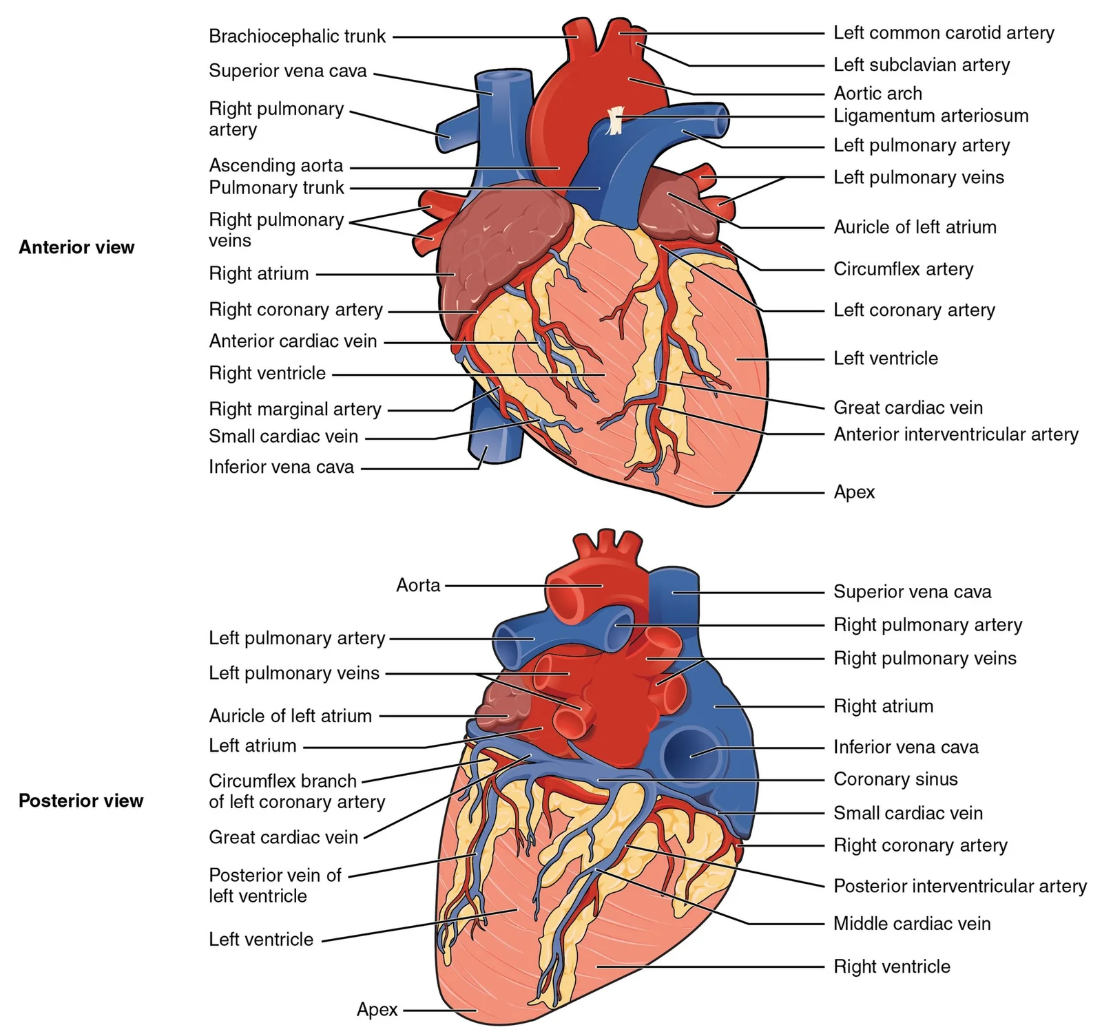
On the surface of the heart, auricles (atrial appendages) are visible as superficial leaf-like extensions near the superior surface of the heart. The heart also features fat-filled grooves called sulci, which house major coronary blood vessels. The deep coronary sulcus is located between the atria and ventricles, while two additional sulci between the left and right ventricles are less deep. [1]
Figure 72005 Surface Anatomy of the Heart
Long description
Inside the pericardium, the surface features of the heart are visible.

Figure 7: Inside the pericardium, the surface features of the heart are visible.
The heart valves ensure one-way blood flow. The atrioventricular valves (tricuspid on the right, mitral on the left) are located between the atria and ventricles, while semilunar valves (pulmonary and aortic) are at the exits of the ventricles. [1]
The coronary circulation is supplied by the right coronary artery, which runs along the coronary sulcus and gives rise to marginal arteries and the posterior interventricular artery.
Figure 8Heart-thorax-gray
Long description
Relationship of the anatomy of the human thorax to the internal viscera ( heart and lungs ) and bones ( sternum and ribs , showing the outline of the heart (in red).

Figure 8: Relationship of the anatomy of the human thorax to the internal viscera ( heart and lungs ) and bones ( sternum and ribs , showing the outline of the heart (in red).
Deep to this lies the parietal layer of the serous pericardium. The visceral layer of the serous pericardium, also known as the epicardium, adheres directly to the heart muscle. In most organs within the body, visceral serous membranes such as the epicardium are microscopic. However, in the case of the heart, it is not a microscopic layer but rather a macroscopic layer, consisting of a simple squamous epithelium called a mesothelium, reinforced with loose, irregular, or areolar connective tissue that attaches to the pericardium. This mesothelium secretes the lubricating serous fluid that fills the pericardial cavity and reduces friction as the heart contracts. [1]
The wall of the heart is composed of three layers of unequal thickness. From superficial to deep, these are the epicardium, the myocardium, and the endocardium. The outermost layer of the wall of the heart is also the innermost layer of the pericardium, the epicardium, or the visceral pericardium discussed earlier. The myocardium forms the bulk of the heart wall and is responsible for contraction. The endocardium lines the interior chambers and covers the heart valves. [1]
The heart is surrounded by the pericardial cavity, which contains serous fluid. Anteriorly, the heart relates to the sternum and costal cartilages. Posteriorly, it relates to the oesophagus and the descending thoracic aorta. Superiorly, the great vessels emerge from the base. Inferiorly, the heart rests on the diaphragm. The right atrium forms the right border of the heart shadow. The left ventricle forms the apex and the left border. The left atrium is the most posterior chamber, lying anterior to the oesophagus. [2]
Figure 9Gray491
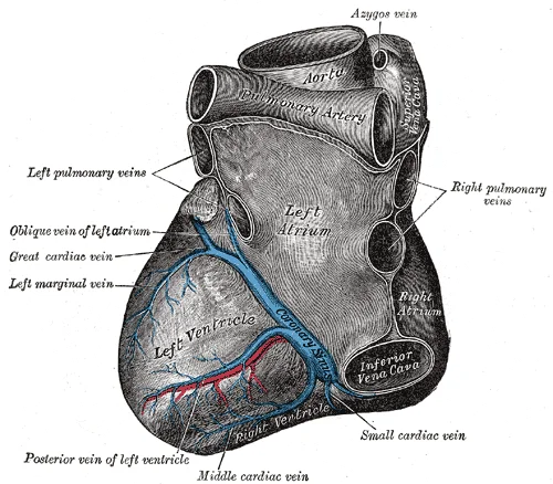
Long description
Base and diaphragmatic surface of heart.

Figure 9: Base and diaphragmatic surface of heart.
The septa divide the heart into chambers. The word septum is derived from the Latin for “something that encloses;” in this case, a septum (plural = septa) refers to a wall or partition that divides the heart into chambers. The septa are physical extensions of the myocardium lined with endocardium. Located between the two atria is the interatrial septum. Normally in an adult heart, the interatrial septum bears an oval-shaped depression known as the fossa ovalis, a remnant of an opening in the fetal heart known as the foramen ovale. The foramen ovale allowed blood in the fetal heart to pass directly from the right atrium to the left atrium, allowing some blood to bypass the pulmonary circuit. Within seconds after birth, a flap of tissue known as the septum primum that previously acted as a valve closes the foramen ovale and establishes the typical cardiac circulation pattern. [1]
Mesoderm is one of the three primary germ layers that differentiates early in development that collectively gives rise to all subsequent tissues and organs. Following chemical signals called factors from the underlying endoderm (another of the three primary germ layers), the cardiogenic area begins to form two strands called the cardiogenic cords. As the cardiogenic cords develop, a lumen rapidly develops within them. At this point, they are referred to as endocardial tubes. The two tubes migrat [2]
It forms from the cardiogenic region near the head and is visible as a prominent heart bulge on the surface of the embryo. [2]
| Layer | Description |
|---|---|
| Epicardium | Visceral pericardium; mesothelium and connective tissue |
| Myocardium | Muscle layer; septa are extensions of this |
| Endocardium | Inner lining; covers valves and septa |
Figure 10Gray internal structure of heart
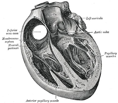
Long description
Internal structure of the human heart. Labelled illustration from Wikimedia Commons, used under its open licence.

Figure 10: Internal structure of the human heart.
The heart wall comprises three distinct layers. The innermost layer is the endocardium, a smooth endothelial lining that covers the heart valves and the inner surfaces of the chambers. It is continuous with the endothelial lining of the large blood vessels entering and leaving the heart. The middle and thickest layer is the myocardium, made largely of cardiac muscle cells. It is built upon a framework of collagenous fibers, plus the blood vessels that supply the myocardium and the nerve fibers that help regulate the heart. The muscle pattern is elegant and complex, as the muscle cells swirl and spiral around the chambers of the heart. They form a figure 8 pattern around the atria and around the bases of the great vessels. Deeper ventricular muscles also form a figure 8 around the two ventricles and proceed toward the apex. More superficial layers of ventricular muscle wrap around both ventricles. This complex swirling pattern allows the heart to pump blood more effectively than a simple linear pattern would. The outermost layer is the epicardium, which is the visceral layer of the serous pericardium. [1]
Figure 112011 Heart Valves
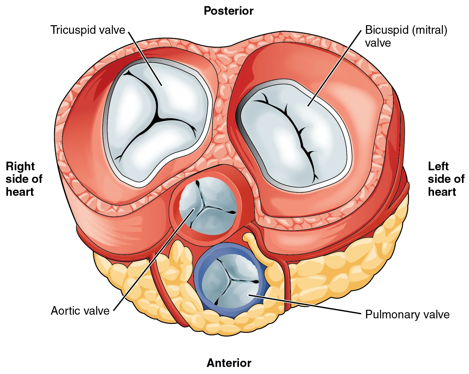
Long description
With the atria and major vessels removed, all four valves are clearly visible, although it is difficult to distinguish the three separate cusps of the tricuspid valve.

Figure 11: With the atria and major vessels removed, all four valves are clearly visible, although it is difficult to distinguish the three separate cusps of the tricuspid valve.
Figure 122006 Heart Musculature
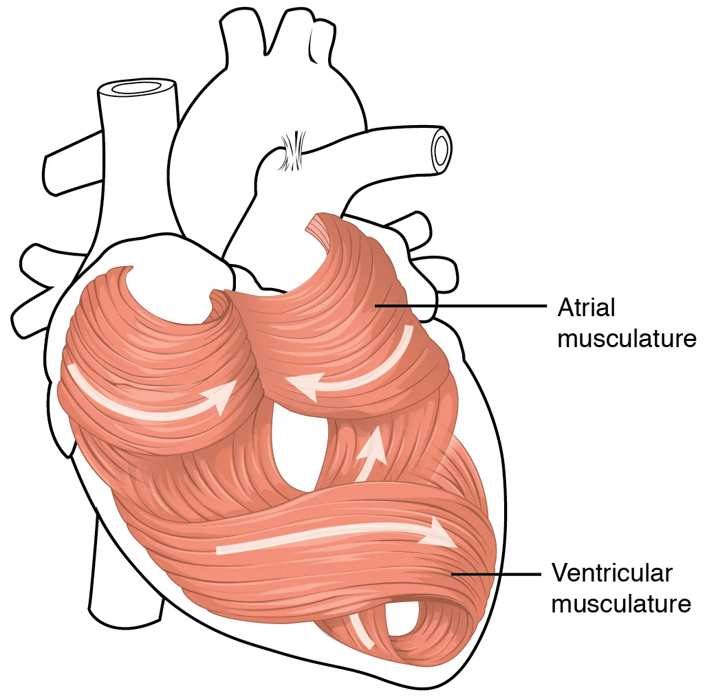
Long description
The swirling pattern of cardiac muscle tissue contributes significantly to the heart’s ability to pump blood effectively.

Figure 12: The swirling pattern of cardiac muscle tissue contributes significantly to the heart’s ability to pump blood effectively.
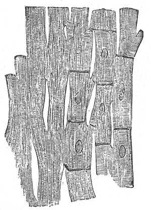
Figure 13Gray499
Long description
Anastomosing muscular fibers of the heart seen in a longitudinal section. On the right the limits of the separate cells with their nuclei are exhibited somewhat diagrammatically.

Figure 13: Anastomosing muscular fibers of the heart seen in a longitudinal section.
Figure 14Gray495
Long description
Base of ventricles exposed by removal of the atria.

Figure 14: Base of ventricles exposed by removal of the atria.
DefinitionMyocardial Architecture
The myocardium consists of cardiac muscle cells arranged in a complex swirling pattern, forming figure 8 shapes around the atria and ventricles to optimize pumping efficiency.
The right ventricle pumps blood into the pulmonary circuit. Its wall is thinner than that of the left ventricle because it pumps against lower resistance. The interior of the right ventricle contains trabeculae carneae, irregular muscular ridges. One prominent trabecula, the moderator band, crosses the cavity to the anterior papillary muscle. It contains part of the right bundle branch of the atrioventricular bundle, ensuring rapid conduction to the papillary muscles. [1]
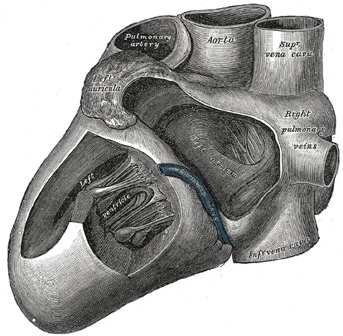
Figure 15Gray496
Long description
Interior of left side of heart. Labelled illustration from Wikimedia Commons, used under its open licence.

Figure 15: Interior of left side of heart.
| Structure | Location / Relation | Function |
|---|---|---|
| Endocardium | Inner lining of chambers | Smooth blood flow |
| Myocardium | Middle muscular layer | Contraction and pumping |
| Coronary Sinus | Posterior right atrium | Drains cardiac veins |
| Moderator Band | Right ventricle | Conducts electrical impulses |
The human heart consists of a pair of atria, which receive blood and pump it into a pair of ventricles, which pump blood into the vessels. The walls of the heart are composed of an outer epicardium, a thick myocardium, and an inner lining layer of endocardium. The heart resides within the pericardial sac and is located in the mediastinal space within the thoracic cavity. The pericardial sac consists of two fused layers: an outer fibrous capsule and an inner parietal pericardium lined with a serous membrane. Between the pericardial sac and the heart is the pericardial cavity, which is filled with lubricating serous fluid. [1]
Each of the major pumping chambers of the heart ejects approximately 70 mL blood per contraction in a resting adult. [1]
The heart comprises four chambers arranged in two receiving atria and two pumping ventricles. These chambers are separated by septa. The interatrial septum divides the right and left atria, while the interventricular septum divides the ventricles. The thickness of the interventricular septum reflects the high-pressure workload of the left ventricle, which pumps blood into the systemic circulation.
The circulation is divided into two distinct but linked circuits: the pulmonary and systemic circuits. The pulmonary circuit transports blood to and from the lungs, where it picks up oxygen and delivers carbon dioxide for exhalation. This circuit operates at lower pressures than the systemic circuit. The systemic circuit transports oxygenated blood to virtually all of the tissues of the body and returns relatively deoxygenated blood and carbon dioxide to the heart to be sent back to the pulmonary circulation. [1]
Blood returns to the left atrium high in oxygen via one of the four pulmonary veins. Blood flows nearly continuously from the pulmonary veins back into the atrium, which acts as the receiving chamber. From here, blood passes through an opening guarded by the mitral valve into the left ventricle. Most blood flows passively into the heart while both the atria and ventricles are relaxed. However, toward the end of the ventricular relaxation period, the left atrium contracts, pumping blood into the ventricle. This atrial contraction accounts for approximately 20 percent of ventricular filling. This 'atrial kick' is particularly important in conditions where ventricular compliance is reduced, such as in hypertensive heart disease or aortic stenosis, where passive filling alone may be insufficient to maintain cardiac output. [1]
The heart is a remarkable pump composed largely of cardiac muscle cells that are incredibly active throughout life. Like all other cells, a cardiomyocyte requires a reliable supply of oxygen and nutrients, and a way to remove wastes, so it needs a dedicated, complex, and extensive coronary circulation. Because of the critical and nearly ceaseless activity of the heart throughout life, this need for a blood supply is even greater than for a typical cell. However, coronary circulation is not continuous; rather, it cycles, reaching a peak when the heart muscle is relaxed and nearly ceasing while it is contracting. [1]
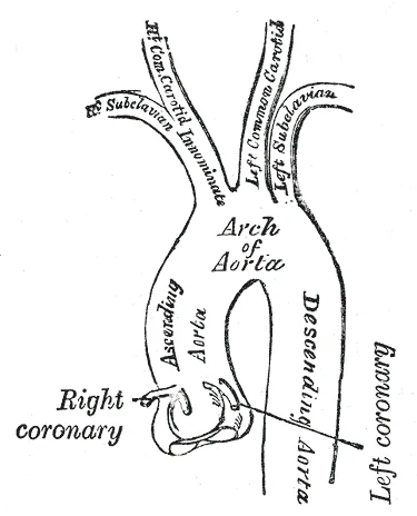
Figure 16Gray506
Long description
The only branches of the ascending aorta are the two coronary arteries which supply the heart; they arise near the commencement of the aorta immediately above the attached margins of the semilunar valves. The branches given off from the arch of the aorta are three in number: the innominate, the left

Figure 16: The only branches of the ascending aorta are the two coronary arteries which supply the heart; they arise near the commencement of the aorta immediately above the attached margins of the semilunar valves.
The innermost layer of the heart wall, the endocardium, is joined to the myocardium with a thin layer of connective tissue. The endocardium lines the chambers where the blood circulates and covers the heart valves. It is made of simple squamous epithelium called endothelium, which is continuous with the endothelial lining of the blood vessels. This endothelial lining ensures smooth blood flow within the chambers and valves, reducing friction and preventing thrombus formation. [1]
The arterial supply is crucial for maintaining the high metabolic demands of the heart. The coronary arteries branch from the aorta and distribute blood to the myocardium. The right coronary artery supplies the right atrium, right ventricle, and parts of the conduction system. The left coronary artery supplies the left atrium, left ventricle, and most of the interventricular septum. Any obstruction in these arteries can lead to myocardial ischaemia, highlighting the importance of a robust and unobstructed coronary circulation. [1]
Figure 17Gray577
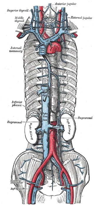
Long description
There are shown the superior and the inferior vena cava with their branches. The heart has been taken away and most of the aorta cut off.

Figure 17: There are shown the superior and the inferior vena cava with their branches.
Inferior to the opening of the superior vena cava, the inferior vena cava enters the posterior portion of the atria, draining blood from areas inferior to the diaphragm, specifically the lower limbs and abdominopelvic region. The coronary sinus, a large coronary vein that drains the heart myocardium, also empties into the right atrium. The right atrium serves as the receiving chamber for blood returning to the heart from the systemic circulation. The atria receive venous blood on a nearly continuous basis, preventing venous flow from stopping while the ventricles are contracting. While most ventricular filling occurs while the atria are relaxed, they do demonstrate a contractile phase and actively pump blood into the ventricles just prior to ventricular contraction. The opening between the atrium and ventricle is guarded by the tricuspid valve. [1] [2]
Figure 18Gray493
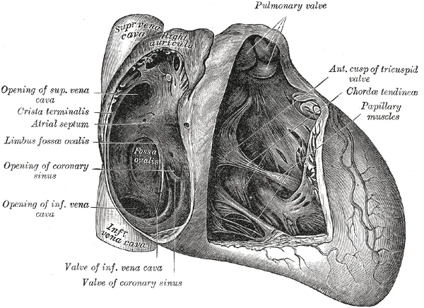
Long description
Interior of right side of heart. Labelled illustration from Wikimedia Commons, used under its open licence.

Figure 18: Interior of right side of heart.
DefinitionRight Atrium
The right atrium receives blood from the systemic circulation via the superior and inferior venae cavae and the coronary sinus. [1]
The superior vena cava is a large vein that carries deoxygenated blood from the upper half of the body to the right atrium. It is formed by the union of the left and right brachiocephalic veins. The inferior vena cava carries deoxygenated blood from the lower half of the body to the right atrium. It is formed by the union of the common iliac veins. The coronary sinus is a venous structure located in the posterior aspect of the coronary sulcus. It collects blood from the cardiac veins and empties into the right atrium. The crista terminalis is a vertical ridge that separates the smooth posterior wall from the rough anterior wall. The fossa ovalis is a depression in the interatrial septum, representing the remnant of the foramen ovale. The tricuspid valve is located between the right atrium and the right ventricle. It prevents backflow of blood into the right atrium during ventricular systole. [1]
| Structure | Origin | Termination |
|---|---|---|
| Superior Vena Cava | Union of brachiocephalic veins | Right Atrium |
| Inferior Vena Cava | Union of common iliac veins | Right Atrium |
| Coronary Sinus | Cardiac veins | Right Atrium |
The venous drainage of the heart is essential for maintaining cardiac function. The right atrium receives blood from the systemic circulation and the coronary circulation. The superior vena cava drains blood from the head, neck, upper limbs, and thoracic region. The inferior vena cava drains blood from the lower limbs and abdominopelvic region. The coronary sinus drains blood from the heart myocardium. The right atrium pumps blood into the right ventricle through the tricuspid valve. The tricuspid valve prevents backflow of blood into the right atrium during ventricular systole. The right atrium receives venous blood on a nearly continuous basis. This prevents venous flow from stopping while the ventricles are contracting. The atria demonstrate a contractile phase and actively pump blood into the ventricles just prior to ventricular contraction. This ensures efficient ventricular filling. [1]
Figure 19Gray968
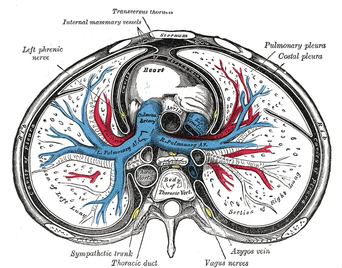
Long description
A transverse section of the thorax, showing the contents of the middle and the posterior mediastinum. The pleural and pericardial cavities are exaggerated since normally there is no space between parietal and visceral pleura and between pericardium and heart.

Figure 19: A transverse section of the thorax, showing the contents of the middle and the posterior mediastinum.
The lymphatics originate from the capillary networks within the myocardium. They converge into larger collecting vessels. These vessels follow the coronary sulcus and the interventricular sulci. The anterior lymphatic vessels drain into the anterior mediastinal lymph nodes. The posterior lymphatic vessels drain into the posterior mediastinal lymph nodes. Some vessels may also drain into the tracheobronchial lymph nodes. Note how the lymphatics run parallel to the epicardial coronary arteries. This anatomical arrangement is crucial for understanding the spread of infection or malignancy.
It is supplied by the posterior interventricular artery in right-dominant hearts. Its lymphatics drain into the posterior mediastinal nodes. The bundle branches run within the interventricular septum. Their lymphatic drainage is less distinct but generally follows the coronary vessels on the surface of the septum. Understanding this drainage is important for interpreting lymphadenopathy in cardiac patients.
DefinitionLymphatic Drainage
The movement of lymph fluid from the interstitial spaces into the lymphatic vessels, eventually returning to the venous circulation.
Instead, it relies on a network of superficial and deep lymphatics. The superficial lymphatics lie in the subepicardial layer. They accompany the coronary arteries and veins. This dual system ensures efficient removal of interstitial fluid. This anatomical distribution explains why cardiac pathology can sometimes present with mediastinal lymphadenopathy.
| Structure | Primary Drainage Pathway | Target Lymph Nodes |
|---|---|---|
| Anterior Heart | Superficial lymphatics along coronary arteries | Anterior mediastinal nodes |
| Posterior Heart | Deep lymphatics along posterior vessels | Posterior mediastinal nodes |
| Sinoatrial Node | Along right coronary artery | Right atrioventricular sulcus nodes |
| Atrioventricular Node | Along posterior interventricular artery | Posterior mediastinal nodes |
Clinical relevance arises when lymphatic obstruction occurs. This can lead to lymphoedema, although this is rare in the heart itself. More commonly, lymphatic spread is a route for metastatic disease. Tumours from the lungs or mediastinum can invade these lymphatic channels. Recognising the anatomical pathways helps in staging and treatment planning. The lymphatic system also plays a role in immune surveillance. It removes debris and pathogens from the cardiac tissue. This function is vital for maintaining cardiac health.
The heart receives its innervation from both autonomic and somatic sources, creating a complex network that regulates cardiac function and mediates pain perception. Understanding this supply requires examining the autonomic components, which control rate and contractility, and the somatic pathways, which are primarily responsible for referred pain.
The autonomic nervous system provides the primary regulatory input to the heart. This system is divided into sympathetic and parasympathetic divisions, which generally exert opposing effects on cardiac activity. The sympathetic nervous system increases heart rate and contractility, while the parasympathetic nervous system, primarily via the vagus nerve, slows the heart rate.
Figure 20Gray970
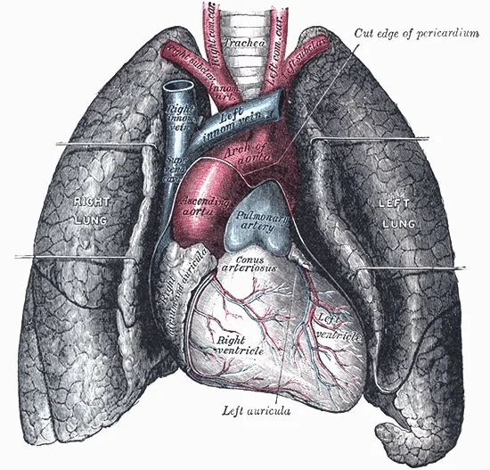
Long description
Front view of heart and lungs. Labelled illustration from Wikimedia Commons, used under its open licence.

Figure 20: Front view of heart and lungs.
The sympathetic trunk lies lateral to the vertebral bodies, with cardiac branches extending medially to join the plexus. The vagus nerve descends through the thorax, giving off cardiac branches that also contribute to the plexus. The plexus then distributes fibers to the sinoatrial and atrioventricular nodes, as well as the myocardium. The sympathetic fibers originate from the upper thoracic spinal cord segments, traveling through the sympathetic chain to reach the heart. The parasympathetic fibers originate in the medulla, traveling via the vagus nerve to the heart.
DefinitionAutonomic Balance
Healthy cardiac function relies on a dynamic balance between sympathetic and parasympathetic outflows, modulating heart rate and contractility in response to physiological demands.
The sympathetic supply arises from the thoracic sympathetic ganglia. Fibers travel to the cardiac plexus, where they synapse or pass through to reach the heart. These fibers primarily innervate the sinoatrial node, atrioventricular node, and myocardial muscle, increasing heart rate and force of contraction.
The parasympathetic supply is provided by the vagus nerve (cranial nerve X). Stimulation of these fibers releases acetylcholine, which slows the heart rate and decreases conduction velocity through the atrioventricular node.
Somatic innervation of the heart is limited but clinically significant due to the phenomenon of referred pain. The heart itself lacks somatic sensory innervation; therefore, pain from cardiac ischemia is perceived as coming from somatic structures. This occurs because visceral afferent fibers from the heart enter the spinal cord at the same levels as somatic afferent fibers from the chest wall and upper limbs.
Exam trapReferred Pain
Cardiac pain is referred to the left arm, jaw, or epigastrium because visceral afferent fibers converge with somatic afferent fibers in the spinal cord, leading the brain to misinterpret the source of the pain.
The visceral afferent fibers travel alongside sympathetic nerves to the upper thoracic spinal segments (T1-T4). When these fibers are stimulated by ischemia, the brain interprets the signal as coming from the dermatomes supplied by these spinal segments.
| Division | Origin | Primary Effect | Key Structures Innervated |
|---|---|---|---|
| Sympathetic | Thoracic sympathetic ganglia | Increase HR and contractility | SA node, AV node, myocardium |
| Parasympathetic | Vagus nerve (CN X) | Decrease HR and conduction | SA node, AV node, atria |
| Somatic Afferent | T1-T4 spinal segments | Pain perception | Referred pain to chest/arm |
This convergence of pathways explains why cardiac pathology often presents with pain in areas distant from the heart itself. Recognizing this pattern is crucial for accurate diagnosis and timely intervention.
The heart is a muscular pump composed of specialised cardiac muscle tissue, distinct from skeletal and smooth muscle. Cardiac myocytes are striated, branched, and typically uninucleate. They are connected by intercalated discs, which contain desmosomes for mechanical adhesion and gap junctions for electrical coupling. This structure allows the heart to function as a functional syncytium, ensuring coordinated contraction. The myocardium is the thick middle layer of the heart wall, responsible for the contractile force. It is surrounded by the endocardium, a smooth endothelial lining, and the epicardium, the outer visceral layer of the serous pericardium. The pericardial cavity between the layers contains lubricating fluid.
DefinitionCardiac Muscle
Striated, branched muscle cells connected by intercalated discs, enabling synchronized contraction of the heart.
Link to the histology page.
These discs facilitate rapid transmission of action potentials via gap junctions. Desmosomes within the discs provide strong mechanical attachment between cells. Cardiac muscle cells have a single central nucleus, unlike the peripheral nuclei of skeletal muscle. Mitochondria are abundant in cardiac myocytes to support continuous aerobic metabolism. The sarcoplasmic reticulum is less developed than in skeletal muscle, relying more on extracellular calcium influx for contraction.
Clinical pearlIntercalated Discs
Structures that link cardiac myocytes, containing gap junctions for electrical coupling and desmosomes for mechanical strength.
The conduction system consists of specialised cardiac muscle cells that generate and transmit electrical impulses. These cells are larger and paler than contractile myocytes. They include the sinoatrial node, atrioventricular node, bundle of His, and Purkinje fibres. The atrioventricular node delays the impulse to allow atrial contraction before ventricular contraction. The bundle of His divides into left and right bundle branches. Purkinje fibres distribute the impulse rapidly through the ventricular walls. [1]
NoteConduction System
Specialised cells that generate and transmit electrical impulses, ensuring coordinated cardiac contraction.
Histological examination of the heart wall reveals three layers. The endocardium lines the chambers and covers the valves. It is composed of endothelium and a thin layer of connective tissue. The myocardium is the thickest layer, composed of cardiac muscle bundles arranged in a spiral pattern. The epicardium is the outermost layer, consisting of mesothelium and underlying connective tissue. It contains blood vessels and nerves. The pericardium consists of the fibrous pericardium and the serous pericardium. The fibrous pericardium is a tough, inelastic sac. The serous pericardium has parietal and visceral layers.
DefinitionMyocardium
The thick middle layer of the heart wall, composed of cardiac muscle tissue responsible for contraction.
Valves are composed of dense connective tissue covered by endocardium. They prevent backflow of blood. The atrioventricular valves (tricuspid and mitral) have chordae tendineae and papillary muscles. These structures prevent valve prolapse during ventricular systole. The semilunar valves (pulmonary and aortic) are pocket-like. They open when ventricular pressure exceeds arterial pressure. Histologically, they lack the muscular support of the AV valves. [1]
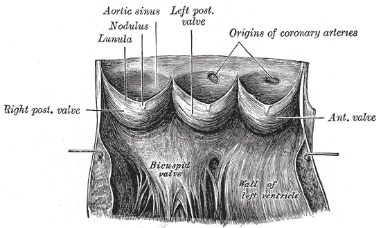
Figure 21Gray497
Long description
Aorta laid open to show the semilunar valves.

Figure 21: Aorta laid open to show the semilunar valves.
Exam trapValve Structure
AV valves have chordae tendineae and papillary muscles; semilunar valves do not.
The coronary arteries supply the myocardium. They arise from the aorta. The right and left coronary arteries branch from the aortic sinuses. They run in the coronary sulci. Cardiac veins parallel the arteries. They drain into the coronary sinus. The coronary sinus empties into the right atrium. This arrangement ensures efficient blood supply and drainage.
DefinitionCoronary Circulation
Arteries arising from the aorta supply the myocardium; veins drain into the coronary sinus.
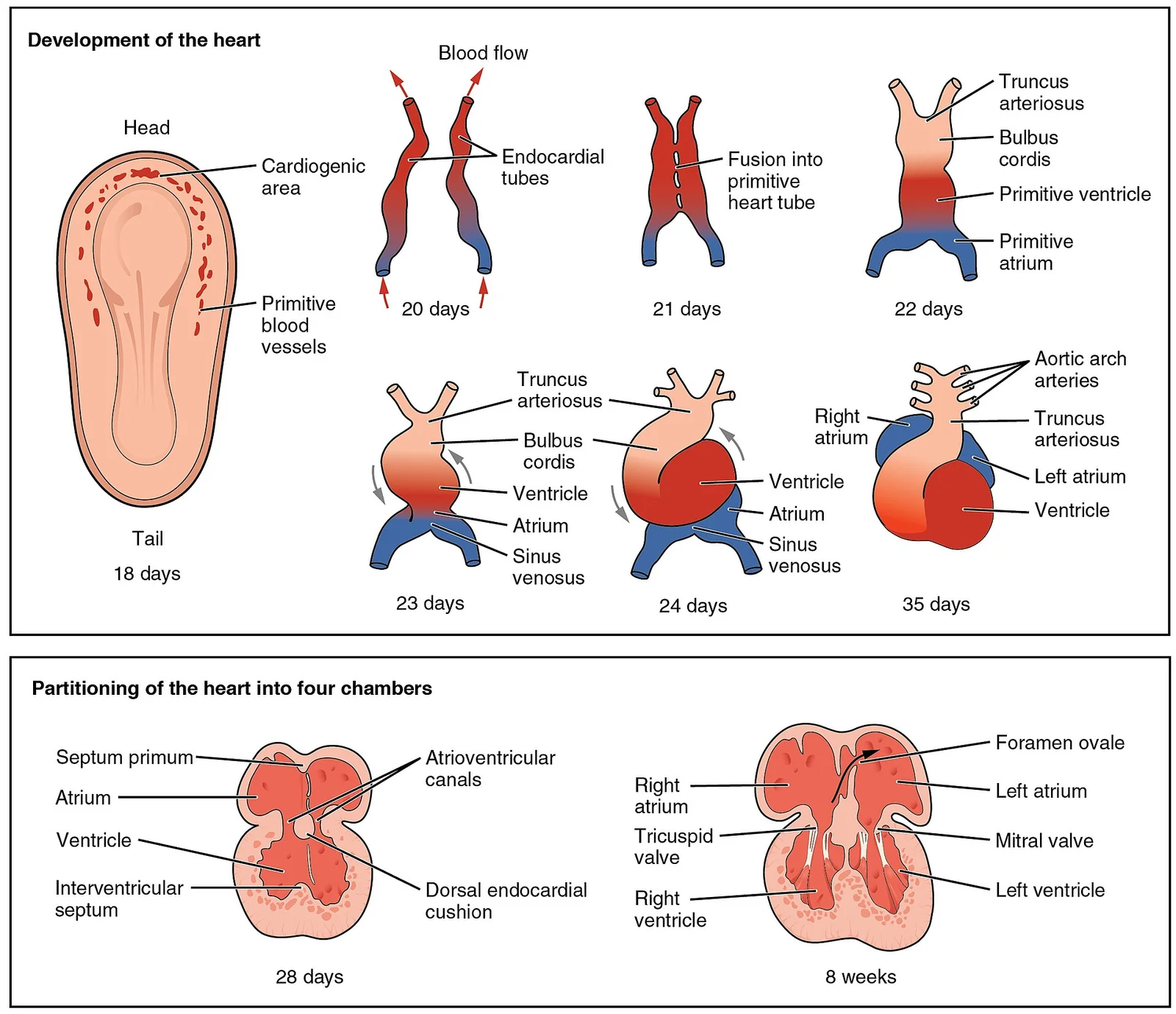
This tube undergoes rightward looping, establishing the basic orientation of the chambers. By the end of the eighth week, the four-chambered structure is largely defined. A critical feature of this development is the formation of the septa. The interatrial septum divides the atria, while the interventricular septum separates the ventricles. This structural integrity is vital because the ventricles generate far greater pressure when they contract, necessitating a robust barrier. Consequently, the interventricular septum is substantially thicker than the interatrial septum. [1] [2]
Figure 222037 Embryonic Development of Heart
Long description
This diagram outlines the embryological development of the human heart during the first eight weeks and the subsequent formation of the four heart chambers.

Figure 22: This diagram outlines the embryological development of the human heart during the first eight weeks and the subsequent formation of the four heart chambers.
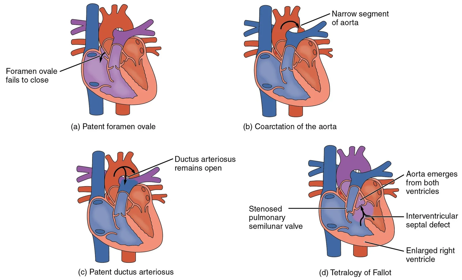
In clinical practice, understanding this embryological timeline is essential for interpreting congenital heart defects. Defects such as atrial septal defects (ASD) or ventricular septal defects (VSD) represent failures in this septation process. While the interatrial septum has a complex developmental history involving the septum primum and secundum, the ventricular septum’s integrity is a key marker of normal postnatal circulation. The transition from fetal to neonatal circulation involves the closure of shunts like the foramen ovale and ductus arteriosus, but the septal structures themselves remain the primary anatomical divisions.
Figure 232009 Congenital Heart Defects
Long description
(a) A patent foramen ovale defect is an abnormal opening in the interatrial septum, or more commonly, a failure of the foramen ovale to close. (b) Coarctation of the aorta is an abnormal narrowing of the aorta. (c) A patent ductus arteriosus is the failure of the ductus arteriosus to close. (d) Tetr

Figure 23: (a) A patent foramen ovale defect is an abnormal opening in the interatrial septum, or more commonly, a failure of the foramen ovale to close.
For a detailed understanding of cardiac embryology, including the specific molecular pathways and timing of chamber formation, please refer to the dedicated embryology page. This resource covers the neural crest contributions and the precise morphological changes that occur week by week.
The heart’s rhythm and force of contraction are regulated by the autonomic nervous system, which maintains a dynamic relative balance between the sympathetic nervous system (SNS) and parasympathetic nervous system (PNS). At rest, PNS activity predominates, resulting in an average heart rate of 75 beats per minute (bpm). This resting rate is significantly slower than the SA node's intrinsic rate, which decreases with age from an average 107 bpm at 20 years to 90 bpm at 50 years. This age-related decline in intrinsic rate is a normal physiological change, but it can impact the heart's reserve capacity in older adults. [3]
The parasympathetic branch can slow the heart to 20 or 30 bpm or briefly stop it. This illustrates the response called accentuated antagonism. Parasympathetic nerves exert their effects more rapidly (<1 s) than sympathetic nerves (>5 s). This rapid onset is crucial for immediate adjustments in heart rate, such as during the baroreceptor reflex in response to sudden postural changes. In contrast, an increase in sympathetic activity is the principal method used to increase HR above the intrinsic level generated by the SA node. Following the onset of sympathetic stimulation, there is a delay of up to 5 s before the stimulation induces a progressive increase in HR. This increase reaches a steady level in 20–30 s if the stimulus is continuous. The slowness of the response to sympathetic stimulation is in direct contrast to vagal stimulation, which is almost instantaneous. However, the effect on HR is longer lasting, and even a short stimulus can affect HR for 5–10 s. [3]
Beyond neural control, the heart functions as an endocrine gland. Atrial myocytes can secrete atrial natriuretic peptide (ANP), a hormone that promotes salt and water excretion, to lower BP and produce vasodilation. Additionally, intrinsic cardiac adrenergic cells can synthesize and secrete catecholamines such as dopamine, NE, and E. [3]
Figure 24Mediastinum anatomy
Long description
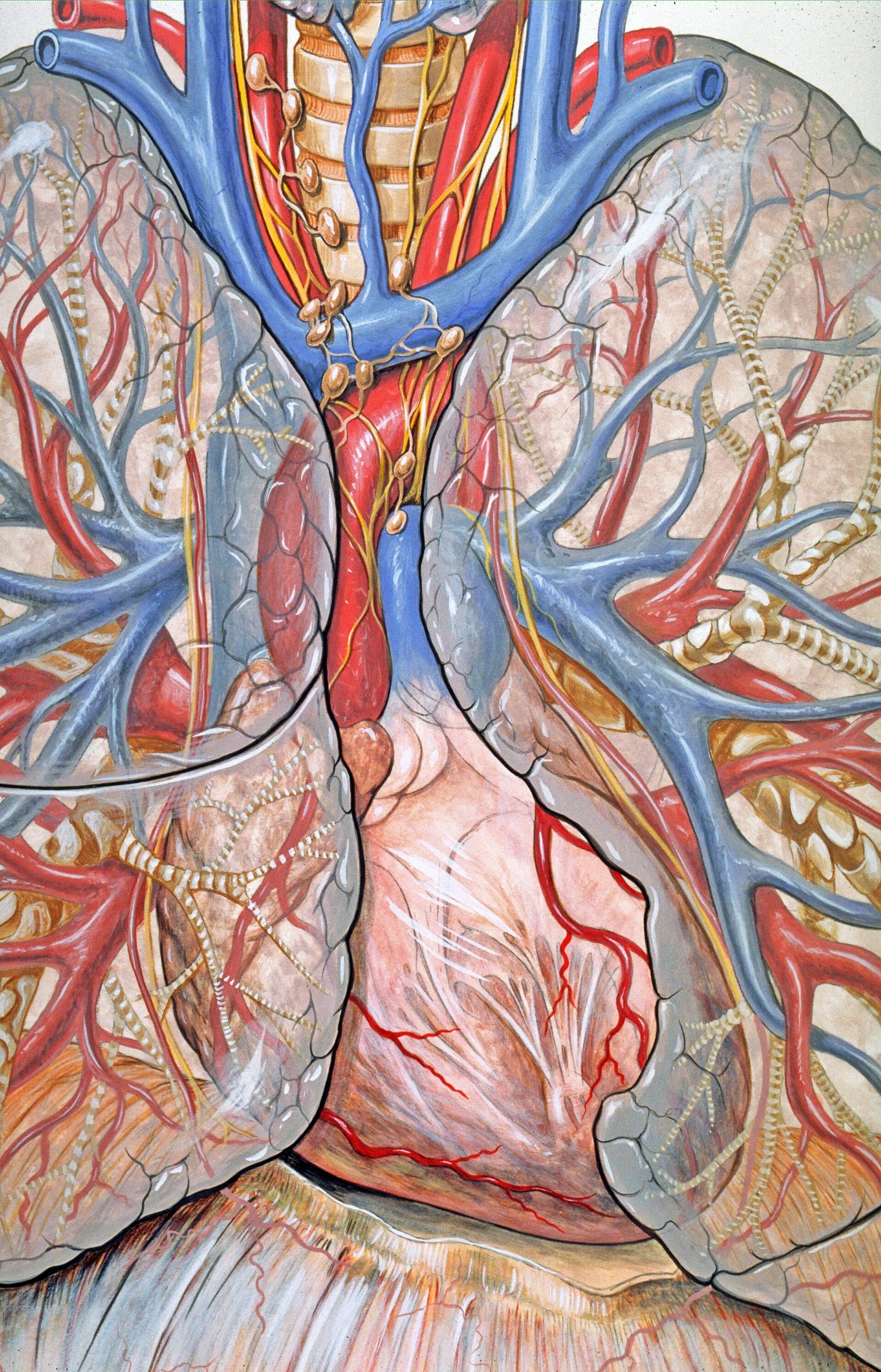
Anatomy of the mediastinum (the central chest and heart area).

Figure 24: Anatomy of the mediastinum (the central chest and heart area).
The heart is a muscular organ situated in the middle mediastinum, enclosed within the pericardial sac. Radiologically, the right border of the cardiac silhouette is formed primarily by the right atrium. This chamber receives deoxygenated blood from the superior and inferior vena cavae. The superior vena cava enters the right atrium from above, while the inferior vena cava enters from below. The left ventricle constitutes the majority of the anterior surface of the heart, which faces the sternum.
The apex of the heart is formed by the left ventricle and points downwards, forwards, and to the left. It typically lies in the fifth intercostal space, just medial to the mid-clavicular line on the left side. This landmark is crucial for clinical palpation and auscultation.
The great vessels emerge from the superior aspect of the heart. The ascending aorta arises from the left ventricle and curves posteriorly to become the aortic arch. The pulmonary trunk arises from the right ventricle and bifurcates into the left and right pulmonary arteries. These vessels are clearly visible on cross-sectional imaging and are critical for assessing pulmonary circulation. [2]
The pericardium surrounds the heart and consists of two layers: the fibrous pericardium and the serous pericardium. The fibrous pericardium is a tough, inelastic connective tissue layer that anchors the heart to surrounding structures. The serous pericardium has two layers: the parietal layer, which lines the fibrous pericardium, and the visceral layer, which adheres to the heart muscle. The space between these layers contains a small amount of serous fluid, which reduces friction during cardiac contractions.
Understanding these radiological landmarks is vital for diagnosing cardiac pathology. Enlargement of specific chambers can alter the cardiac silhouette, providing clues to underlying conditions. For instance, left ventricular hypertrophy may result in a leftward displacement of the apex, while right atrial enlargement can widen the right heart border. Accurate interpretation of these images requires a solid grasp of normal anatomical relationships.
DefinitionCardiac Silhouette
The outline of the heart as seen on a chest radiograph, formed by the borders of the cardiac chambers and great vessels.
| Structure | Radiological Border |
|---|---|
| Right Atrium | Right heart border |
| Left Ventricle | Left heart border and apex |
| Left Atrium | Base, posterior |
| Ascending Aorta | Superior mediastinum, right |
| Pulmonary Trunk | Superior mediastinum, left |
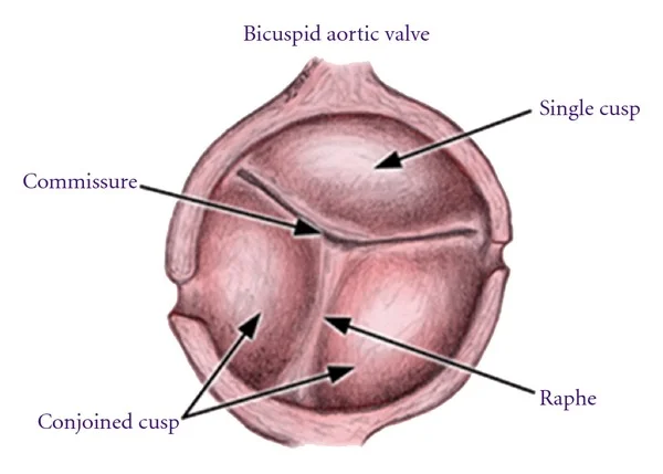
Figure 25The basic anatomy of the bicuspid aortic valve
Long description
Diagram of a bicuspid aortic valve. Labelled illustration from Wikimedia Commons, used under its open licence.

Figure 25: Diagram of a bicuspid aortic valve.
Understanding these spatial relationships is vital for identifying where structural failures, such as chordal rupture or papillary muscle dysfunction, might occur. [1]
The walls of the ventricle are lined with trabeculae carneae, which are ridges of cardiac muscle covered by endocardium. These muscular ridges provide structural support and increase the surface area for contraction. The left ventricle serves as the major pumping chamber for the systemic circuit, ejecting blood into the aorta through the aortic semilunar valve. The aortic valve, located at the base of the aorta, normally consists of three flaps. When the ventricle relaxes, blood attempting to flow back from the aorta fills these cusps, causing the valve to close and producing an audible sound. Similarly, the pulmonary semilunar valve, or pulmonary valve, emerges from the right ventricle at the base of the pulmonary trunk. It is comprised of three small flaps of endothelium reinforced with connective tissue. Unlike the atrioventricular valves, there are no papillary muscles or chordae tendineae associated with the pulmonary valve. Its closure is also produced by the filling of its pocket-like flaps with backflowing blood. [1]
The mitral valve, also known as the bicuspid valve, is located at the opening between the left atrium and left ventricle. Structurally, it consists of two cusps, which are attached by chordae tendineae to two papillary muscles projecting from the ventricular wall. In clinical settings, the term mitral valve is preferred over bicuspid valve. The conduction system's proximity to these structures is critical; for instance, the moderator band's role in conduction highlights the integration of mechanical and electrical functions within the ventricular architecture. Disruption to these pathways or structures can lead to significant clinical sequelae, including heart block or valvular incompetence, which are central to the applied anatomy of the heart. [1]
- The heart has four chambers: two atria (right and left) and two ventricles (right and left), with the right atrium receiving deoxygenated blood from the superior and inferior venae cavae. [1]
- Blood flows from systemic capillaries into venules, then larger veins, and finally into the superior and inferior venae cavae, which return deoxygenated blood to the right atrium.
- The right atrium pumps blood into the right ventricle, which then sends it through the pulmonary valve into the pulmonary artery for oxygenation in the lungs. [1]
- The pulmonary circuit carries deoxygenated blood from the right ventricle to the lungs and returns oxygenated blood to the left atrium via the pulmonary veins. [1]
- The systemic circuit carries oxygenated blood from the left ventricle through the aortic valve into the aorta, distributing it throughout the body before returning to the right atrium.
- The heart valves ensure unidirectional blood flow: tricuspid between right atrium and ventricle, pulmonary valve between right ventricle and pulmonary artery, mitral between left atrium and ventricle, and aortic valve between left ventricle and aorta.
- The coronary arteries arise from the aortic root and supply the heart muscle itself, with the right coronary artery typically arising from the anterior aortic sinus and supplying the right atrium, right ventricle, and parts of the left ventricle, while the left coronary artery arises from the left posterior aortic sinus and supplies the left atrium and left ventricle. [1]
- The cardiac cycle consists of systole (contraction) and diastole (relaxation), with systole accounting for approximately one-third of the cycle.
- The heart's electrical conduction system begins with the sinoatrial node in the right atrium, then to the atrioventricular node at the junction of the atria and ventricles, and through the bundle of His to the Purkinje fibers. [1]
- A common trap is confusing the pulmonary and systemic circuits - remember that the pulmonary circuit carries deoxygenated blood to the lungs, while the systemic circuit carries oxygenated blood to the body.
- The interventricular septum separates the right and left ventricles, while the interatrial septum separates the right and left atria, with defects in these septa being common congenital abnormalities.
- The chordae tendineae and papillary muscles prevent the atrioventricular valves from prolapsing into the atria during ventricular contraction, and their rupture can lead to severe valvular regurgitation.
The heart is a muscular organ located in the mediastinum, between the lungs, and tilted to the left with the apex pointing inferiorly. The heart consists of four chambers: two atria (right and left) and two ventricles (right and left). The right atrium receives deoxygenated blood from the superior and inferior venae cavae and the coronary sinus. The left atrium receives oxygenated blood from the pulmonary veins.
The right ventricle pumps blood through the pulmonary valve into the pulmonary artery, which carries blood to the lungs for oxygenation. The left ventricle pumps blood through the aortic valve into the aorta, which distributes oxygenated blood to the systemic circulation. The interventricular septum separates the right and left ventricles, while the interatrial septum separates the right and left atria. [1]
The heart valves ensure unidirectional blood flow. The atrioventricular valves include the tricuspid valve (right side) and the mitral valve (left side). The semilunar valves include the pulmonary valve and the aortic valve. The chordae tendineae and papillary muscles prevent the atrioventricular valves from prolapsing into the atria during ventricular contraction. [1]
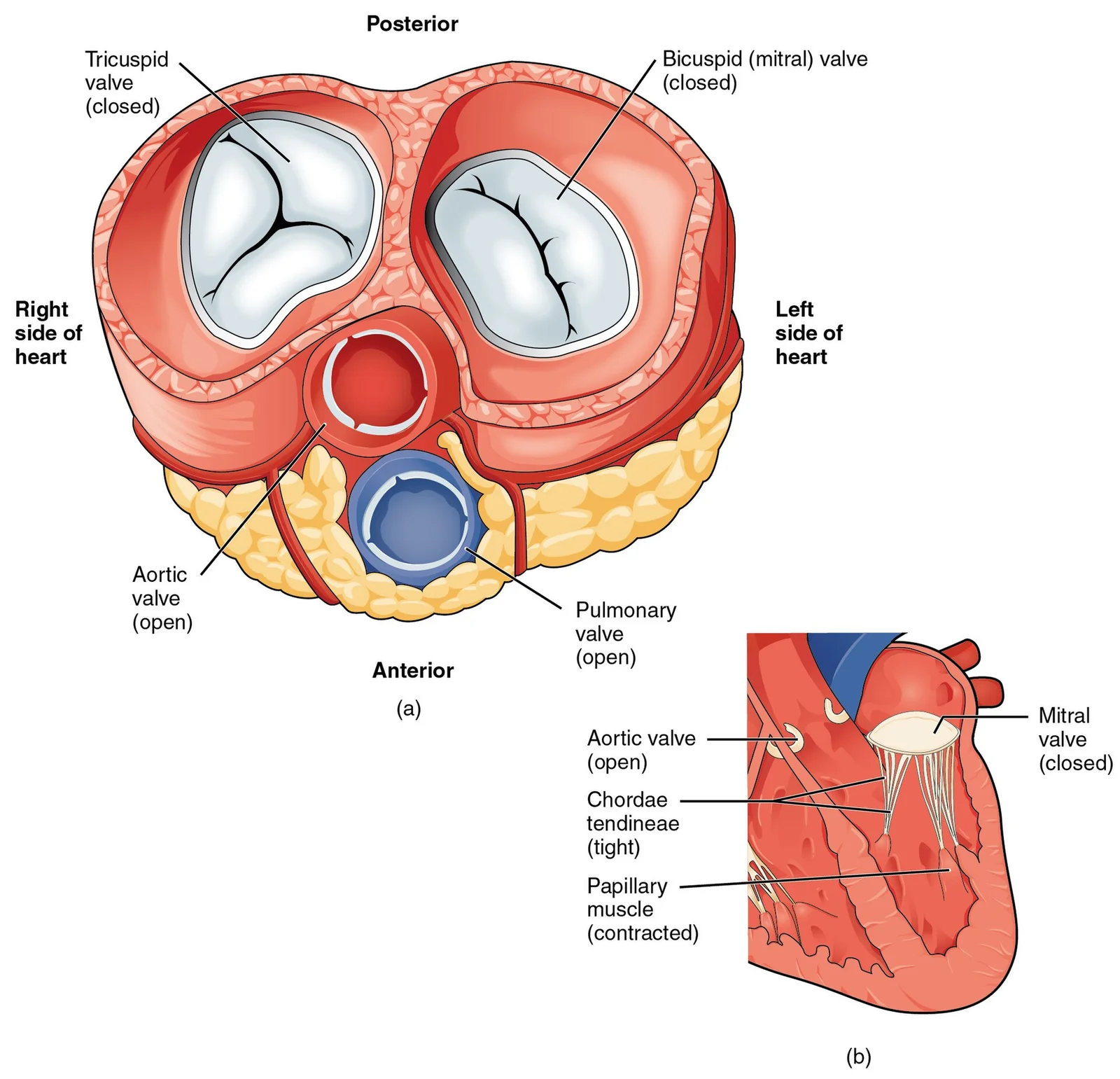
Figure 262013 Blood Flow Contracted Ventricles
Long description
(a) A transverse section through the heart illustrates the four heart valves during ventricular contraction. The two atrioventricular valves are closed, but the two semilunar valves are open. The atria and vessels have been removed. (b) A frontal view shows the closed mitral (bicuspid) valve that pr

Figure 26: (a) A transverse section through the heart illustrates the four heart valves during ventricular contraction.
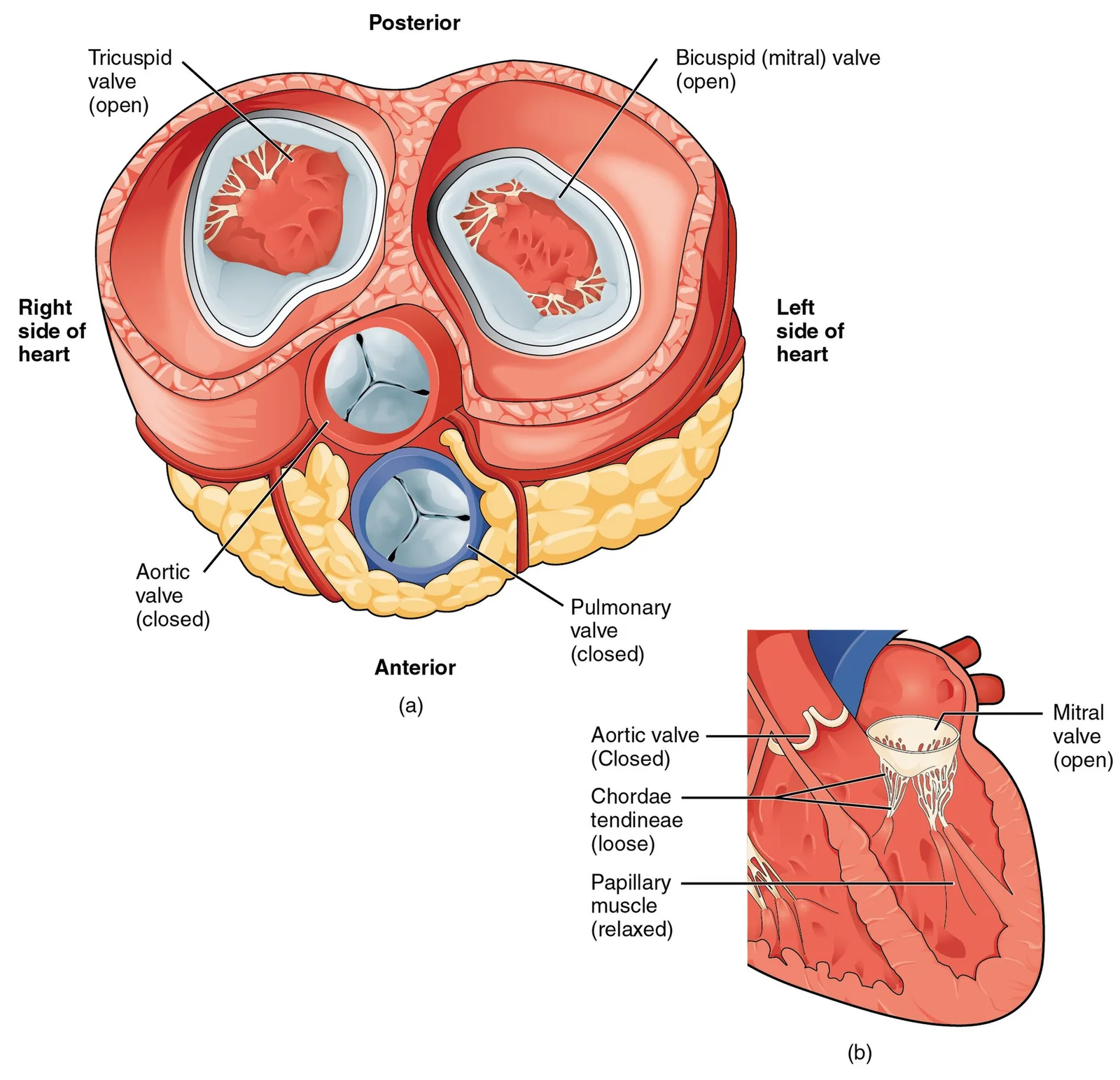
Figure 272012 Blood Flow Relaxed Ventricles
Long description
(a) A transverse section through the heart illustrates the four heart valves. The two atrioventricular valves are open; the two semilunar valves are closed. The atria and vessels have been removed. (b) A frontal section through the heart illustrates blood flow through the mitral valve. When the mitr

Figure 27: (a) A transverse section through the heart illustrates the four heart valves.
The coronary arteries supply the heart muscle with oxygenated blood.
The heart's electrical conduction system coordinates contractions. The sinoatrial node, located in the right atrium, initiates the electrical impulse. The impulse spreads through the atria and reaches the atrioventricular node, located at the junction of the atria and ventricles. From there, the impulse travels through the bundle of His and Purkinje fibers to cause ventricular contraction. [1]
Students frequently misinterpret the autonomic control of the heart rate, leading to significant errors in clinical reasoning and examination answers. The first major pitfall is assuming that the heart rate is controlled by a single dominant system at all times. In a healthy individual, the heart rate estimated at any given time represents the net effect of the neural output of the parasympathetic (vagus) nerves, which slow heart rate, and the sympathetic nerves, which accelerate it. At rest, both sympathetic and parasympathetic nerves are tonically active with the vagal effects dominant. Therefore, heart rate reflects the relative activity of the sympathetic and parasympathetic systems. Students must remember that the more important question is whether the relative balance is appropriate for the context the person is engaged in at any given moment. [3]
A second common error involves the interpretation of heart rate variability (HRV) spectral components, specifically the low-frequency (LF) band. In ambulatory 24-h HRV recordings, it has been suggested that the LF band also reflects sympathetic activity and the LF/HF ratio has been controversially reported as an assessment of the balance between sympathetic and parasympathetic activity. However, a number of researchers have challenged this perspective and have persuasively argued that in resting conditions, the LF band reflects baroreflex activity and not cardiac sympathetic innervation [3]
The third pitfall is the misunderstanding of the autonomic balance hypothesis. The autonomic balance hypothesis assumes that the sympathetic nervous system and parasympathetic nervous system competitively regulate sinoatrial node firing, where increased sympathetic nervous system activity is paired with decreased parasympathetic nervous system activity. While some orthostatic challenges can produce reciprocal changes in sympathetic nervous system activation and vagal withdrawal, psychological stressors can also result in independent changes in sympathetic nervous system or parasympathetic nervous system activity. It is now generally accepted that both branches of the autonomic nervous system can be simultaneously active. Therefore, the relationship between the sympathetic nervous system and parasympathetic nervous system in generating LF power appears to be complex, non-linear, and dependent upon the experimental manipulation employed. [3]
Exam trapLF/HF Ratio
The interpretation of the LF/HF ratio is controversial due to the issues regarding the LF band described above. It is often assumed that a low LF/HF ratio reflects greater parasympathetic activity relative to sympathetic activity due to energy conservation and engaging in “tend-and-befriend” behaviors. However, this ratio is often shifted due to reductions in LF power. [3]
The fourth mistake is confusing different HRV time-domain measures. The pNN50 is the percentage of adjacent NN intervals that differ from each other by more than 50 ms. It is correlated with the RMSSD and HF power. However, the RMSSD typically provides a better assessment of respiratory sinus arrhythmia (especially in older subjects) and most researchers prefer it to the pNN50. Using pNN50 as the primary marker for vagal tone is less accurate than using RMSSD. [3]
The fifth error relates to the SDNN index. The SDNN index is the mean of the standard deviations of all the NN intervals for each 5-min segment of a 24-h HRV recording. Therefore, this measurement only estimates variability due to the factors affecting HRV within a 5-min period. It is calculated by first dividing the 24-h record into 288 5-min segments and then calculating the standard deviation of all NN intervals contained within each segment. The SDNN Index is the average of these 288 values. The SDNN index is believed to primarily measure autonomic influence on HRV. This measure tends to correlate with VLF power over a 24-h period. Students often mistake SDNN for a pure measure of overall variability rather than a specific estimate of autonomic influence within short segments. [3]
DefinitionResonance Frequency
They suggest that when people breathe at this rate, which varies in adults from 4.5 to 6.5 breaths per minute, they “exercise” the baroreflex. They have shown that during this paced period, HR and BP oscillations are 180° out of phase, and HRV amplitude is maximized. [3]
The sixth pitfall involves the clinical application of breathing techniques. Resonance frequency breathing is typically used in the context of HRVB training. Several months of steady practice can reset the baroreflex gain so that it is sustained, even when clients are not receiving feedback. Increased baroreflex gain is analogous to a more sensitive thermostat, allowing the body to regulate BP and gas exchange more effectively. Students must understand that this is a trained physiological response, not an immediate reflex. [3]
NoteNeurocardiology
As discussed above, neurocardiology research has established that heart-brain interactions are remarkably complex. Patterns of baroreceptor afferent activity modulate CNS activity over time periods that range from milliseconds to minutes. The intrinsic cardiac ganglia demonstrate both short- and long-term memory. [3]
This knowledge is used at the bedside to assess autonomic tone and cardiovascular stability. Clinicians evaluate whether the heart rate is higher during the daytime and when dealing with challenging tasks, and lower at night, during sleep or when not engaged in challenging duties or activities. This contextual assessment helps determine if the autonomic response is appropriate. In the operating theatre, understanding that both branches of the autonomic nervous system can be simultaneously active is crucial for managing anaesthesia and surgical stress. Surgeons must recognise that psychological stressors can result in independent changes in sympathetic or parasympathetic activity, complicating the prediction of heart rate responses.
On imaging and in diagnostic testing, HRV analysis is employed to evaluate autonomic function. The SDNN index is calculated by dividing the 24-h record into 288 5-min segments and then calculating the standard deviation of all NN intervals contained within each segment. The SDNN Index is the average of these 288 values. The SDNN index is believed to primarily measure autonomic influence on HRV. This measure tends to correlate with VLF power over a 24-h period. In examinations, students are tested on the distinction between time-domain and frequency-domain measures. The HR Max – HR Min is the average difference between the highest and lowest HRs during each respiratory cycle. This measure is especially used for assessment in paced breathing protocols and is highly correlated with the SDNN and RMSSD. [3]
Autonomic Assessment
- Assess HR ContextIs HR appropriate for activity?
- Question: Appropriate?
Yes
- Monitor Resting Tone
No
- Evaluate Autonomic Balance
In examinations, candidates must correctly identify the components of HRV. The pNN50 is the percentage of adjacent NN intervals that differ from each other by more than 50 ms. It is correlated with the RMSSD and HF power. However, the RMSSD typically provides a better assessment of respiratory sinus arrhythmia (especially in older subjects) and most researchers prefer it to the pNN50. Understanding that the LF/HF ratio is controversial is also a key examination point. The interpretation of the LF/HF ratio is controversial due to the issues regarding the LF band described above. However, once the mechanisms are understood as well as the importance of the recording context, the controversy is resolved.
Clinical pearlBaroreflex Exercise
They suggest that when people breathe at this rate, which varies in adults from 4.5 to 6.5 breaths per minute, they “exercise” the baroreflex. They have shown that during this paced period, HR and BP oscillations are 180° out of phase, and HRV amplitude is maximized.
The clinical utility extends to understanding brain-heart interactions. Thayer et al. contend that dynamic connections between the amygdala and medial prefrontal cortex, which evaluate threat and safety, help regulate HRV through their connections with the NST. They propose that vagally-mediated HRV is linked to higher-level executive functions and that HRV reflects the functional capacity of the brain structures that support working memory and emotional and physiological self-regulation. They hypothesize that vagally-mediated HRV is positively correlated with prefrontal cortical performance and the ability to inhibit unwanted memories and intrusive thoughts. In their model, when the CAN decreases prefrontal cortical activation, HR increases and HRV decreases. The prefrontal cortex can be taken “offline” when individuals perceive that they are threatened. [3]
DefinitionIntrinsic Cardiac Ganglia
The intrinsic cardiac ganglia demonstrate both short- and long-term memory. This affects afferent activity rhythms produced by both mechanical variables (e.g., pressure and HR) that occur over milliseconds (single cycles) and hormonal variables that fluctuate over periods ranging from seconds to minutes. [3]
In practice, the ability to reset the baroreflex gain is utilised in therapeutic settings. Resonance frequency breathing is typically used in the context of HRVB training. Several months of steady practice can reset the baroreflex gain so that it is sustained, even when clients are not receiving feedback. Increased baroreflex gain is analogous to a more sensitive thermostat, allowing the body to regulate BP and gas exchange more effectively. This knowledge is tested in examinations regarding non-pharmacological interventions for hypertension and anxiety. Students must also recognise that HRV reflects the relative activity of the sympathetic and parasympathetic systems. Therefore, HR reflects the relative activity of the sympathetic and parasympathetic systems. With the more important question being, is the relative balance (HR) appropriate for the context the person is engaged in at any given moment. In other words, is HR higher during the daytime and when dealing with challenging tasks, and lower at night, during sleep or when not engaged in challenging duties or activities. [3]
- Heart rate is the net effect of parasympathetic slowing and sympathetic acceleration.
- At rest, vagal effects are dominant but both systems are tonically active.
- LF band in resting conditions reflects baroreflex activity, not just sympathetic tone.
- Both autonomic branches can be simultaneously active, challenging simple balance hypotheses.
- RMSSD is preferred over pNN50 for assessing respiratory sinus arrhythmia.
- SDNN index estimates autonomic influence within 5-min segments of 24-h recordings.
- Resonance breathing (4.5-6.5 breaths/min) maximises HRV amplitude.
- Vagal HRV correlates with prefrontal cortical performance and self-regulation.
Answer each one in your head first, then open the answer.
What is the dominant autonomic influence on heart rate at rest?
Show answerHide answer
Vagal effects are dominantWhat does the LF band reflect in resting conditions?
Show answerHide answer
Baroreflex activity and not cardiac sympathetic innervationWhich measure is preferred over pNN50 for RSA?
Show answerHide answer
RMSSDHow many 5-min segments are used in SDNN index calculation?
Show answerHide answer
288What is the typical adult resonance breathing rate?
Show answerHide answer
4.5 to 6.5 breaths per minute
- Betts JG, Young KA, Wise JA, et al. Heart Anatomy. OpenStax, Anatomy and Physiology 2e (CC BY-NC-SA 4.0). Accessed .
- Betts JG, Young KA, Wise JA, et al. Development of the Heart. OpenStax, Anatomy and Physiology 2e (CC BY-NC-SA 4.0). Accessed .
- Shaffer F, McCraty R, Zerr CL. A healthy heart is not a metronome: an integrative review of the heart's anatomy and heart rate variability. Frontiers in psychology 2014;5:1040. doi:10.3389/fpsyg.2014.01040. Accessed .
Figure credits
- Figure 1: Henry Vandyke Carter, via Wikimedia Commons, Public domain, Public domain.
- Figure 2: OpenStax College, via Wikimedia Commons, CC BY 3.0, CC BY 3.0. resized.
- Figure 3: OpenStax College, via Wikimedia Commons, CC BY 3.0, CC BY 3.0. resized.
- Figure 4: Henry Vandyke Carter, via Wikimedia Commons, Public domain, Public domain.
- Figure 5: OpenStax College, via Wikimedia Commons, CC BY 3.0, CC BY 3.0. resized.
- Figure 6: Henry Vandyke Carter, via Wikimedia Commons, Public domain, Public domain.
- Figure 7: OpenStax College, via Wikimedia Commons, CC BY 3.0, CC BY 3.0. resized.
- Figure 8: This file has source information, but it either links directly to the file or is a generic base URL, or is not an Internet source for a file that was likely found on the Internet. Source information should be provided so that the copyright status can be verified by others. It is requested that a better source be provided to make determination of the copyright information easier. Please provide a URL to an HTML page that contains this file. See Commons:Licensing#License_information for more information. Please remove this template once better source information is provided. Notify the uploader with:, via Wikimedia Commons, Public domain, Public domain.
- Figure 9: Henry Vandyke Carter, via Wikimedia Commons, Public domain, Public domain.
- Figure 10: Henry Vandyke Carter, via Wikimedia Commons, Public domain, Public domain.
- Figure 11: OpenStax College, via Wikimedia Commons, CC BY 3.0, CC BY 3.0.
- Figure 12: OpenStax College, via Wikimedia Commons, CC BY 3.0, CC BY 3.0.
- Figure 13: Henry Vandyke Carter, via Wikimedia Commons, Public domain, Public domain.
- Figure 14: Henry Vandyke Carter, via Wikimedia Commons, Public domain, Public domain.
- Figure 15: Henry Vandyke Carter, via Wikimedia Commons, Public domain, Public domain.
- Figure 16: Henry Vandyke Carter, via Wikimedia Commons, Public domain, Public domain.
- Figure 17: Henry Vandyke Carter, via Wikimedia Commons, Public domain, Public domain.
- Figure 18: Henry Vandyke Carter, via Wikimedia Commons, Public domain, Public domain.
- Figure 19: Henry Vandyke Carter, via Wikimedia Commons, Public domain, Public domain.
- Figure 20: Henry Vandyke Carter, via Wikimedia Commons, Public domain, Public domain.
- Figure 21: Henry Vandyke Carter, via Wikimedia Commons, Public domain, Public domain.
- Figure 22: OpenStax College, via Wikimedia Commons, CC BY 3.0, CC BY 3.0. resized.
- Figure 23: OpenStax College, via Wikimedia Commons, CC BY 3.0, CC BY 3.0. resized.
- Figure 24: Patrick J. Lynch, medical illustrator, via Wikimedia Commons, CC BY 2.5, CC BY 2.5. resized.
- Figure 25: Ify Mordi and Nikolaos Tzemos, via Wikimedia Commons, CC BY 3.0, CC BY 3.0.
- Figure 26: OpenStax College, via Wikimedia Commons, CC BY 3.0, CC BY 3.0. resized.
- Figure 27: OpenStax College, via Wikimedia Commons, CC BY 3.0, CC BY 3.0. resized.
Educational content for medical students and doctors. It is not a substitute for clinical judgement or local guidelines.