Pericardium
The pericardium is a fibroelastic sac enclosing the heart and the roots of the great vessels within the thoracic cavity.
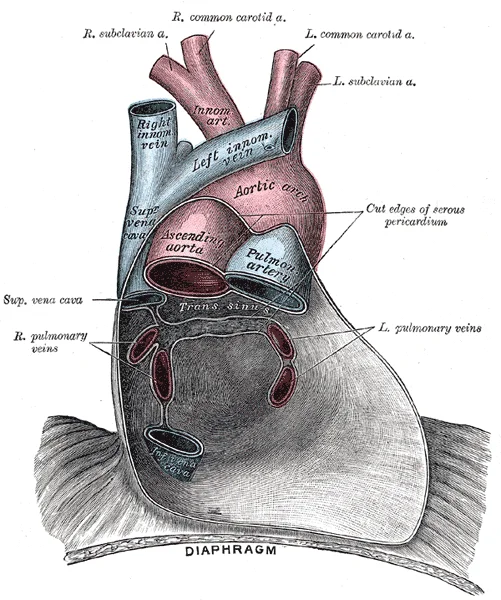
Figure 1Gray489
Long description
Posterior wall of the pericardial sac, showing the lines of reflection of the serous pericardium on the great vessels.

Figure 1: Posterior wall of the pericardial sac, showing the lines of reflection of the serous pericardium on the great vessels.
The pericardium is the membrane that directly surrounds the heart and defines the pericardial cavity. It consists of two distinct sublayers: the sturdy outer fibrous pericardium and the inner serous pericardium. The fibrous pericardium is composed of dense connective tissue that protects the heart and maintains its position within the thorax. The serous pericardium is divided into the parietal pericardium, which is fused to the fibrous layer, and the visceral pericardium, or epicardium, which is fused to the heart wall. The pericardial cavity, containing lubricating serous fluid, lies between the epicardium and the parietal pericardium.
DefinitionPericardial Sinuses
The pericardial sinuses are specific reflections of the serous pericardium around the great vessels, creating the transverse and oblique sinuses.
Pericardial effusion refers to the accumulation of fluid within the pericardial sac. While physiologic fluid levels range from 25 to 50 mL, volumes exceeding 50 mL result in an echo-free space persisting throughout the cardiac cycle. Small effusions initially appear over the posterobasal left ventricle, but as volume increases, the fluid spreads anteriorly, laterally, and behind the left atrium. Large effusions may lead to a "swinging heart" appearance. [2]
| Condition | Mechanism | Clinical Consequence |
|---|---|---|
| Pericardial Effusion | Fluid accumulation | Potential for tamponade |
| Cardiac Tamponade | High intrapericardial pressure | Cardiogenic shock |
Cardiac tamponade is a life-threatening compression of the heart caused by the accumulation of fluid, pus, blood, or gas. When the pericardium reaches its stretch limit, ongoing fluid accumulation increases intrapericardial pressure, which impedes cardiac filling and impairs function. This state results in cardiogenic shock and circulatory collapse. Clinicians must correlate echocardiographic findings, such as right ventricular or atrial collapse and IVC plethora, with clinical signs like hypotension or pulsus paradoxus. While large effusions are often associated with tamponade, tamponade can develop without a large effusion or swinging heart if the fluid accumulation is rapid. Pericardiocentesis is indicated when the effusion is large, symptomatic, or accompanied by tamponade. [2]
Management of Pericardial Effusion
- Detect effusionUse echocardiography
- Question: Is tamponade present?
Yes
- PericardiocentesisEmergency intervention
No
- Assess size and etiologyMonitor clinically
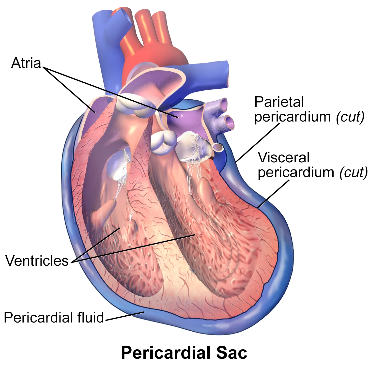
Figure 2Blausen 0724 PericardialSac
Long description
Pericardial Sac. See a full animation of this medical topic.

Figure 2: Pericardial Sac.
The heart is separated from other mediastinal structures by a tough membrane known as the pericardium, or pericardial sac, and sits in its own space called the pericardial cavity[1]. The dorsal surface of the heart lies near the bodies of the vertebrae, and its anterior surface sits deep to the sternum and costal cartilages[1]. The great veins, the superior and inferior venae cavae, and the great arteries, the aorta and pulmonary trunk, are attached to the superior surface of the heart, called the base[1].
DefinitionPericardium
The pericardium is a tough membrane that separates the heart from other mediastinal structures, enclosing it in the pericardial cavity[1].
The pericardial sac consists of two fused layers: an outer fibrous capsule and an inner parietal pericardium lined with a serous membrane[1]. Between the pericardial sac and the heart is the pericardial cavity, which is filled with lubricating serous fluid[1]. This fluid is secreted by the mesothelium, a simple squamous epithelium that reinforces the visceral serous membrane, or epicardium[1].
Inside the pericardium, the surface features of the heart are visible, including the four chambers[1]. A superficial leaf-like extension of the atria near the superior surface of the heart, one on each side, is called an auricle[1]. Auricles are relatively thin-walled structures that can fill with blood and empty into the atria or upper chambers of the heart[1]. You may also hear them referred to as atrial appendages[1].
Also prominent is a series of fat-filled grooves, each of which is known as a sulcus (plural = sulci), along the superior surfaces of the heart[1]. Major coronary blood vessels are located in these sulci[1]. The deep coronary sulcus is located between the atria and ventricles[1]. Located between the left and right ventricles are two additional sulci that are not as deep as the coronary sulcus[1].
The wall of the heart is composed of three layers of unequal thickness[1]. From superficial to deep, these are the epicardium, the myocardium, and the endocardium[1]. The outermost layer of the wall of the heart is also the innermost layer of the pericardium, the epicardium, or the visceral pericardium discussed earlier[1]. The human heart consists of a pair of atria, which receive blood and pump it into a pair of ventricles, which pump blood into the vessels[1]. The right atrium receives systemic blood relatively low in oxygen and pumps it into the right ventricle, which pumps it into the pulmonary circuit[1].
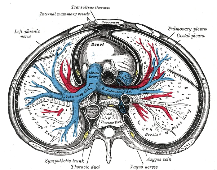
Figure 3Gray968
Long description
A transverse section of the thorax, showing the contents of the middle and the posterior mediastinum. The pleural and pericardial cavities are exaggerated since normally there is no space between parietal and visceral pleura and between pericardium and heart.

Figure 3: A transverse section of the thorax, showing the contents of the middle and the posterior mediastinum.
The heart is situated between the lungs, enclosed within the pericardial sac. The anterior surface faces the sternum and costal cartilages, while the posterior surface rests against the vertebral column and oesophagus. The inferior surface, or diaphragmatic surface, lies upon the central tendon of the diaphragm. The apex points inferiorly, anteriorly, and to the left. The base faces posteriorly, superiorly, and to the right. The pericardium consists of two layers: the fibrous pericardium, which is the tough outer layer, and the serous pericardium, which is divided into parietal and visceral layers. The parietal layer lines the fibrous pericardium, while the visceral layer, also known as the epicardium, adheres directly to the heart muscle. The pericardial cavity, containing a small amount of serous fluid, lies between these two layers. The heart is deflected such that the right side is anterior and the left side is posterior. The apex lies just to the left of the sternum between the junction of the fourth and fifth ribs near their articulation with the costal cartilages. This slight deviation creates a depression in the medial surface of the superior lobe of the left lung, called the cardiac notch. The shape of the heart is similar to a pinecone, rather broad at the superior surface and tapering to the apex. A typical heart is approximately the size of your fist: 12 cm (5 in) in length, 8 cm (3.5 in) wide, and 6 cm (2.5 in) in thickness. The human heart consists of four chambers: The left side and the right side each have one atrium and one ventricle. Each of the upper chambers, the right atrium (plural = atria) and the left atrium, acts as a receiving chamber and contracts to push blood into the lower chambers, the right ventricle and the left ventricle. The ventricles serve as the primary pumping chambers of the heart, propelling blood to the lungs or to the rest of the body. The middle and thickest layer is the myocardium, made largely of cardiac muscle cells. It is built upon a framework of collagenous fibers, plus the blood vessels that supply the myocardium and the nerve fibers that help regulate the heart. It is the contraction of the myocardium that pumps blood through the heart and into the major arteries. The muscle pattern is elegant and complex, as the muscle cells swirl and spiral around the chambers of the heart. More superficial layers of ventricular muscle wrap around both ventricles. This complex swirling pattern allows the heart to pump blood more effectively than a simple linear pattern would. The word septum is derived from the Latin for “something that encloses;” in this case, a septum (plural = septa) refers to a wall or partition that divides the heart into chambers. The septa are physical extensions of the myocardium lined with endocardium. Located between the two atria is the interatrial septum. Normally in an adult heart, the interatrial septum bears an oval-shaped depression known as the fossa ovalis, a remnant of an opening in the fetal heart known as the foramen ovale. The foramen ovale allowed blood in the fetal heart to pass directly from the right atrium to the left atrium, allowing some blood to bypass the pulmonary circuit. Within seconds after birth, a flap of tissue known as the septum primum that previously acted as a valve closes the foramen ovale and establishes the typical cardiac circulation pattern. Since these openings and valves structurally weaken the atrioventricular septum, the remaining tissue is heavily reinforced with dense connective tissue called the cardiac skeleton, or skeleton of the heart. It includes four rings that surround the openings between the atria and ventricles, and the openings to the pulmonary trunk and aorta, and serve as the point of attachment for the heart valves. The cardiac skeleton also provides an important boundary in the heart electrical conduction system. A transverse section through the heart slightly above the level of the atrioventricular septum reveals all four heart valves along the same plane. The valves ensure unidirectional blood flow through the heart. Between the right atrium and the right ventricle is the right atrioventricular valve, or tricuspid valve. It typically consists of three flaps, or leaflets, made of endocardium reinforced with additional connective tissue. The flaps are connected by chordae tendineae to the papillary muscles, which control the opening and closing of the valves. [1]
DefinitionPericardium
The pericardium is a double-walled sac that contains the heart and the roots of the great vessels, consisting of the fibrous pericardium and the serous pericardium.
| Classification | Diastolic Separation | Fluid Volume |
|---|---|---|
| Minimal | < 5 mm | 50–100 mL |
| Small | 5–10 mm | 100–250 mL |
| Moderate | 10–20 mm | 250–500 mL |
| Large | > 20 mm | > 500 mL |
For circumferential pericardial effusions, any pericardial effusion with less than 5 mm of pericardial separation in diastole (corresponding to a fluid volume of 50 to 100 mL) is defined as minimal; 5 to 10 mm of separation as small (corresponding to a fluid volume of 100 to 250 mL); 10 to 20 mm of separation as moderate (corresponding to a fluid volume of 250 to 500 mL); and greater than 20 mm separation as large (corresponding to a fluid volume greater than 500 mL)[1]. This classification may be useful in daily clinical practice[1]. Percutaneous needle pericardiocentesis has been the most useful therapeutic procedure for the early management of large, symptomatic pericardial effusion or cardiac tamponade, and it continues to be used as a diagnostic procedure in some patients with asymptomatic pericardial effusion, such as chronic idiopathic effusion[1]. Before, during, and after the percutaneous pericardiocentesis, transthoracic echocardiography guidance is essential[1]. If the pericardiocentesis is performed at bedside without echocardiographic guidance, the risk of threatening complications like bleeding or shock have been reported to be as high as 20%[1].
The internal architecture of the heart is defined by the precise arrangement of chambers, septa, and valves that facilitate efficient circulation. To understand the pericardium’s protective role, one must first appreciate the complex internal environment it encloses. The heart operates via a dual circulation pattern, with pairs of chambers pumping blood into the pulmonary and systemic circuits. This functional requirement drives the development of specialized internal structures designed to ensure unidirectional flow and structural integrity. [1]
The heart is divided into four chambers by muscular walls known as septa. The most critical of these for understanding internal flow is the atrioventricular septum, which separates the atria from the ventricles. This septum is characterised by four distinct openings. Two of these openings allow blood to move from the atria into the ventricles, while the other two permit flow from the ventricles into the pulmonary trunk and aorta. The interventricular septum, visible in standard anatomical views, separates the left and right ventricles. The interatrial septum is often obscured in anterior views because its location is covered by the aorta and pulmonary trunk. [1]
Located within the openings of the atrioventricular septum are specialized structures called valves, which ensure one-way blood flow. The valves between the atria and ventricles are generically known as atrioventricular valves. Specifically, the right side features the tricuspid valve, while the left side has the bicuspid (mitral) valve. At the exits of the ventricles, leading to the pulmonary trunk and aorta, are the semilunar valves. These valves prevent backflow during ventricular relaxation, maintaining the efficiency of the cardiac cycle. [1]
The internal surfaces of the atria differ significantly. The bulk of the right atrium’s internal surface is smooth, except for the depression of the fossa ovalis, which lies medially. The anterior surface of the right atrium demonstrates prominent ridges of muscle called pectinate muscles. These muscles are also present in the right auricle. In contrast, the left atrium lacks pectinate muscles except within its auricle. Immediately superior and slightly medial to the opening of the inferior vena cava on the posterior surface is the opening of the coronary sinus. This thin-walled vessel drains most coronary veins, returning systemic blood from the heart. [1]
The ventricular walls are lined with trabeculae carneae, which are ridges of cardiac muscle covered by endocardium. In the right ventricle, a band of cardiac muscle known as the moderator band reinforces the thin walls. This band arises from the inferior portion of the interventricular septum and crosses the interior space to connect with the inferior papillary muscle. It plays a crucial role in cardiac conduction. [1]
The heart is a remarkable pump composed largely of cardiac muscle cells that are incredibly active throughout life. Like all other cells, a cardiomyocyte requires a reliable supply of oxygen and nutrients, and a way to remove wastes, so it needs a dedicated, complex, and extensive coronary circulation. And because of the critical and nearly ceaseless activity of the heart throughout life, this need for a blood supply is even greater than for a typical cell. However, coronary circulation is not continuous; rather, it cycles, reaching a peak when the heart muscle is relaxed and nearly ceasing while it is contracting. [1]
Coronary arteries supply blood to the myocardium and other components of the heart. The first portion of the aorta after it arises from the left ventricle gives rise to the coronary arteries. There are three dilations in the wall of the aorta just superior to the aortic semilunar valve. The third sinus, the right posterior aortic sinus, typically does not give rise to a vessel. [1]
The right coronary artery proceeds along the coronary sulcus and distributes blood to the right atrium, portions of both ventricles, and the heart conduction system. Normally, one or more marginal arteries arise from the right coronary artery inferior to the right atrium. The marginal arteries supply blood to the superficial portions of the right ventricle. On the posterior surface of the heart, the right coronary artery gives rise to the posterior interventricular artery, also known as the posterior descending artery. It runs along the posterior portion of the interventricular sulcus toward the apex of the heart, giving rise to branches that supply the interventricular septum and portions of both ventricles.
The right coronary artery is a key structure in this supply. It arises from the anterior aortic sinus. It courses along the coronary sulcus. It gives off marginal arteries inferior to the right atrium. These marginal arteries supply the superficial portions of the right ventricle. It gives rise to the posterior interventricular artery on the posterior surface. This artery is also known as the posterior descending artery. It runs along the posterior portion of the interventricular sulcus. It moves toward the apex of the heart. It gives rise to branches that supply the interventricular septum. These branches also supply portions of both ventricles.
It supplies the left atrium and left ventricle. It gives off the anterior interventricular artery. This artery runs along the anterior portion of the interventricular sulcus. It supplies the anterior portion of the interventricular septum. It supplies portions of both ventricles.
| Structure | Origin | Course | Supply |
|---|---|---|---|
| Marginal Arteries | Right coronary artery | Inferior to right atrium | Superficial portions of right ventricle |
| Posterior Interventricular Artery | Right coronary artery | Posterior interventricular sulcus | Interventricular septum, portions of both ventricles |
The pericardium itself receives its blood supply from these coronary branches. The fibrous pericardium is supplied by pericardiophrenic arteries. The phrenic nerves accompany these vessels. They provide sensory innervation to the fibrous pericardium. The visceral pericardium is supplied by the coronary arteries. These arteries run in the epicardial fat. They follow the sulci of the heart. They are epicardial coronary arteries.
The right coronary artery distributes blood to the right atrium. It distributes blood to portions of both ventricles. It distributes blood to the heart conduction system. The posterior interventricular artery runs toward the apex. It gives rise to branches that supply the interventricular septum. These branches supply portions of both ventricles. The figure presents views of the coronary circulation from both the anterior and posterior views.
The pericardium requires a dedicated venous outflow to manage the continuous return of blood from the surrounding tissues. This drainage system operates in parallel with the cardiac chambers, ensuring that fluid accumulation does not impede the heart's mechanical function. The venous architecture is not merely a passive conduit but an integral part of the pericardial physiology, closely mirroring the arterial supply.
The primary route for pericardial venous drainage involves the pericardiophrenic veins. These vessels accompany the pericardiophrenic artery and the phrenic nerve. They drain the fibrous pericardium and the adjacent parietal pleura. The superior portions of these veins empty into the brachiocephalic veins. This connection ensures that blood from the upper pericardial region returns directly to the superior vena cava system. The inferior portions of the pericardiophrenic veins drain into the azygos vein or the hemiazygos vein.
In addition to the pericardiophrenic veins, the muscularis pericardii contributes to drainage. These connections provide an alternative route for venous return, particularly from the anterior and inferior aspects of the pericardium. The anastomoses between these systems ensure redundancy, preventing congestion even if one pathway is compromised.
The clinical significance of this drainage pattern lies in its relationship with central venous pressure. Elevated central venous pressure, as seen in conditions like cardiac tamponade, can impede pericardial venous outflow. This obstruction leads to fluid accumulation within the pericardial space. The pericardium's ability to accommodate this fluid depends on the compliance of the fibrous layer and the efficiency of the venous drainage. Understanding this anatomy is crucial for interpreting signs of venous congestion, such as jugular venous distension, which reflects the backup of pressure into the systemic veins.
DefinitionPericardiophrenic Veins
Veins accompanying the pericardiophrenic artery and phrenic nerve, draining the fibrous pericardium into the brachiocephalic and azygos systems.
| Structure | Course | Termination |
|---|---|---|
| Pericardiophrenic Veins | Accompany artery and nerve | Brachiocephalic (superior), Azygos (inferior) |
| Muscularis Pericardii | Small channels in muscular layer | Internal thoracic, Superior epigastric |
The lymphatic drainage of the pericardium is a critical anatomical pathway that facilitates the removal of interstitial fluid and immune surveillance within the fibrous and serous layers. Understanding this drainage is essential for comprehending the spread of pathology, such as metastatic disease or infectious processes, from the heart to regional lymph nodes.
The fibrous pericardium, the outermost tough layer, drains primarily into the anterior mediastinal lymph nodes. These nodes are situated in the anterior mediastinum, anterior to the pericardium and posterior to the sternum. From the anterior mediastinal nodes, lymph flows into the tracheobronchial nodes. This pathway ensures that any inflammatory or neoplastic cells from the fibrous layer are filtered before entering the systemic circulation via the thoracic duct or right lymphatic duct.
The serous pericardium, comprising the parietal and visceral layers, has a more complex drainage pattern. The parietal layer, lining the fibrous pericardium, drains similarly to the fibrous layer, contributing to the anterior mediastinal and tracheobronchial nodes. This distinction is clinically significant because diseases affecting the epicardium may present with different nodal involvement compared to those affecting the pericardial sac itself. The lymphatic capillaries in the visceral layer are numerous and permeable, allowing for efficient fluid absorption from the myocardial surface.
Disruption or obstruction of these lymphatic pathways can lead to pericardial effusion, where fluid accumulates in the pericardial cavity. This condition can compromise cardiac function by restricting ventricular filling, a phenomenon known as cardiac tamponade. Recognising the anatomical routes of drainage helps clinicians predict the spread of malignancies, such as lung cancer or breast cancer, which may metastasize to the pericardium via these lymphatic channels. Additionally, infections like tuberculosis can spread to the pericardium through lymphatic dissemination from adjacent mediastinal nodes.
The pericardium receives its innervation from both somatic and autonomic sources, a dual supply that explains the complex nature of pericardial pain. The fibrous pericardium and the parietal layer of the serous pericardium are sensitive to pain, whereas the visceral layer (epicardium) is insensitive. This distinction is crucial for understanding clinical presentations.
The somatic innervation is provided by the phrenic nerves. These nerves arise from the cervical spinal roots C3, C4, and C5. They descend through the thorax, passing anterior to the root of the lung and posterior to the superior vena cava. The phrenic nerves provide sensory fibres to the fibrous pericardium and the parietal layer of the serous pericardium. Because these nerves originate from the cervical spine, irritation of the pericardium can result in referred pain to the shoulder region, specifically the supraclavicular area, which shares the same dermatomal origin. This phenomenon is known as referred pain.
The autonomic innervation is more complex, involving both sympathetic and parasympathetic fibres. Sympathetic fibres arise from the thoracic sympathetic trunk. These fibres travel to the cardiac plexus and then distribute to the pericardium. They are primarily vasomotor and secretomotor, regulating blood flow and glandular secretion within the pericardial tissues. Parasympathetic fibres are derived from the vagus nerves (cranial nerve X). These fibres also contribute to the cardiac plexus before reaching the pericardium. The autonomic nerves are largely concerned with the regulation of the pericardial vessels and smooth muscle elements, rather than direct sensation.
The viewer should note that the nerves are embedded in the epicardial fat and lie on the surface of the parietal pericardium. The labels identify the phrenic nerve, the fibrous pericardium, and the adjacent great vessels. Understanding this spatial relationship is vital for surgical procedures to avoid iatrogenic nerve damage.
Clinical pearlReferred Pain
Pain from the parietal pericardium is referred to the shoulder due to shared cervical spinal roots (C3-C5) with the phrenic nerve.
The clinical significance of this nerve supply is evident in pericarditis. Inflammation of the parietal pericardium stimulates the somatic sensory fibres of the phrenic nerve. The patient experiences sharp, pleuritic chest pain that may radiate to the trapezius ridge. This radiation is a classic sign of pericardial irritation. In contrast, visceral pericardial inflammation does not cause pain because it lacks somatic innervation.
| Structure | Innervation | Type |
|---|---|---|
| Fibrous Pericardium | Phrenic Nerves | Somatic |
| Parietal Serous Pericardium | Phrenic Nerves | Somatic |
| Visceral Serous Pericardium | Autonomic Nerves | Autonomic |
The autonomic fibres, while not directly painful, modulate the physiological state of the pericardium. Sympathetic stimulation can cause vasoconstriction of the pericardial vessels. Parasympathetic stimulation has the opposite effect. These autonomic influences are subtle but important for maintaining pericardial homeostasis.
The pericardium is a fibroelastic sac that encloses the heart and the roots of the great vessels. It consists of two primary layers: the fibrous pericardium and the serous pericardium. The fibrous pericardium is the tough, outer connective tissue layer that anchors the heart to the diaphragm and the sternum, preventing over-distension. The serous pericardium is further divided into the parietal layer, which lines the inner surface of the fibrous pericardium, and the visceral layer (epicardium), which adheres directly to the myocardium. Between these two serous layers lies the pericardial cavity, a potential space containing a thin film of serous fluid that reduces friction during cardiac contraction.
DefinitionPericardial Layers
The pericardium is a double-walled sac comprising an outer fibrous layer and an inner serous layer, the latter of which is subdivided into parietal and visceral components.
The histology of the pericardium is essential for understanding its role in cardiac protection and lubrication. For a detailed microscopic breakdown of these tissue layers, please refer to the histology page.
- Fibrous pericardium: Composed of dense, irregular connective tissue providing structural rigidity.
- Serous pericardium: A mesothelial lining that secretes lubricating fluid.
- Epicardium: The visceral layer containing coronary vessels and adipose tissue.
| Layer | Composition | Function |
|---|---|---|
| Fibrous | Dense connective tissue | Anchoring and protection |
| Parietal | Mesothelium | Lining of fibrous sac |
| Visceral | Mesothelium and fat | Epicardial surface |
- The pericardial cavity normally contains 15 to 50 ml of serous fluid.
- The fibrous pericardium is innervated by the phrenic nerves, which also provide sensory innervation to the parietal pericardium.
- The visceral pericardium is insensitive to pain, unlike the parietal layer.
- The right and left coronary arteries are the first to branch off the aorta and arise from two of the three sinuses located near the base of the aorta and are generally located in the sulci. [1]
- Cardiac veins parallel the small cardiac arteries and generally drain into the coronary sinus. [1]
Pericardial layers
- Fibrous PericardiumOuter protective layer
- Parietal SerousInner lining of fibrous sac
- Pericardial CavityFluid-filled space
- Visceral SerousEpicardium on myocardium
The pericardium develops from the splanchnic and somatic mesoderm, forming a double-layered sac that encloses the heart. Initially, the heart tube forms within the pericardial cavity, which is lined by mesothelium. As the heart undergoes looping and septation, the pericardial cavity expands to accommodate the growing cardiac structures.
A critical aspect of cardiac development involves the formation of the septa that divide the heart into four chambers. This structural integrity is essential for maintaining the separation of oxygenated and deoxygenated blood in the systemic and pulmonary circulations.
The pericardial cavity itself forms as a potential space between the parietal and visceral layers of the serous pericardium. This space contains a small amount of serous fluid, which reduces friction during cardiac contractions. The development of the pericardium is closely linked to the rotation and positioning of the heart within the thoracic cavity. Any abnormalities in this developmental process can lead to congenital heart defects or pericardial diseases. For instance, failure of the septa to close properly can result in septal defects, allowing abnormal shunting of blood between chambers.
Understanding the embryological origins of the pericardium is crucial for appreciating its anatomical relationships and clinical significance. The fibrous pericardium provides protection and prevents overdistension of the heart, while the serous pericardium facilitates smooth movement. The interventricular septum, being a robust structure, plays a vital role in maintaining the high-pressure gradients necessary for effective cardiac output. Its thickness and integrity are direct consequences of the hemodynamic demands placed on the ventricles during fetal and postnatal life.
The pericardium encloses the heart, forming a protective fibrous sac. The right atrium forms the right border of the cardiac silhouette. It serves as the receiving chamber for blood returning from the systemic circulation. The superior and inferior venae cavae empty into the superior and posterior portions of the right atrium. The left atrium is centrally located and forms the majority of the posterior heart border. It receives oxygenated blood from the lungs via four pulmonary veins. The left ventricle forms the apex and the left border of the heart. Its muscular wall is significantly thicker than that of the right ventricle to generate higher pressure for systemic circulation. The right ventricle is anterior and forms the majority of the sternocostal surface. It pumps deoxygenated blood to the lungs. It drains into the coronary sinus, which lies in the atrioventricular sulcus on the posterior surface. Understanding these relations is crucial for interpreting radiological images. The heart's position is central, slightly tilted to the left. The aorta and pulmonary trunk emerge from the superior aspect. This volume ensures adequate perfusion of tissues. Radiological anatomy helps distinguish physiological adaptation from pathology. The pericardium's fibrous layer is dense and inelastic. The serous layer has parietal and visceral layers. The pericardial cavity contains a small amount of lubricating fluid.
The pericardium is a fibrous sac enclosing the heart, critical for limiting cardiac dilation and preventing infection spread. Its clinical significance arises when fluid accumulates, causing tamponade, or when adjacent structures are compressed. Understanding the spatial relationships is vital for interventions such as pericardiocentesis.
Pericardiocentesis is the needle aspiration of pericardial fluid, indicated in cardiac tamponade or diagnostic evaluation. These factors include the amount and location of the effusion, hemodynamics on echocardiography, sufficient margins of the echo free space so as to prevent laceration, clinical indication and urgency, underlying etiologies, and bleeding tendency.
The needle enters between the xiphisternum and left costal margin, directed towards the left shoulder. This path avoids the lung and traverses the diaphragm to reach the pericardial space.
Ortner syndrome, or cardiovocal syndrome, involves hoarseness due to left recurrent laryngeal nerve palsy. This nerve loops under the aortic arch, adjacent to the pericardium. Enlargement of the left atrium or pericardial effusion can compress the nerve.
DefinitionOrtner Syndrome
Hoarseness caused by compression of the left recurrent laryngeal nerve by cardiovascular structures, often the left atrium or a dilated pulmonary artery.
| Approach | Landmark | Angle/Direction | Risk |
|---|---|---|---|
| Left Parasternal | Left 5th/6th ICS | Close to sternum | Pneumothorax |
| Subxyphoid | Xiphisternum/Costal margin | 15-30 degrees to skin | Liver injury |
- The pericardium is a fibroelastic sac consisting of an outer fibrous layer and an inner serous layer. * The serous pericardium is divided into a parietal layer lining the fibrous sac and a visceral layer (epicardium) covering the heart. * The pericardial space normally contains a small amount of serous fluid to reduce friction during cardiac contraction. * Cardiac tamponade is a clinical emergency where fluid accumulation restricts diastolic filling.
DefinitionPericardial space
The potential space between the parietal and visceral layers of the serous pericardium, which may become a pathological space during effusion.
| Structure | Clinical significance |
|---|---|
| Fibrous pericardium | Provides structural support and prevents overfilling |
| Serous pericardium | Secretes lubricating fluid for the heart |
| Pericardial effusion | Abnormal fluid accumulation requiring measurement |
| Right atrium | Site of early diastolic collapse in tamponade |
Assessment of Pericardial Effusion
- Clinical suspicionSigns of tamponade
- Question: Perform Echocardiogram
Effusion present
- Measure dimensionAnnotate location and size
No effusion
- Consider other causesExclude non-pericardial pathology
Students often mistake the pericardium for a simple, inert sac, failing to appreciate its role in restricting cardiac over-distension. A common error is assuming the pericardial space contains a large volume of fluid; in health, it contains only a small amount of serous fluid to facilitate friction-free cardiac movement. Another frequent pitfall is confusing the fibrous pericardium with the serous pericardium. The fibrous pericardium is the tough, outer connective tissue layer that anchors the heart to the diaphragm and great vessels, whereas the serous pericardium consists of a parietal layer lining the fibrous sac and a visceral layer (epicardium) adherent to the heart.
Many students incorrectly assume that cardiac tamponade is an "all-or-none" phenomenon. Instead, it must be understood as a spectrum of findings. Furthermore, learners often overlook that echocardiography has limitations; for instance, severe hypovolemia or large pleural effusions can cause diastolic chamber collapse, potentially mimicking tamponade. Conversely, conditions that increase right-sided cardiac pressures, such as pulmonary hypertension or pulmonary thromboembolism, may prevent the expected diastolic chamber collapse, masking the diagnosis. Finally, students often fail to distinguish between the anatomical layers when considering surgical or procedural access, such as pericardiocentesis. [2]
Exam trapThe Tamponade Spectrum
Cardiac tamponade is not an all-or-none phenomenon, but rather a spectrum of findings. [2]
Knowledge of the pericardium is essential for clinical assessment and intervention. At the bedside, clinicians use physical examination to detect signs of tamponade, while imaging is used to confirm the presence of pericardial effusion. When performing echocardiography, clinicians must systematically evaluate the heart from multiple angles, including the apical, subxyphoid, and left para-sternal views, to gain insight into the effusion. [2]
In the operating theatre or during emergency procedures, the pericardium serves as a landmark for safe intervention. When performing pericardiocentesis, the clinician must choose sites with the biggest echo-free space for safe needle entry, marking the site clearly. During the procedure, the operator must feel for the "pop" moment at the puncture and observe negatively drained fluid in the syringe to confirm successful entry into the pericardial space. [2]
Pericardial Assessment
- Clinical SuspicionAssess for signs of tamponade
- Question: Imaging Required?
Yes
- Perform 2D EchocardiographyUse apical, subxyphoid, and left para-sternal views
No
- Monitor patient
In examinations, students are expected to differentiate between the layers of the pericardium and understand the clinical implications of pericardial disease. Understanding the anatomy of the pericardial sac is vital for interpreting why certain disease states, such as pulmonary hypertension, might alter the typical echocardiographic presentation of tamponade.
| Layer | Characteristics |
|---|---|
| Fibrous Pericardium | Tough, outer layer; anchors the heart |
| Parietal Serous Pericardium | Lines the inner surface of the fibrous sac |
| Pericardial Cavity | Contains serous fluid for friction-free movement |
| Visceral Serous Pericardium | Also known as the epicardium; adheres to the heart |
- The pericardium consists of an outer fibrous layer and an inner serous layer.
- The serous pericardium has parietal and visceral (epicardium) components.
- Cardiac tamponade is a spectrum of findings rather than a binary diagnosis.
- Echocardiography is the primary imaging modality for evaluating effusions.
- Hypovolemia can mimic tamponade by causing diastolic chamber collapse.
- Pulmonary hypertension can mask tamponade signs by preventing chamber collapse.
- Pericardiocentesis requires identifying the largest echo-free space for safety.
Answer each one in your head first, then open the answer.
What are the two main layers of the pericardium?
Show answerHide answer
Fibrous pericardium and serous pericardium.Is cardiac tamponade an all-or-none phenomenon?
Show answerHide answer
No, it is a spectrum of findings.Which condition might prevent diastolic chamber collapse in tamponade?
Show answerHide answer
Pulmonary hypertension or pulmonary thromboembolism.What is the visceral layer of the serous pericardium called?
Show answerHide answer
The epicardium.How is successful needle entry confirmed during pericardiocentesis?
Show answerHide answer
Feeling the pop moment and observing negatively drained fluid.
- Betts JG, Young KA, Wise JA, et al. Heart Anatomy. OpenStax, Anatomy and Physiology 2e (CC BY-NC-SA 4.0). Accessed .
- Jung HO. Pericardial effusion and pericardiocentesis: role of echocardiography. Korean circulation journal 2012;42(11):725-734. doi:10.4070/kcj.2012.42.11.725. Accessed .
Figure credits
Educational content for medical students and doctors. It is not a substitute for clinical judgement or local guidelines.