Mediastinum
Anatomical description of the mediastinal compartments, boundaries, and key structures including the thymus.
The mediastinum is the central compartment of the thoracic cavity, situated between the two pleural sacs. It extends from the thoracic inlet superiorly to the diaphragm inferiorly. Anteriorly, it is bounded by the sternum, while posteriorly it rests against the vertebral column. Laterally, it is limited by the mediastinal pleura of each lung. This space houses the heart, great vessels, trachea, oesophagus, thymus, and major nerves. It is traditionally divided into superior and inferior sections by a transverse plane passing through the sternal angle and the fourth thoracic vertebra. The inferior mediastinum is further subdivided into anterior, middle, and posterior regions based on anatomical relations.
Figure 1Gray967
Long description
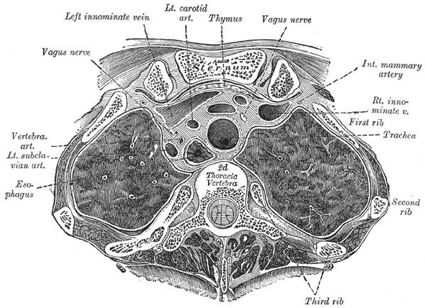
Transverse section through the upper margin of the second thoracic vertebra. (Braune.)

Figure 1: Transverse section through the upper margin of the second thoracic vertebra (Braune.).
The thymus is a lobulated lymphoid organ located in the anterior mediastinum. It lies posterior to the sternum and anterior to the pericardium. In children, the thymus is relatively large and plays a crucial role in T-cell maturation. It consists of two lobes connected by an isthmus. The organ is encapsulated and contains cortical and medullary regions. With age, the thymus undergoes involution. This process involves the replacement of functional lymphoid tissue with adipose tissue. By adulthood, the thymus is significantly smaller and largely replaced by fat. This involution is a normal physi
DefinitionThymic Involution
The replacement of thymic lymphoid tissue with adipose tissue that occurs with age, leading to a smaller and less active organ in adults.
The boundaries of the anterior mediastinum include the sternum anteriorly, the pericardium posteriorly, and the mediastinal pleura laterally. The thymus resides within this space. Its structure is distinct from the surrounding fat and vascular structures. Recognizing its normal appearance helps in identifying pathological changes. The organ's position relative to the great vessels is consistent, aiding in surgical planning and diagnostic imaging. The involution process is gradual and complete by middle age. This change reduces the organ's immunological function but does not typically cause clinical symptoms. [4]
| Feature | Description |
|---|---|
| Location | Anterior mediastinum, posterior to sternum |
| Structure | Bilobed, encapsulated, cortical and medullary regions |
| Involution | Replacement of lymphoid tissue with adipose tissue |
| Adult Appearance | Smaller, fatty, reduced immunological function |
The mediastinum is not a jointed structure; it is a central compartment of the thoracic cavity bounded by the sternum anteriorly, the vertebral column posteriorly, and the mediastinal pleura laterally. Consequently, there are no synovial joints, ligaments, or movements within the mediastinum itself. The term 'joints' in this context is anatomically incorrect for the mediastinal space. However, the mediastinum houses structures that interact with adjacent jointed regions, such as the sternocostal joints anteriorly and the costovertebral joints posteriorly. Pathologies within the mediastinum can mimic joint-related pain or restrict movement of the thoracic cage.
For instance, mediastinal masses can cause chest pain and dyspnea due to compression of adjacent structures. Large mature cystic teratomas, which are tumors containing ectodermal, mesodermal, and endodermal derivatives, are usually asymptomatic but may cause chest pain, dyspnea, and cough when they grow large. These tumors are predominantly located in the anterior mediastinum, although approximately 3-8% occur in the posterior mediastinum. On chest radiograph, they appear as a large anterior mediastinal mass extending to one side of the midline. CT imaging reveals multiple-tissue elements within the cystic mass, with fatty and cystic components present in half of the cases. Sometimes, a fat-fluid level within the mass strongly suggests the diagnosis of a mature teratoma. [5]
In the posterior mediastinum, esophageal duplication cysts are another consideration. The majority of these cysts occur in infants or children and are usually located adjacent to or within the esophageal wall in the lower posterior mediastinum. Ectopic gastric mucosa within the cyst may cause hemorrhage, perforation, or infection. CT or MRI features are similar to bronchogenic cysts, except for their proximity to the esophagus. [5]
Pancreatic fluid can also migrate to the mediastinum due to its aggressiveness, leading to mediastinal pseudocysts. These collections are found most commonly in the lesser sac but uncommonly in the mediastinum. A mediastinal pseudocyst almost always is located in the lower part of the posterior mediastinum. CT shows an encapsulated fluid collection with low attenuation in the posterior mediastinum, associated with compression or displacement of the esophagus or splaying of the diaphragmatic crura. [5]
Cardiac involvement by hydatid disease is rare, comprising 0.02 - 0.2% of all hydatid cases. The left ventricle is affected most often (50-60%), while the ventricular septum (10-20%), right ventricle (5-15%), pericardium (10-15%), and right or left atrium (5-8%) may be involved. MRI is the method of choice for specific diagnosis and exact assessment of hydatid cysts and their correlation with cardiac structures. [5]
Mediastinal hematoma may occur as a sequela of anterior mediastinal vein rupture after trauma or coronary artery catheterization. Ninety percent of hematomas have areas of high attenuation during the early period. Subacute or chronic hematomas may appear as low-attenuation masses. [5]
Synovial sarcoma, though very rarely encountered, could arise in the mediastinum. Less than 10–20% of synovial sarcomas arise in extra-extremity locations. Soft tissue sarcomas account for less than 0.01% of all malignant thoracic neoplasms. Around 17% of new cases of soft tissue sarcomas arise in thoracic locations, including the mediastinum, with an approximate incidence of 6 per million populations. The most common site of progression for these tumors was locally within the mediastinum. Of 15 patients with available data, 6 (40%) had isolated progression in the primary mediastinal tumor site. [6]
NoteAnatomical Clarification
The mediastinum contains no joints. Pain or movement restrictions are due to compression of adjacent structures or invasion of surrounding tissues.
The mediastinum is the central compartment of the thoracic cavity, situated between the two pleural sacs. It extends from the thoracic inlet superiorly to the diaphragm inferiorly. Understanding its surface anatomy requires correlating external palpable landmarks with internal radiological appearances.
Figure 2Gray972
Long description
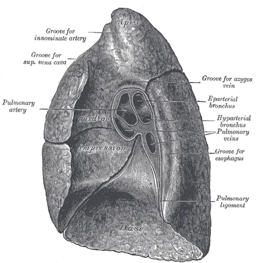
Mediastinal surface of right lung. Labelled illustration from Wikimedia Commons, used under its open licence.

Figure 2: Mediastinal surface of right lung.
The sternum forms the anterior boundary. The manubrium lies at the level of the second rib, while the sternal angle marks the fourth thoracic vertebra. This angle is a critical landmark for the superior mediastinum.
Radiological appearances help distinguish these compartments. CT and MRI show the mediastinal structures clearly. Simple cysts without solid nodularity are likely thymic cysts. Mature cystic teratomas in the anterior mediastinum show multiple tissue elements. Fatty and cystic components are present in half of these cases. A fat-fluid level strongly suggests the diagnosis. In the posterior mediastinum, esophageal duplication cysts are located adjacent to or within the esophageal wall. Ectopic gastric mucosa may cause hemorrhage. [4] [1]
Clinical pearlKey Landmark
The sternal angle marks the bifurcation of the trachea and the level of the fourth thoracic vertebra, separating the superior and middle mediastinum.
| Compartment | Boundaries | Key Structures | Common Lesions |
|---|---|---|---|
| Prevascular | Sternum to pericardium | Thymus, great vessels | Thymic cysts, teratomas |
| Visceral | Pericardium to vertebral line | Heart, esophagus, trachea | Esophageal duplication cysts |
| Paravertebral | Lateral to visceral | Sympathetic chain, aorta | Lateral meningoceles, cisterna chyli |
The mediastinum is the central compartment of the thoracic cavity, bounded laterally by the mediastinal pleura of the lungs. Its boundaries are defined by the bony structures of the thorax, providing a rigid framework that protects the vital organs within. Understanding these osseous landmarks is essential for localising pathology and interpreting imaging studies accurately. [3]
Figure 3Gray969
Long description
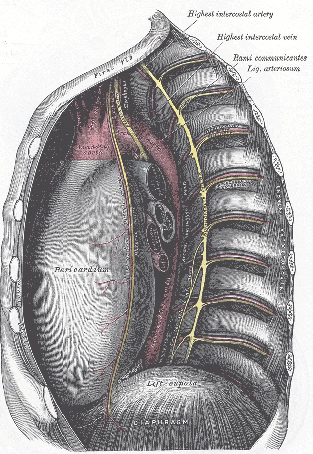
The middle and posterior mediastina. Left side.

Figure 3: The middle and posterior mediastina (left side).
The sternum forms the anterior boundary. It consists of the manubrium, the body, and the xiphoid process. The manubrium articulates with the clavicles and the first pair of ribs. The body articulates with the second to seventh costal cartilages. The xiphoid process is the inferior tip.
The mediastinum is the central compartment of the thoracic cavity, situated between the two pleural sacs. It extends from the thoracic inlet superiorly to the diaphragm inferiorly, and from the sternum anteriorly to the vertebral column posteriorly. To understand its anatomy, we divide it into distinct fascial compartments. This division allows for systematic study and clinical localization of pathology.
Figure 4Gray968
Long description
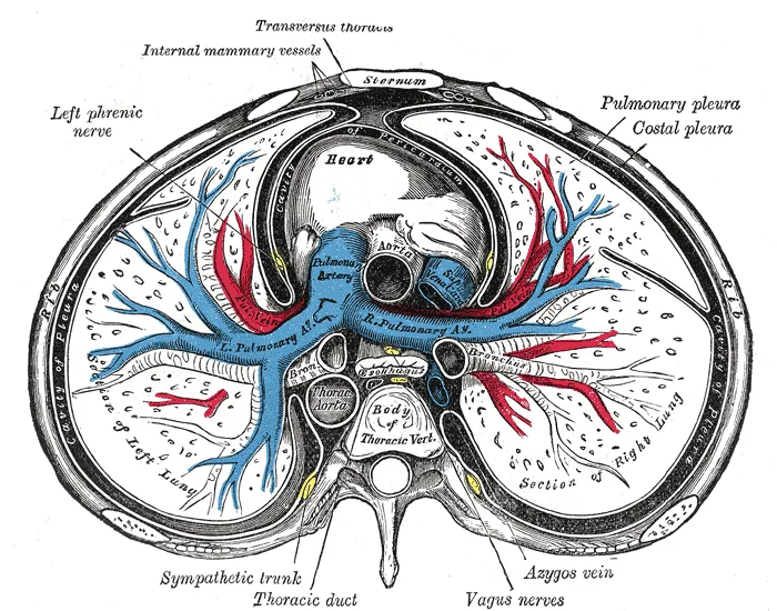
A transverse section of the thorax, showing the contents of the middle and the posterior mediastinum. The pleural and pericardial cavities are exaggerated since normally there is no space between parietal and visceral pleura and between pericardium and heart.

Figure 4: A transverse section of the thorax, showing the contents of the middle and the posterior mediastinum.
The superior mediastinum lies above the sternal angle and extends to the thoracic inlet. It contains the thymus, great vessels, trachea, and oesophagus. The anterior mediastinum is located between the sternum and the pericardium. It is narrow and contains loose connective tissue and lymph nodes. The middle mediastinum contains the pericardium and the heart. It is bounded by the anterior and posterior mediastinum. The posterior mediastinum lies between the pericardium and the vertebral column. It contains the descending aorta, azygos vein, and thoracic duct.
NoteImaging Context
Each imaging modality has strengths and weaknesses in mediastinal imaging—computed tomography (CT) offers increased spatial resolution at the cost of poorer soft tissue differentiation and requiring ionizing radiation, while magnetic resonance imaging (MRI) offers superior soft tissue contrast/characterization at significantly greater cost.
Important pericardial recesses to mention include the posterior pericardial recess (blind ending extension of the oblique sinus) which is located immediately above the left atrium to the right of midline, and the pulmonary venous recesses which surround the pulmonary veins near their attachment to the left atrium. [4]
The mediastinum is a central thoracic compartment bounded by the sternum anteriorly, the vertebral column posteriorly, and the pleura laterally. It is divided into superior and inferior regions by the sternal angle, with the inferior mediastinum further subdivided into anterior, middle, and posterior compartments. Understanding the muscular boundaries and contents is essential for localising pathology.
Figure 5Gray973
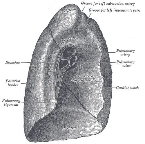
Long description
Mediastinal surface of left lung. Labelled illustration from Wikimedia Commons, used under its open licence.

Figure 5: Mediastinal surface of left lung.
The diaphragm forms the inferior floor, separating the thoracic cavity from the abdomen. Anteriorly, the sternum and costal cartilages define the anterior boundary. Posteriorly, the thoracic vertebrae and intervertebral discs form the posterior wall.
| Structure | Origin | Insertion | Action | Nerve Supply |
|---|---|---|---|---|
| Diaphragm | Sternum, ribs, lumbar vertebrae | Central tendon | Respiration | Phrenic nerve |
| Pericardium | Fibrous sheath | N/A | Protects heart | N/A |
Neurenteric cysts are persistent connections between the spinal canal and bowel. Bronchogenic cysts are abnormal ventral budding/branching of tracheobronchial tree lined by pseudostratified columnar epithelium. Esophageal cysts are outpouchings arising from the esophagus. Neurenteric cysts are more commonly found in the paravertebral compartment, while bronchogenic and esophageal cysts originate in the visceral compartment. [4]
Mediastinal cysts of foregut origin constitute up to 9% of all primary mediastinal masses. Mediastinal cysts result from embryologic aberrations and develop on abnormal budding of the primitive foregut and early tracheobronchial tree. The spectrum of foregut cysts includes bronchogenic cysts, esophageal duplication cysts, and neurenteric cysts. [1]
Mediastinal cystic lesions are typically well-marginated, rounded or oval shaped lesions filled with fluid and lined with epithelium. While most mediastinal cystic masses are benign (duplication cysts, pericardial cysts, thymic cysts, teratomas, meningoceles, lymphangiomas, etc.), several tumors may initially present with a cystic component or undergo cystic degeneration, including lymphoma, thymoma, teratoma and nodal metastases. [4]
Thymic cysts have been described as rare lesions, though reports have found them to represent up to 28.6% of mediastinal cysts and 3.7% of all mediastinal masses. Congenital thymic cysts are rarer than acquired thymic cysts, which tend to have a thicker wall and be multilocular secondary to inflammation or hemorrhage. Thymic cysts can coexist with thymic hyperplasia and thymic neoplasms. Thymic cysts are often difficult to differentiate from other mediastinal lesions. Typical imaging characteris [4]
Paravertebral compartment lesions are most likely neurogenic in origin. Cystic lesions of neurogenic origin include meningocele, cystic schwannoma, and neurenteric cysts. [4]
MRI may directly demonstrate a heterogenous mediastinal mass containing a variable mixture of fat, fluid, soft tissue, and calcification. Rupture of the cystic tumor into the pleura or pericardial space is a rare complication causing clinical symptoms. [1]
In conclusion, a number of mediastinal masses contain areas with CT attenuation values lower than those of chest wall musculature and higher than those of subcutaneous fat and therefore, these areas appear as cystic or cyst-like low-attenuation masses on CT. Compared with single detector helical CT, MDCT has greater accuracy for detecting mediastinal lesions. MDCT also allows more detailed evaluation of the entire thorax in a breath- hold without any loss of resolution and more consistent contra [1]
Coronal and sagittal MR images that may be obtained in conjunction with transaxial images provide the exact spatial relationship between tumor and other mediastinal structures. However, MRI also carries the same limitations as CT with inflammatory or inhomogeneous low-attenuation mediastinal lesions. Consequently, there may be some overlap in characteristics among various mediastinal masses and the correct diagnosis can only be made surgically. [1]
Transthoracic mediastinal ultrasonography can be effective in the characterization of mediastinal masses. B-flow imaging allows the visualization of flow and microbubble contrast can be safely administered to all patients (contrast-enhanced ultrasound, CEUS). The European Federation of Societies for Ultrasound in Medicine and Biology includes CEUS in its armamentarium for mediastinal imaging. An additional benefit of ultrasonographic imaging is its ability to be used for procedural guidance. [4]
The mediastinum receives its blood supply from multiple sources, reflecting its complex anatomical relationships with the heart, lungs, and great vessels. Understanding this vascular network requires examining the specific arteries that traverse or border the mediastinal compartments.
The superior mediastinum is primarily supplied by branches of the aortic arch and its major trunks. The brachiocephalic trunk arises from the right side of the aortic arch and quickly divides into the right common carotid and right subclavian arteries. The left common carotid artery and left subclavian artery arise directly from the aortic arch. These vessels provide the primary inflow to the head, neck, and upper limbs, but also give off small branches to the adjacent mediastinal structures. The thymus and trachea receive minor contributions from the inferior thyroid arteries, which are branches of the thyrocervical trunk arising from the subclavian artery.
The anterior mediastinum, containing the thymus and loose connective tissue, is supplied by the internal thoracic arteries. These arteries descend posterior to the costal cartilages. As they course downwards, they give off pericardiophrenic branches that supply the pericardium and the anterior mediastinal structures. The internal thoracic arteries also give off anterior intercostal arteries, which supply the upper intercostal spaces and contribute to the collateral circulation around the lungs.
The middle mediastinum, housing the heart and pericardium, is supplied by the pericardiophrenic arteries and the anterior intercostal arteries. The posterior mediastinum, containing the oesophagus and descending aorta, receives its supply from the oesophageal branches of the descending thoracic aorta and the posterior intercostal arteries.
DefinitionMediastinal Supply
The mediastinum is supplied by branches from the aortic arch, internal thoracic arteries, and descending thoracic aorta, ensuring perfusion to all its contained structures.
The mediastinum is drained by a complex network of veins that ultimately return blood to the superior vena cava and the azygos system. Understanding this drainage requires visualising the spatial relationships within the thoracic cavity. This illustration depicts the venous architecture in an anterior view. The superior mediastinal veins are shown draining into the brachiocephalic veins. The azygos vein is visible on the right side, arching over the root of the right lung to enter the superior vena cava. The hemiazygos vein is seen on the left, crossing to join the azygos vein. These structures lie posterior to the heart and great vessels. The image highlights the collateral pathways that allow blood to bypass obstructions in the superior vena cava.
| Feature | T1-Weighted | T2-Weighted |
|---|---|---|
| Serpentine venous channels | Intermediate signal intensity | High signal intensity |
| Phleboliths | Low-signal intensity | Low-signal intensity |
| Uncomplicated cystic lesions | Hypointense | Hyperintense |
| Hemorrhagic cysts | Variable | Variable with fluid-fluid levels |
The mediastinum contains a complex network of autonomic and somatic nerves that regulate cardiac, respiratory, and oesophageal function. Understanding their origin, course, and relations is essential for interpreting pathology and surgical outcomes.
The vagus nerves give rise to the pulmonary and oesophageal plexuses, which then reform into anterior and posterior vagal trunks. The phrenic nerves provide motor supply to the diaphragm and sensory innervation to the pericardium and mediastinal pleura. The sympathetic trunks contribute to the cardiac and pulmonary plexuses via splanchnic nerves.
DefinitionVagus Nerve
The tenth cranial nerve providing parasympathetic motor and sensory supply to the thoracic viscera.
Clinical pearlPhrenic Nerve
The phrenic nerve arises from C3-C5 and provides motor innervation to the diaphragm.
Both nerves carry sensory fibres from the pericardium and mediastinal pleura. [3] [4]
Exam trapSympathetic Chain
The sympathetic trunks lie posterior to the mediastinal pleura, not within the lung parenchyma.
The sympathetic trunks run vertically along the heads of the ribs, posterior to the mediastinal pleura. They give off cardiac branches that form the deep and superficial cardiac plexuses. These plexuses regulate heart rate and contractility. The greater, lesser, and least splanchnic nerves arise from the sympathetic trunks and pass through the diaphragm to innervate abdominal viscera. [1]
| Nerve | Origin | Course | Relations | Termination |
|---|---|---|---|---|
| Right Vagus | Cranial nerve X | Behind SVC, right main bronchus | Posterior to lung root | Posterior vagal trunk |
| Left Vagus | Cranial nerve X | Between carotid/subclavian, aortic arch | Anterior to lung root | Anterior vagal trunk |
| Right Phrenic | C3-C5 | Right SVC, right atrium | Anterior to lung root | Diaphragm |
| Left Phrenic | C3-C5 | Aortic arch, left ventricle | Anterior to lung root | Diaphragm |
| Sympathetic Trunk | Thoracic ganglia | Along rib heads | Posterior to pleura | Splanchnic nerves |
NoteClinical Relevance
Compression of the vagus nerve can cause hoarseness due to recurrent laryngeal nerve involvement.
Lesions of these nerves can lead to significant clinical syndromes. Compression of the recurrent laryngeal nerve, a branch of the vagus, can cause vocal cord paralysis and hoarseness. Damage to the phrenic nerve results in diaphragmatic paralysis and respiratory compromise. Understanding the anatomical relationships is crucial for surgical planning and avoiding iatrogenic injury.
The lymphatic drainage of the mediastinum is a complex network that ultimately converges towards the thoracic duct and the right lymphatic duct, ensuring the return of interstitial fluid and immune surveillance for the central thoracic organs. Understanding this pathway requires a systematic approach, dividing the mediastinum into regions that drain into specific nodal groups before reaching the major collecting trunks.
The anterior mediastinal nodes are situated in front of the pericardium and behind the sternum. They receive lymph from the thymus and the upper part of the anterior diaphragm. These nodes drain into the paratracheal nodes. The middle mediastinal nodes are located around the heart and the roots of the great vessels. They receive lymph from the pericardium and the heart itself. These nodes also drain into the tracheobronchial nodes. The posterior mediastinal nodes lie along the oesophagus and the descending aorta. They receive lymph from the lower part of the oesophagus and the posterior diaphragm. These nodes drain into the bronchomediastinal trunks. The left bronchomediastinal trunk typically drains into the thoracic duct, while the right bronchomediastinal trunk drains into the right lymphatic duct. The thoracic duct ascends through the posterior mediastinum, crossing from the right to the left side at the level of T5. It then passes behind the left lung root and empties into the junction of the left internal jugular and left subclavian veins. The right lymphatic duct is shorter and drains the right upper quadrant of the thorax into the junction of the right internal jugular and right subclavian veins.
The anterior mediastinal nodes are small lymph nodes located in the anterior mediastinum. They are positioned between the sternum and the pericardium. These nodes receive lymphatic vessels from the thymus gland, which lies directly anterior to them. They also receive drainage from the upper anterior diaphragm. The efferent vessels from these nodes pass posteriorly to join the paratracheal nodes. This connection is clinically significant because pathology in the thymus, such as thymoma, can spread to these nodes and subsequently to the paratracheal chain.
The middle mediastinal nodes are situated around the heart and the roots of the great vessels. They include the pericardial nodes and the tracheobronchial nodes. The pericardial nodes drain the fibrous and serous pericardium. The tracheobronchial nodes are located at the bifurcation of the trachea and along the main bronchi. They receive lymph from the lungs and the bronchi. These nodes are crucial in the drainage of pulmonary pathology. Efferent vessels from the middle mediastinal nodes converge to form the bronchomediastinal trunks.
The posterior mediastinal nodes are located along the oesophagus and the descending thoracic aorta. They receive lymph from the lower oesophagus and the posterior aspect of the diaphragm. These nodes drain into the bronchomediastinal trunks. This arrangement ensures that lymph from the posterior mediastinal structures is efficiently returned to the venous circulation.
The mediastinum is the central compartment of the thoracic cavity, situated between the two pleural sacs. It extends from the thoracic inlet superiorly to the diaphragm inferiorly, and from the sternum anteriorly to the vertebral column posteriorly. To understand its complex anatomy, we divide it into distinct regions. However, the four-compartment model is more clinically useful for localising pathology. This model divides the mediastinum into the superior, anterior, middle, and posterior mediastinum.
Figure 6Gray1032
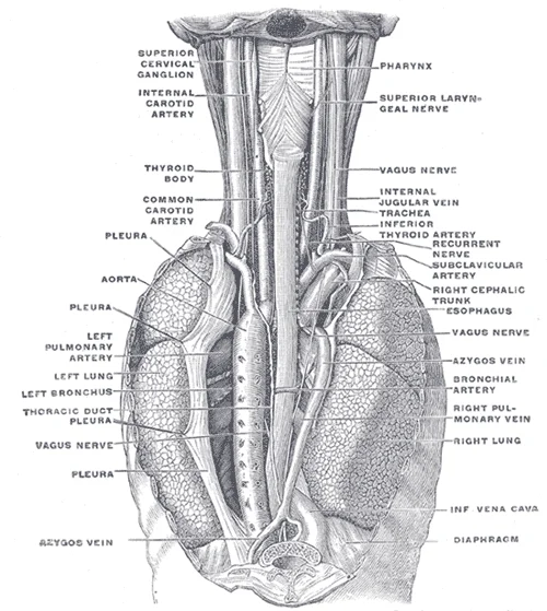
Long description
The position and relation of the esophagus in the cervical region and in the posterior mediastinum. Seen from behind. (Poirier and Charpy.)

Figure 6: The position and relation of the esophagus in the cervical region and in the posterior mediastinum (seen from behind).
In the superior mediastinum, we find the thymus remnant, the great vessels including the aortic arch and its branches, the trachea, and the oesophagus. The anterior mediastinum, located behind the sternum and in front of the pericardium, contains loose connective tissue and the thymus. The middle mediastinum houses the pericardium and the heart. The posterior mediastinum, lying behind the pericardium and in front of the vertebral column, contains the descending thoracic aorta, the azygos vein, the thoracic duct, and the oesophagus.
DefinitionThymus
The thymus is a primary lymphoid organ located in the anterior and superior mediastinum, crucial for T-cell maturation. It is most prominent in childhood and undergoes involution with age.
Mediastinal masses can arise in any of these compartments, and their location often hints at their origin. For instance, pericardial cysts are functionally mesothelial cysts of the pericardium, most commonly found in the cardiophrenic angles (right greater than left)[1]. These cysts account for up to 6% of all mediastinal masses and 33% of mediastinal cysts[1]. They are typically single, simple cysts—well-defined, low-density lesions on CT (30–40 HU), homogeneous T1 hypo/T2 hyper (unless proteinaceous)[1]. [4]
In contrast, neurenteric cysts are rare posterior mediastinal lesions that may be connected to the meninges through a midline defect in one or more vertebral bodies. This abnormality may be associated with vertebral anomalies, such as hemivertebrae, butterfly vertebra, or spina bifida. Enteric cysts are usually single, smooth, and unilocular. They occur primarily in the lower cervical and thoracic regions. The fluid within the cyst may be nearly identical to CSF or may be milky, cream-colored, y [1]
| Compartment | Common Masses | Key Features |
|---|---|---|
| Anterior | Thymoma, Teratoma, Thyroid goitre, Lymphoma | Thymic lesions may show microscopic fat on MRI[1]. |
| Middle | Pericardial cyst, Lymphadenopathy | Pericardial cysts are low-density on CT (30–40 HU)[1]. |
| Posterior | Neurogenic tumours, Neurenteric cysts, Oesophageal lesions | Neurenteric cysts may have vertebral anomalies. |
Imaging is critical for characterising these masses. Magnetic resonance imaging is a key problem-solving tool for the characterization of mediastinal cystic masses[1]. Unlike CT, MRI can more clearly characterize soft tissue lesions and identify infiltrative disease[1]. Diffusion weighted imaging can identify lesions mimicking cysts, as cellular content/debris will cause diffusion restriction in comparison to simple cysts, which will not[1]. Gadolinium contrast further allows characterization of lesion vascularity and enhancement[1]. In and out of phase imaging allows identification of microscopic fat which is commonly seen in normal or hyperplastic thymic tissue[1]. [4]
NoteImaging Choice
While CT is the modality of choice for lung parenchyma, MRI is preferred for soft tissue characterisation of mediastinal masses, especially after an indeterminate CT[1].
Enhancing septations, indicative of increased vascularity, may be a herald of malignancy[1]. Dual-energy CT with intravenous contrast can help differentiate cystic from solid lesions with material decomposition techniques using digital subtraction of iodine (intravenous contrast)[1]. This allows differentiation of tissue enhancement from hemorrhagic/proteinaceous fluid as well as differentiation of calcium from contrast accumulation[1]. Although there is scarcity of [4]
Figure 7Mediastinum anatomy
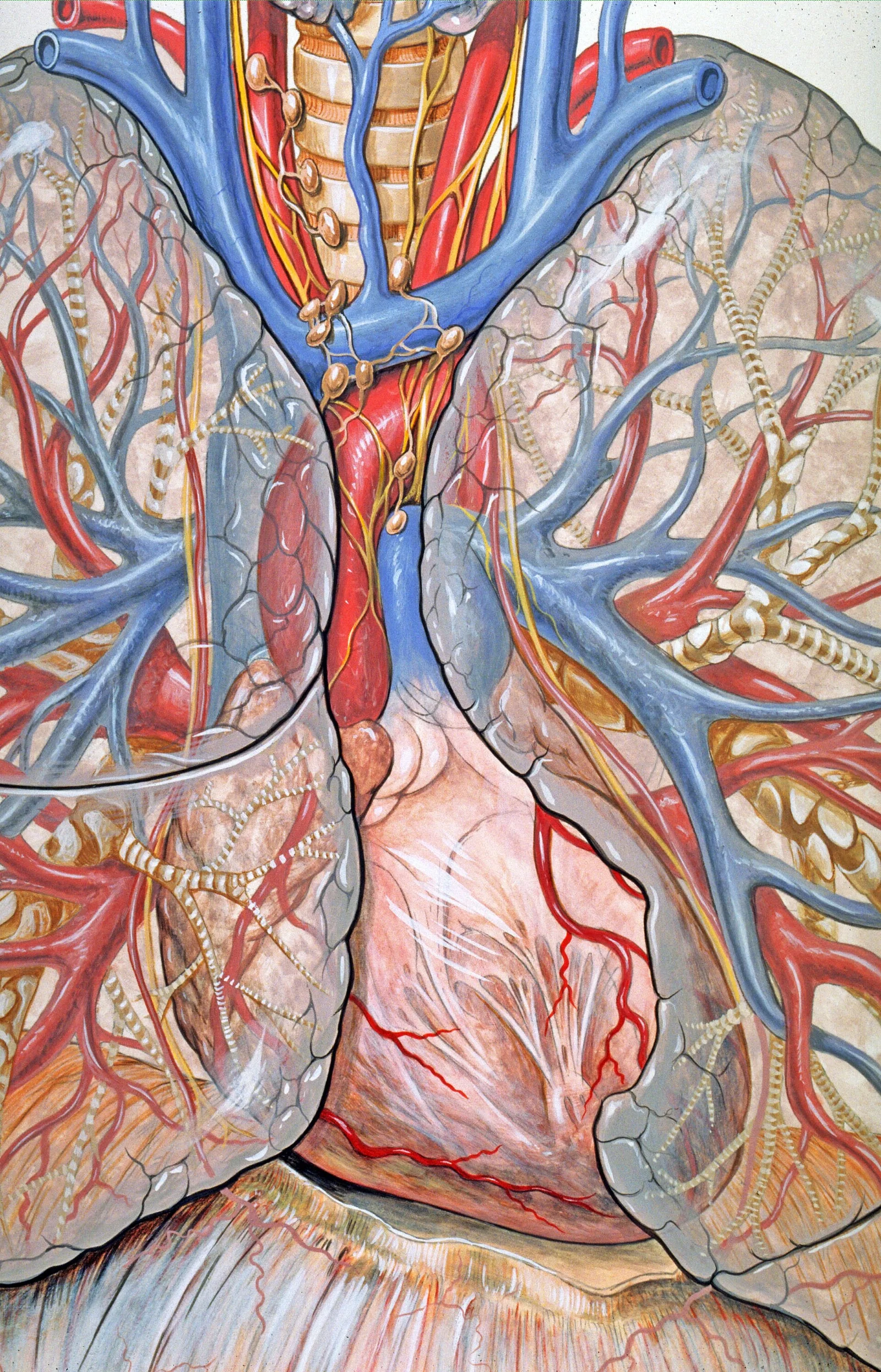
Long description
Anatomy of the mediastinum (the central chest and heart area).

Figure 7: Anatomy of the mediastinum (the central chest and heart area).
Plain chest X-ray provides a basic overview but lacks the detail required for precise characterization of mediastinal masses. It serves primarily as an initial screening tool rather than a definitive diagnostic modality for complex pathology.
Computed tomography (CT) is the workhorse modality for the characterization of virtually all mediastinal cystic lesions. It offers high spatial resolution and is often the first-line modality used in identification and characterization. CT allows reliable differentiation of cystic masses from solid tumors. However, CT is of limited value in the evaluation of pericardial and cardiac hydatid cysts due to motion artifacts. Contrast-enhanced CT is particularly useful for identifying enhancing vascul [1][4]
Magnetic resonance imaging (MRI) appears to be the method of choice for specific diagnosis and exact assessment of hydatid cysts and their correlation with cardiac structures. MRI has been shown to be particularly sensitive for the detection of cerebral, abdominal and vertebral metastases. Strengths of MRI include differentiation between cystic and solid lesions, evaluation of simple versus complex cystic lesions, and evaluation for restricted diffusion. MRI is a critical problem-solving tool, u [1][2][4]
Ultrasound can be utilized without risk to the patient but is limited by operator ability and depth of target lesion. Ultrasonography is useful in real-time characterization and procedural guidance. [4]
Clinical pearlModality Selection
CT is the first-line modality for mediastinal cysts, while MRI is the critical problem-solving tool for complex cases and cardiac involvement.
| Modality | Primary Role | Key Advantage | Limitation |
|---|---|---|---|
| CT | First-line characterization | High spatial resolution | Motion artifacts in cardiac regions |
| MRI | Problem-solving | High soft tissue contrast | Longer scan time |
| Ultrasound | Real-time guidance | No radiation | Operator dependent |
NoteMalignancy Signs
Findings suggestive of malignancy include enhancing solid or nodular components, thick vascular septations, and diffusion restriction. [4]
The mediastinum is the central compartment of the thoracic cavity, bounded laterally by the mediastinal pleura. It extends from the thoracic inlet superiorly to the diaphragm inferiorly. Clinically, it is divided into anterior, middle, and posterior regions to localise pathology. Understanding these boundaries is essential for interpreting imaging and planning surgical interventions.
The anterior mediastinum lies behind the sternum and contains the thymus remnants and fat. The middle mediastinum houses the pericardium and heart. The posterior mediastinum is situated anterior to the vertebral column and contains the oesophagus and descending aorta. The phrenic nerves run vertically along the lateral aspects of the pericardium. The vagus nerves pass posterior to the root of the lung. Recognising these relationships allows clinicians to predict the spread of infection or malignancy. For instance, a mass in the anterior mediastinum may compress the superior vena cava, leading to facial swelling. A lesion in the posterior mediastinum may impinge on the sympathetic chain, causing Horner’s syndrome.
Injuries to the mediastinum often result from blunt or penetrating trauma. The heart and great vessels are particularly vulnerable. Aortic rupture typically occurs at the ligamentum arteriosum due to deceleration forces. Nerve palsies can arise from surgical injury or tumour compression. Recurrent laryngeal nerve palsy causes hoarseness and is associated with left-sided mediastinal masses. The phrenic nerve injury leads to diaphragmatic paralysis, evident on fluoroscopy as paradoxical movement.
Surface landmarks guide clinical procedures. This landmark is crucial for identifying the level of the aortic arch and the bifurcation of the trachea. The mid-clavicular line is used to locate the apex of the heart. The parasternal line is relevant for assessing the right ventricle.
Procedures such as mediastinoscopy allow direct visualisation of lymph nodes. This is vital for staging lung cancer. The anterior approach provides access to prevascular nodes. Complications include bleeding and nerve damage. Ultrasound-guided biopsy is increasingly used for safer access to posterior mediastinal lesions.
| Structure | Location | Clinical Significance |
|---|---|---|
| Thymus | Anterior | Tumours may cause myasthenia gravis |
| Pericardium | Middle | Effusions cause cardiac tamponade |
| Oesophagus | Posterior | Obstruction leads to dysphagia |
| Aorta | Posterior | Aneurysms may compress the trachea |
The mediastinum is the central compartment of the thoracic cavity, situated between the two pleural sacs. It extends from the thoracic inlet superiorly to the diaphragm inferiorly. Anteriorly, it is bounded by the sternum, and posteriorly by the vertebral column. The inferior mediastinum is further subdivided into anterior, middle, and posterior sections.
This region contains the thymus, great vessels, trachea, and oesophagus. The thymus is a lymphoid organ that is prominent in children and atrophies in adults. It lies anterior to the brachiocephalic veins and the superior vena cava. The aortic arch and its branches emerge from this space. The vagus and phrenic nerves also traverse this compartment.
Located between the sternum and the pericardium, this space is small in adults. It contains loose connective tissue, lymph nodes, and remnants of the thymus. Pathological masses here are often thymomas or lymphomas.
This is the largest subdivision, containing the pericardium and the heart. The roots of the great vessels, including the superior and inferior vena cava, enter and exit here. The phrenic nerves run along the lateral aspects of the pericardium.
Situated behind the pericardium and in front of the vertebral column, this area houses the descending thoracic aorta and the azygos vein. The oesophagus and thoracic duct also pass through this space. The sympathetic trunks lie against the heads of the ribs.
Mediastinal masses can compress adjacent structures, leading to symptoms such as dyspnoea or superior vena cava syndrome. Lymphadenopathy in this region may indicate malignancy or infection. Imaging is crucial for diagnosis. While CT is standard, MRI offers advantages for soft tissue contrast. For instance, 3D gradient-echo sequences can detect small nodules with high sensitivity. However, MRI is not yet routine for lung screening due to motion artefacts. Automated detection tools for MRI have been explored. [2][4]
Students frequently misinterpret the signal intensity of mediastinal cysts on MRI, leading to diagnostic errors. A common mistake is assuming all cysts are simple fluid. Bronchogenic cysts often contain proteinaceous fluid, mucus, or blood debris, which increases their density above that of water. Another error is ignoring the variable signal patterns on T1-weighted MRI, which depend on the specific cystic contents such as protein or mucoid material. Students must also recognise that while T2-weighted MRI typically shows high signal intensity for cysts, rare cases may show a fluid–fluid level or peripheral calcification. [1]
A second pitfall involves the interpretation of attenuation values on CT. When a cystic mass has attenuation slightly higher than water, it may represent elevated protein content or a combination of fluid, solid tissue, and fat. Assuming uniform fluid density can lead to missing complex lesions. On MRI, lesions with signal similar to or greater than muscle on T1-weighted images require careful differentiation from solid tumours. [1]
Diffusion-weighted imaging (DWI) is another area prone to error. Cystic lesions do not demonstrate diffusion restriction unless they are infected. A restricting lesion must therefore represent either a solid lesion or an infected cyst. Students often confuse T2 shine-through with true restriction. Comparison with apparent diffusion coefficient (ADC) maps is essential, as simple fluid is bright on the ADC map, whereas truly restricting lesions are dark. [4]
Misclassification of thymic lesions is also common. Acquired thymic cysts are benign and characteristically multiloculated. However, some cystic thymomas have overlapping imaging characteristics with benign cysts. Lesions with less than fluid signal on T2WI and non-enhancement should undergo follow-up imaging to ensure stability. Assuming all anterior mediastinal masses are thymomas without considering cystic variants is a significant error. [4]
Mature cystic teratomas and dermoid cysts are characterised by multiple tissue densities within a single mass. These features may not be readily apparent on CT imaging. MR imaging demonstrating macro or microscopic fat can aid in diagnosis. Multiple fat-fluid levels are highly specific for these lesions. Gross fat is often easier to identify than microscopic fat. [4]
Pericardial abscesses are sometimes mistaken for simple pericardial effusions. Pericardial abscesses are characterised by a rim-enhancing fluid collection that demonstrates T1 intermediate or low signal and T2 intermediate or high signal. Diffusion restriction is characteristic of abscesses. A history of recent pericardial intervention is beneficial in confirming the diagnosis. [4]
Müllerian cysts are rare paravertebral cystic lesions in the mid-thoracic region. They are typically found in women with concomitant gynaecologic pathology. These lesions are singular, well-defined fluid attenuation lesions without enhancement that do not communicate with other structures. Surgical excision is often the preferred treatment given a presumed risk of malignant transformation. [4]
Knowledge of mediastinal anatomy and pathology is critical at the bedside for interpreting imaging studies. Clinicians use MRI to visualise enlarged lymph nodes and skeletal lesions using transverse T2-weighted STIR sequences[1]. This sequence helps differentiate cystic from solid lesions. Alternatively, T2-weighted STIR can be replaced by a fat saturated T2-weighted FSE with PROPELLER acquisition to reduce motion artefacts in patients with severe dyspnoea[1]. For viewing 3D data obtained with breath-hold 3D-GRE, a 3D viewer and reconstruction of 6–8 mm maximum intensity projection reformations is recommended[1]. [2]
In the operating theatre, surgeons rely on imaging to plan excisions. For Müllerian cysts, surgical excision is often the preferred treatment given a presumed risk of malignant transformation. Surgeons must identify the paravertebral location in the T3–T8 region to avoid nerve damage. For pericardial abscesses, the presence of a rim-enhancing fluid collection guides drainage procedures.
On imaging, differentiation between benign and malignant lesions is paramount. The potential of perfusion methods for monitoring chemotherapy has not been investigated so far[1]. However, absent or very low contrast uptake might have a high negative predictive value for the exclusion of malignancy of a lung nodule[1]. This helps in deciding whether to biopsy or observe. [2]
In examinations, students must distinguish between various cyst types. Bronchogenic cysts show high signal intensity on T2-weighted MRI, suggesting a cystic lesion. However, T1-weighted images may show variable patterns due to protein or haemorrhage. Students must recall that acquired thymic cysts are benign and multiloculated. They must also know that mature cystic teratomas contain multiple tissue densities. [1]
Understanding the origin of lesions aids diagnosis. Talaromycosis enters via inhalation, with lungs being the primary organ of infection. At the host–pathogen interface, conidia adhere to bronchial epithelial cells by attaching to extra-cellular matrix laminin. Although phagocytosis occurs, the pathogen evades macrophage killing by producing superoxide dismutase and catalase-peroxidase. [3]
Mediastinal Cyst Characterisation
- Identify Cystic LesionCT or MRI finding
- Question: T1 Signal > Muscle?
Yes
- Check T2 SignalHigh signal suggests cyst
No
- Consider Simple FluidLow T1, High T2
- Question: Diffusion Restriction?
Yes
- Infected Cyst or SolidCheck ADC map
No
- Benign CystSimple fluid
- Final DiagnosisCorrelate with clinical history
Bronchogenic cysts often have high T1 signal due to protein or blood. T2 shine-through can mimic diffusion restriction on DWI. ADC maps are essential to distinguish true restriction from T2 shine-through. Acquired thymic cysts are benign and typically multiloculated. Mature cystic teratomas show multiple tissue densities and fat-fluid levels. Pericardial abscesses show rim enhancement and diffusion restriction. Müllerian cysts are rare, paravertebral, and occur in women aged 40–60. STIR sequences help visualise lymph nodes and skeletal lesions.
Answer each one in your head first, then open the answer.
What causes high T1 signal in bronchogenic cysts?
Show answerHide answer
Calcium, proteinaceous fluid, mucus, and/or blood debris.How do you distinguish true diffusion restriction from T2 shine-through?
Show answerHide answer
By comparing DWI images with ADC maps; simple fluid is bright on ADC.What is the typical appearance of acquired thymic cysts?
Show answerHide answer
They are benign lesions that characteristically are multiloculated.Where are Müllerian cysts typically located?
Show answerHide answer
In the T3–T8 region as rare paravertebral cystic lesions.What imaging feature is highly specific for mature cystic teratomas?
Show answerHide answer
Multiple fat-fluid levels.
- Odev K, Arıbaş BK, Nayman A, et al. Imaging of Cystic and Cyst-like Lesions of the Mediastinum with Pathologic Correlation. Journal of clinical imaging science 2012;2:33. doi:10.4103/2156-7514.97750. Accessed .
- Biederer J, Hintze C, Fabel M. MRI of pulmonary nodules: technique and diagnostic value. Cancer imaging : the official publication of the International Cancer Imaging Society 2008. Accessed .
- Narayanasamy S, Dougherty J, van Doorn HR, Le T. Pulmonary Talaromycosis: A Window into the Immunopathogenesis of an Endemic Mycosis. Mycopathologia 2021. Accessed .
- Shah A, Rojas CA. Imaging modalities (MRI, CT, PET/CT), indications, differential diagnosis and imaging characteristics of cystic mediastinal masses: a review. Mediastinum (Hong Kong, China) 2023. Accessed .
- Odev K, Arıbaş BK, Nayman A, et al. Imaging of Cystic and Cyst-like Lesions of the Mediastinum with Pathologic Correlation. Journal of clinical imaging science 2012;2:33. doi:10.4103/2156-7514.97750. Accessed .
- Salah S, Salem A. Primary synovial sarcomas of the mediastinum: a systematic review and pooled analysis of the published literature. ISRN oncology 2014;2014:412527. doi:10.1155/2014/412527. Accessed .
Figure credits
- Figure 1: Henry Vandyke Carter, via Wikimedia Commons, Public domain, Public domain.
- Figure 2: Henry Vandyke Carter, via Wikimedia Commons, Public domain, Public domain.
- Figure 3: Henry Vandyke Carter, via Wikimedia Commons, Public domain, Public domain.
- Figure 4: Henry Vandyke Carter, via Wikimedia Commons, Public domain, Public domain.
- Figure 5: Henry Vandyke Carter, via Wikimedia Commons, Public domain, Public domain.
- Figure 6: Henry Vandyke Carter, via Wikimedia Commons, Public domain, Public domain.
- Figure 7: Patrick J. Lynch, medical illustrator, via Wikimedia Commons, CC BY 2.5, CC BY 2.5. resized.
Educational content for medical students and doctors. It is not a substitute for clinical judgement or local guidelines.