Lungs and bronchial tree
Anatomical structure of the lungs, bronchial tree, and pleurae.
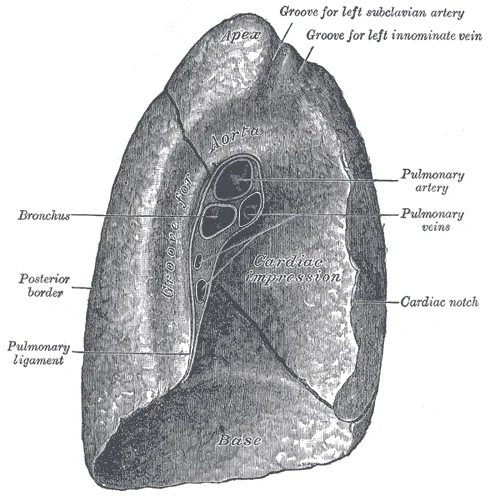
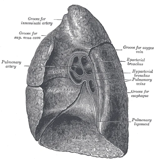
The lungs are the primary organs of the respiratory system, responsible for the exchange of oxygen and carbon dioxide with atmospheric air[1]. This gas exchange occurs across a vast epithelial surface area of approximately 70 square meters, which is highly permeable to gases[1]. Structurally, the lungs are pyramid-shaped, paired organs connected to the trachea via the right and left bronchi[1]. On their inferior surface, they are bordered by the dome-shaped diaphragm muscle[1]. The lungs are enclosed by the pleurae, serous membranes attached to the mediastinum[1]. The right lung is shorter and wider than the left, occupying a larger volume[1]. The left lung features a cardiac notch, an indentation that accommodates the heart[1]. The apex is the superior region, while the base lies near the diaphragm[1]. The costal surface borders the ribs, and the mediastinal surface faces the midline[1].
Figure 1Gray973
Long description
Mediastinal surface of left lung. Labelled illustration from Wikimedia Commons, used under its open licence.

Figure 1: Mediastinal surface of left lung.
Figure 2Gray972
Long description
Mediastinal surface of right lung. Labelled illustration from Wikimedia Commons, used under its open licence.

Figure 2: Mediastinal surface of right lung.
Figure 3Gray970
Long description
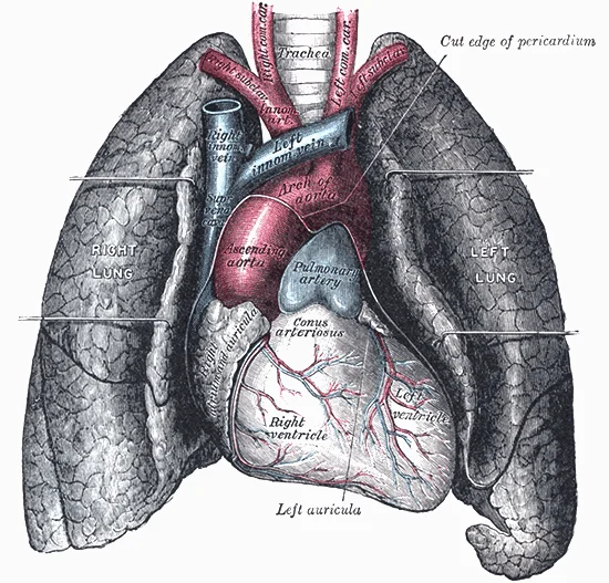
Front view of heart and lungs. Labelled illustration from Wikimedia Commons, used under its open licence.

Figure 3: Front view of heart and lungs.
DefinitionLung Lobes
The right lung has three lobes: superior, middle, and inferior. The left lung has two lobes: superior and inferior.
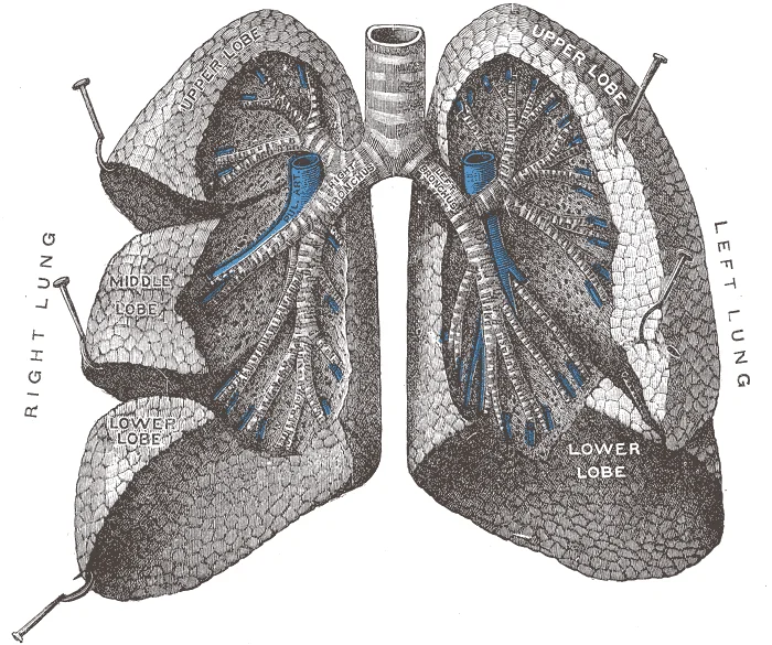
Each lung is divided into lobes by fissures[1]. The right lung consists of the superior, middle, and inferior lobes[1]. The left lung consists of the superior and inferior lobes[1]. Each lobe is further divided into bronchopulmonary segments[1]. Each segment receives air from its own tertiary bronchus and blood from its own artery[1]. This segmentation allows diseased segments to be surgically removed with minimal impact on neighbouring tissue[1]. A pulmonary lobule is a subdivision where bronchi branch into bronchioles[1]. Each lobule receives a large bronchiole with multiple branches[1].
DefinitionPleurae
The pleura consists of the visceral layer lining the lung and fissures, and the parietal layer connecting to the thoracic wall, mediastinum, and diaphragm.
The pleurae surround each lung within its cavity[1]. The right and left pleurae are separated by the mediastinum[1]. The visceral pleura is superficial to the lungs and lines the fissures[1]. The parietal pleura connects to the thoracic wall, mediastinum, and diaphragm[1]. These layers connect at the hilum, enclosing the pleural cavity[1].
DefinitionInnervation
Autonomic control via parasympathetic and sympathetic systems regulates airway dilation and constriction.
Airway dilation and constriction are controlled by the parasympathetic and sympathetic nervous systems[1]. The parasympathetic system causes bronchoconstriction, while the sympathetic system stimulates bronchodilation[1]. Reflexes like coughing and gas regulation result from this autonomic control[1]. The pulmonary plexus forms on the lung root at the hilum[1]. Nerves follow the bronchi, branching to innervate muscle fibers, glands, and blood vessels[1].
| Feature | Right Lung | Left Lung |
|---|---|---|
| Lobes | Superior, Middle, Inferior | Superior, Inferior |
| Volume | Larger | Smaller |
| Special Feature | None | Cardiac Notch |
Figure 4Illu bronchi lungs
Long description
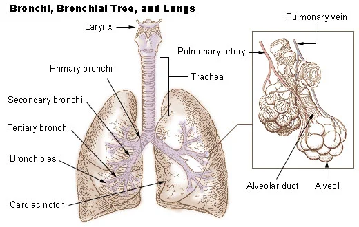
Illu bronchi lungs.jpg. Labelled illustration from Wikimedia Commons, used under its open licence.

Figure 4: Illu bronchi lungs.jpg.
The lungs are enclosed by the pleura, a membrane composed of visceral and parietal pleural layers. The space between these two layers is the pleural cavity. The mesothelial cells of the pleural membrane create pleural fluid, which serves as a lubricant to reduce friction during breathing and as an adhesive to adhere the lungs to the thoracic wall. This adhesive characteristic causes the lungs to enlarge when the thoracic wall expands during ventilation, allowing the lungs to fill with air. The pleurae also create a division between major organs that prevents interference due to the movement of the organs, while preventing the spread of infection. [1]
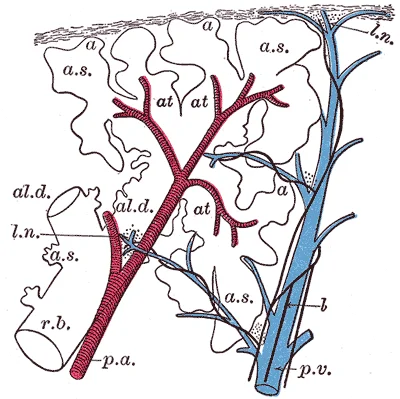
The major function of the lungs is to perform gas exchange, which requires blood from the pulmonary circulation. This blood supply contains deoxygenated blood and travels to the lungs where erythrocytes pick up oxygen to be transported to tissues throughout the body. The pulmonary artery branches multiple times as it follows the bronchi, and each branch becomes progressively smaller in diameter. One arteriole and an accompanying venule supply and drain one pulmonary lobule. As they near the alveoli, the pulmonary arteries become the pulmonary capillary network. The pulmonary capillary network consists of tiny vessels with very thin walls that lack smooth muscle fibers. [1]
Figure 5Gray975
Long description
Schematic longitudinal section of a primary lobule of the lung (anatomical unit); r. b., respiratory bronchiole; al. d., alveolar duct; at., atria; a. s., alveolar sac; a, alveolus or air cell; p. a.: pulmonary artery: p. v., pulmonary vein; l., lymphatic; l. n., lymph node. (Miller.)

Figure 5: Schematic longitudinal section of a primary lobule of the lung (anatomical unit); r (b., respiratory bronchiole; al).
Figure 6Gray962
Long description
Bronchi and bronchioles. The lungs have been widely separated and tissue cut away to expose the air-tubes. (Testut.)

Figure 6: Bronchi and bronchioles.
Figure 7Gray974
Long description
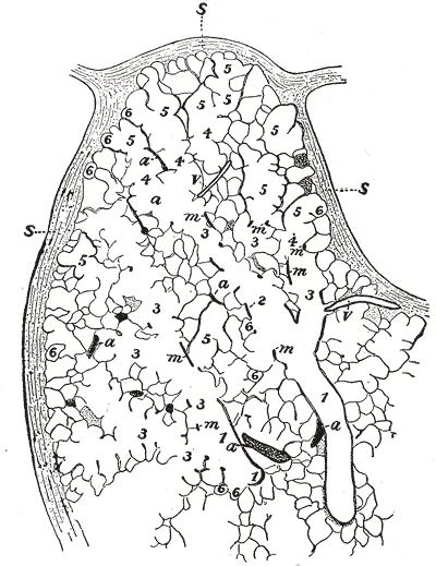
Part of a secondary lobule from the depth of a human lung, showing parts of several primary lobules. 1, bronchiole; 2, respiratory bronchiole; 3, alveolar duct; 4, atria; 5, alveolar sac; 6, alveolus or air cell: m, smooth muscle; a, branch pulmonary artery; v, branch pulmonary vein; s, septum betwe

Figure 7: Part of a secondary lobule from the depth of a human lung, showing parts of several primary lobules.
The lungs are continually exposed to a multitude of microorganisms, some of which are able to persist and colonize the respiratory tract. These microbes that reside in the lower airways constitute the lung microbiome, which is distinct in composition from the microbiota observed in the oral and nasal cavities. The surface of the lungs presents a continuous layer of epithelial cells that constitute a physical and biological barrier for inhaled substances and pathogens. The specialized respiratory epithelium is required for maintenance of immune homeostasis, and epithelial dysfunction is involved in the development of many inflammatory disorders of the airways and lungs. [2]
Basal cells, which are candidate stem cells in the conducting airways, are found in highest concentration in the trachea, and numbers diminish further down the bronchial tree to the bronchioles. Upon damage these cells upregulate p63, a basal cell-restricted transcription factor, which has been implicated in epithelial mesenchymal transition. It has been shown that basal epithelial cells overlying fibroblastic foci in IPF acquire increased reactivity, determined by p63 expression, and gain a partial mesenchymal phenotype. This provides further evidence of the involvement of bronchial-derived cells in the pathogenesis of IPF. [2]
Human β-defensin 1, 2, 3 and 4 are largely expressed on airway epithelial cells. [2]
Collectively, it is likely that the dialogue between specific bacterial communities and host is a dynamic phenomenon. The installation and location of the bacterial communities within the lungs is affected by the cellular components within the lungs as well as the abiotic environment. Specifically, loss of barrier function can influence the composition of the respiratory microbiota by allowing the entry of pathogens and other particles. However, it is evident that more information is needed to elucidate the nature of the dialogue occurring between the respiratory microbiota and epithelial cells in the lungs.
Similar to the gut, the airway microbiota in healthy lungs is dominated by the short-chain fatty acid-producing bacteria. [2]
Figure 8Lungs anatomy
Long description
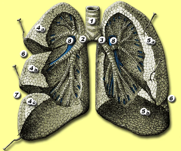
Anatomy of the lungs - based on image 962 from online edition of Gray's Anatomy The lungs 1 = trachea 2 = right bronchus 3 = left bronchus 4 = right lobe: superior (4a), middle (4b), inferior (4c) lobes 5 = left lobe: superior (5a) and inferior (5b) lobes 6 on the right = horizontal fissure 6 on the

Figure 8: Anatomy of the lungs - based on image 962 from online edition of Gray's Anatomy The lungs 1 = trachea 2 = right bronchus 3 = left bronchus 4 = right lobe: superior (4a), middle (4b), inferior (4c) lobes 5 = left lobe: superior (5a) and inferior (5b) lobes 6 on the right = horizontal fissure 6 on the.
The mediastinal surface of each lung faces the midline structures, while the costal surface abuts the inner chest wall.
The lungs are enclosed within the pleural cavities. The right lung is divided into three lobes: superior, middle, and inferior. The left lung has two lobes: superior and inferior. The oblique fissure separates the superior from the inferior lobe in both lungs. The horizontal fissure is present only in the right lung, separating the superior and middle lobes. The bronchial tree begins with the trachea, which bifurcates at the carina, typically at the level of the sternal angle or T4/T5 vertebrae. The right main bronchus gives off the right upper lobe bronchus before continuing to the middle and lower lobes. The left main bronchus gives off the left upper lobe bronchus, which further divides. The basal cells are progenitor cells that act as resident stem cells for the trachea and proximal airways. These cells are capable of self‐renewal as well as repopulating the pseudostratified epithelium during homeostasis and after injury. The tracheal region and large airways harbour stromal cells such as interstitial fibroblasts that aid in the regulation of the regenerative response of the airway epithelium after injury. [2]
Figure 9Gray961
Long description
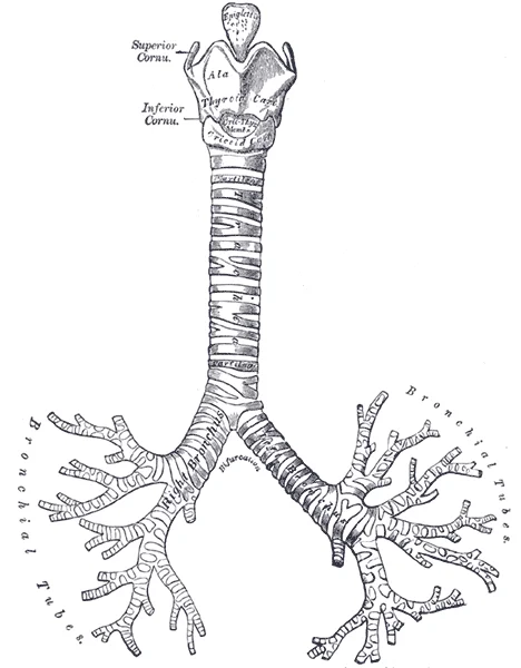
Front view of cartilages of larynx, trachea, and bronchi.

Figure 9: Front view of cartilages of larynx, trachea, and bronchi.
DefinitionPleura
The serous membrane enclosing the lungs, consisting of visceral and parietal layers.
The relations of the lungs are defined by their proximity to the heart, great vessels, esophagus, and diaphragm. The anterior surface of the right lung is related to the right atrium and superior vena cava. The anterior surface of the left lung is related to the left ventricle and the pericardium. The posterior surface is related to the vertebral column and the sympathetic trunks. The inferior surface rests on the diaphragm. The bronchial tree branches within the lung parenchyma, following the vascular supply. The pulmonary arteries accompany the bronchi, while the pulmonary veins run between the lobes. The lymph nodes are located at the hilum and along the bronchial tree. The epithelial surfaces of the lung, including the proximal and distal airways, are now considered to play a pivotal role in maintenance of immune homeostasis. The epithelium acts as the first line of defence against inhaled viral, bacterial and fungal pathogens. [2]
Figure 10Gray971
Long description
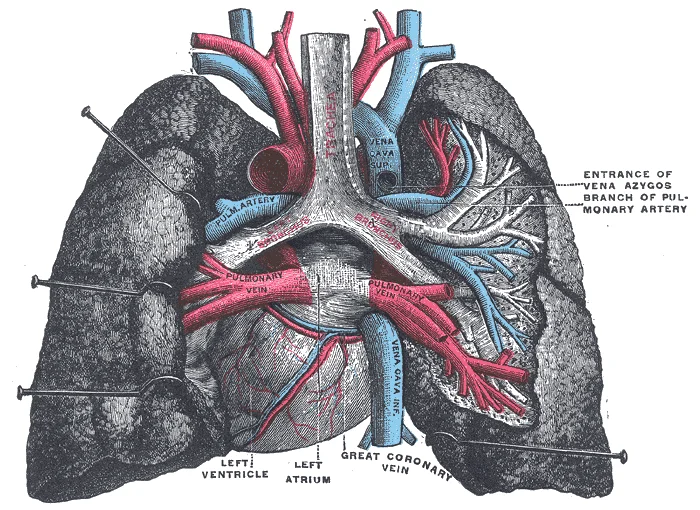
Pulmonary vessels, seen in a dorsal view of the heart and lungs. The lungs have been pulled away from the median line, and a part of the right lung has been cut away to display the air-ducts and bloodvessels. (Testut.)

Figure 10: Pulmonary vessels, seen in a dorsal view of the heart and lungs.
The interior of the lungs and bronchial tree is defined by the intricate relationship between the conducting airways and the vascular network responsible for gas exchange. The respiratory tract functions primarily to exchange oxygen and carbon dioxide, a process facilitated by its complex internal architecture. This internal environment is not uniform; it spans different anatomical sites with varied epithelial cell compositions reflecting distinct functions in the conducting airways versus the alveolar regions. [2]
DefinitionRespiratory Membrane
The interface where gas exchange occurs, formed where the capillary wall meets the alveolar wall.
Internally, the capillaries branch extensively to follow the bronchioles and the detailed structure of the alveoli. It is at this specific juncture that the capillary wall meets the alveolar wall, creating the respiratory membrane. This membrane is the critical site for the diffusion of gases. Once blood is oxygenated within these capillaries, it drains from the alveoli via multiple pulmonary veins. These veins are the primary vascular exit route, leaving the lungs through the hilum. [1]
| Structure | Location/Relation | Function |
|---|---|---|
| Capillaries | Follow bronchioles and alveoli | Site of gas exchange |
| Respiratory Membrane | Junction of capillary and alveolar walls | Diffusion interface |
| Pulmonary Veins | Exit via hilum | Drain oxygenated blood |
| Mucus Layer | Lining conducting airways | Defence and microbial niche |
This layer is supported by a periciliary layer covering the cilia, promoting healthy interactions between microorganisms and epithelial cells. In health, this mucus layer provides effective defence against epithelial injury. However, excessive mucus production is associated with increased susceptibility to viral infections and respiratory allergies. [2]
Exam trapMucus Obstruction
Excessive mucus hinders cilia transport, favouring pathogenic bacteria that thrive in obstructed niches. [2]
Microbial elimination is an active process achieved through mucociliary clearance, cough, and host immune defences. The environmental conditions necessary for microbial growth within the respiratory tract, such as pH, temperature, nutrient availability, oxygen tension, and activation of host inflammatory cells, are heterogenous. [2]
NoteRegional Variation
Considerable regional variation in microbial communities can be observed in a single healthy lung due to differing environmental conditions.
In chronic pulmonary diseases, increased mucus production promotes bacterial growth, leading to zones with low oxygen concentration and high temperature. These physiological characteristics, including temperature, mucus, pulmonary surfactant, and oxygen tension, have a major impact on the establishment and persistence of bacterial communities. Signalling through Toll-like receptors (TLRs) is key in early inflammatory responses to bacteria through the activation and expression of host cytokines, chemokines, and antimicrobial peptides. TLR2, TLR3, TLR5, and TLR6 are the most highly expressed in human bronchial epithelial cells. [2]
Internal Defence Mechanisms
- StartAir enters lungs
- Question: Is mucus layer intact?
Yes
- Mucociliary clearance removes pathogens
No
- Pathogens penetrate epithelium
- TLRs activate immune response
Defects in barrier structure and function can lead to increased susceptibility to infection. During chronic disease, there is evidence that barrier function is impaired, with disruption of tight junctions and increased epithelial permeability. This loss of barrier function leads to the entry of pathogens and other particles. Compromised barrier function is intimately linked to triggering of abnormal repair and remodelling. [2]
The vascular architecture of the lungs and bronchial tree is dual in nature, comprising the pulmonary and bronchial circulations. This arrangement supports both gas exchange and the metabolic needs of the lung tissue itself. The pulmonary arteries carry deoxygenated blood from the right ventricle to the alveolar capillaries for oxygenation. In contrast, the bronchial arteries supply oxygenated blood to the supporting structures of the airways and lungs. Understanding the origin, course, and relations of these vessels is fundamental to interpreting clinical imaging and pathology.
The right pulmonary artery courses horizontally behind the ascending aorta and superior vena cava to reach the right hilum. The left pulmonary artery arches over the left main bronchus. The bronchial arteries typically arise from the descending thoracic aorta. On the right side, they often originate as a single trunk or from the intercostal arteries. On the left, they usually arise as two distinct vessels. These arteries descend along the oesophagus and enter the lung hila. They run in close proximity to the bronchi, following their branching pattern.
The bronchial circulation is part of the systemic circuit. It supplies the visceral pleura, connective tissue, and the walls of the bronchi down to the terminal bronchioles. The blood returns via the bronchial veins. Some bronchial venous blood drains into the azygos system. However, a significant portion drains directly into the pulmonary veins. This mixing of oxygenated and deoxygenated blood contributes to the physiological shunt.
Microaspiration is commonly observed in chronic lung disease, and gastro‐oesophageal reflux disease (GORD) has emerged as a frequent co‐morbidity in chronic respiratory disorders. Although GORD is typically confined to the lower oesophagus, in some individuals it can be associated with pulmonary microaspiration of gastric contents. The occurrence of aspiration can lead to increased levels of bile acid in the lungs, which can favour the establishment of microbial biofilms, composed of microbial cells that form structured communities embedded in an extracellular matrix. Bacterial and fungal biofilms have emerged as a mechanism of virulence for opportunistic pathogens including Pseudomonas aeruginosa and staphylococcal species. A hallmark of biofilms is their remarkable antimicrobial tolerance, which allows them to persist in the host and evade the host immune system. This pathological process underscores the importance of the bronchial blood supply in immune surveillance and tissue repair. [2]
Understanding this dual venous drainage is critical for interpreting gas exchange and potential shunts.
Typically, two pulmonary veins drain each lung: the superior and inferior pulmonary veins. These vessels run within the loose connective tissue of the pulmonary ligament before entering the posterior wall of the left atrium. Unlike arteries, they lack valves. The right superior pulmonary vein passes anterior to the right pulmonary artery and posterior to the superior vena cava. The left superior pulmonary vein crosses anterior to the descending aorta. The inferior pulmonary veins are shorter and more horizontal in their course. They enter the left atrium below the level of the superior pulmonary veins. This anatomical arrangement ensures efficient return of oxygenated blood to the left side of the heart.
Most of this blood drains into the azygos system. The right bronchial vein empties directly into the azygos vein. The left bronchial vein typically drains into the hemiazygos vein or directly into the azygos vein via a communicating branch. However, a significant portion of bronchial venous blood does not follow this path. Instead, it drains into the pulmonary veins. This mixing is responsible for the slight difference in oxygen tension between arterial blood and blood in the left atrium.
There are extensive anastomoses between the pulmonary and bronchial circulations. These connections allow for collateral circulation. In conditions where pulmonary blood flow is obstructed, bronchial vessels can dilate to supply the lung tissue. Conversely, in bronchiectasis or tumours, bronchial veins may enlarge and become prone to bleeding. The drainage pattern explains why pulmonary emboli do not typically originate from bronchial veins, whereas bronchial artery embolisation is a treatment for haemoptysis. The anatomical relationship of these veins to the bronchi is close, meaning inflammation in the airways can affect venous drainage.
The lymphatic system of the lungs and bronchial tree follows a predictable pathway from the peripheral airways to the deep cervical nodes. Understanding this drainage is crucial for interpreting the spread of pulmonary pathology.
The lymphatic vessels of the trachea and bronchi run alongside the blood vessels and nerves. They begin in the submucosa and deep connective tissue. These vessels accompany the bronchial tree from the segmental level up to the main bronchi. The lymph from the trachea drains primarily into the pretracheal and paratracheal nodes. These nodes are located along the sides of the trachea. From here, lymph flows into the bronchomediastinal trunks.
Figure 11Gray964
Long description
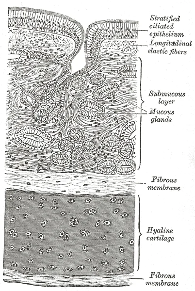
Transverse section of trachea. Labelled illustration from Wikimedia Commons, used under its open licence.

Figure 11: Transverse section of trachea.
Each lobe of the lung has its own lymphatic drainage pattern. The lymphatics of the upper, middle, and lower lobes converge towards the hilum. The oblique and horizontal fissures contain lymphatic channels that connect the adjacent lobes. These channels allow for collateral drainage between lobes. This plexus lies just beneath the visceral pleura.
The bronchopulmonary segments are the functional units of the lung. Each segment has its own segmental bronchus and artery. The lymphatics follow the segmental bronchi. They drain into the intersegmental lymph nodes. These nodes are located at the boundaries between segments. The lymph from all segments eventually converges at the hilum of the lung. The hilar lymph nodes are situated at the root of the lung. They receive lymph from the lung parenchyma, bronchi, and visceral pleura. These nodes are the first major filtering station for pulmonary lymph.
The lymphatic vessels run in close association with the pulmonary vessels and bronchi. They are embedded in the connective tissue sheaths that surround these structures. The nerves of the lung also accompany the lymphatics. The autonomic nervous system regulates the tone of the lymphatic vessels. However, the primary driver of lymph flow is the pressure gradient and the contraction of surrounding tissues.
The lymph from the hilar nodes drains into the bronchomediastinal trunks. The right bronchomediastinal trunk usually drains into the right lymphatic duct. The left bronchomediastinal trunk usually drains into the thoracic duct. These ducts empty into the venous system at the junction of the internal jugular and subclavian veins. This pathway ensures that lymph from the lungs returns to the systemic circulation.
The trachea is central. The main bronchi branch laterally. The hilar nodes are located at the root of each lung. The bronchomediastinal trunks ascend towards the neck.
DefinitionHilar Nodes
The hilar lymph nodes are the first major filtering station for pulmonary lymph, receiving drainage from the lung parenchyma, bronchi, and visceral pleura.
| Structure | Primary Drainage Site |
|---|---|
| Trachea | Pretracheal and paratracheal nodes |
| Bronchi | Hilar lymph nodes |
| Lung Lobes | Hilar lymph nodes |
| Visceral Pleura | Hilar lymph nodes |
| Hilar Nodes | Bronchomediastinal trunks |
The innervation of the lungs and bronchial tree is complex, involving both autonomic and somatic components that govern physiological function and pain perception respectively. Understanding this dual supply is critical for interpreting clinical symptoms such as referred pain.
The autonomic nervous system regulates bronchial smooth muscle tone, glandular secretion, and vascular diameter. This regulation is mediated by the pulmonary plexuses, which are located anterior and posterior to the root of each lung.
The parasympathetic supply originates from the vagus nerves (cranial nerve X). Fibres from the vagus nerve contribute to the pulmonary plexuses. These preganglionic parasympathetic fibres synapse with postganglionic neurons located within or near the bronchial walls. The postganglionic fibres then innervate the bronchial smooth muscle and mucous glands. The primary effect of parasympathetic stimulation is bronchoconstriction and increased glandular secretion. This mechanism is mediated via muscarinic receptors.
The sympathetic supply arises from the sympathetic trunk. Preganglionic fibres originate from the upper thoracic spinal segments (typically T1–T4 or T5). These fibres ascend to the cervical and upper thoracic sympathetic ganglia, where they synapse. Postganglionic fibres then travel to the pulmonary plexuses. The primary effect of sympathetic stimulation is bronchodilation and vasoconstriction. This effect is mediated via beta-2 adrenergic receptors on the smooth muscle. It is important to note that while sympathetic fibres are present, the bronchial smooth muscle lacks direct sympathetic innervation in the same dense pattern as other organs; rather, circulating catecholamines play a significant role in bronchodilation. [1]
Somatic innervation is responsible for pain sensation. The visceral pleura and the lung parenchyma themselves are insensitive to pain. Pain from the lungs is typically referred.
The parietal pleura, which lines the thoracic cavity and covers the diaphragm, is sensitive to pain. This sensitivity is mediated by somatic nerves. The costal and mediastinal parietal pleura are innervated by the intercostal nerves. Pain from this region is felt in the corresponding dermatomes of the chest wall. The diaphragmatic parietal pleura is innervated by the phrenic nerve (C3–C5). Irritation of the diaphragmatic pleura, such as from subphrenic abscesses or liver pathology, results in referred pain to the shoulder tip. This is because the phrenic nerve shares spinal cord segments (C3–C5) with the supraclavicular nerves, which supply the skin over the shoulder.
The trachea and bronchi are innervated by the vagus nerve and the sympathetic trunk. However, pain from the central airways is often poorly localised. In contrast, inflammation of the parietal pleura causes sharp, localised pain that worsens with respiration.
Recognition of referred pain patterns aids in diagnosis. Shoulder pain in a patient with respiratory symptoms should prompt consideration of diaphragmatic irritation. Conversely, chest wall pain that is localised and pleuritic suggests parietal pleural involvement.
Link to the histology page.
The airway epithelium represents a physical barrier to the external environment acting as the first line of defence against potentially harmful environmental stimuli including microbes and allergens. This structure is not merely passive; lung epithelial cells are increasingly recognized as active effectors of microbial defence, contributing to both innate and adaptive immune function in the lower respiratory tract. [2]
In the lower respiratory tract, the respiratory epithelium provides a highly effective barrier acting as the first line of defence against potentially harmful environmental stimuli including microbes and allergens. It constitutes the first site of interaction with inhaled environmental compounds, and is designed to facilitate effective mucociliary clearance of particles and microbes. [2]
Multi‐ciliated cells have cilia on the apical surface that beat co‐ordinately to shuttle inhaled particulates and mucus out of the airways whilst goblet and secretory cells trap inhaled particulates and microorganisms. This coordinated action ensures that inhaled debris is removed before it can cause infection or inflammation. [2]
These transmembrane receptors can be activated independently of the adaptive immune response.
DefinitionPseudostratified Epithelium
A single layer of cells that appears to be multiple layers because nuclei are at different heights, but all cells contact the basement membrane.
Clinical pearlMucociliary Escalator
The combined action of ciliary beating and mucus secretion creates a transport system that moves trapped particles from the lower airways to the pharynx for swallowing or expectoration.
| Cell Type | Primary Function |
|---|---|
| Multi‐ciliated cells | Beat co‐ordinately to shuttle inhaled particulates and mucus out of the airways |
| Goblet cells | Secrete mucus to trap inhaled particulates and microorganisms |
| Basal cells | Serve as stem cells for regeneration of the epithelium |
| Neuroendocrine cells | Secrete surfactant and participate in local immune regulation |
Modern molecular techniques have uncovered the complexity of the lower respiratory tract microbiome. The interaction between the microbiota and the airway epithelium is key to understanding how stable immune homeostasis is maintained. These cells express an ample repertoire of pattern recognition receptors with specificity for conserved microbial and host motifs. This recognition allows the epithelium to distinguish between harmless environmental compounds and potential pathogens, initiating appropriate immune responses without causing unnecessary inflammation. [2]
The structural integrity of the epithelium is crucial for maintaining this barrier function. Damage to the epithelium can compromise the mucociliary clearance mechanism, leading to increased susceptibility to respiratory infections. Understanding the histological composition and functional roles of these cells is essential for grasping the pathophysiology of various respiratory diseases.
The respiratory system originates from the laryngotracheal groove, an outpouching of the ventral foregut endoderm. This primitive structure appears during the fourth week of embryonic development. The groove elongates to form the respiratory diverticulum, also known as the lung bud. This bud grows ventrally into the septum transversum, a mass of mesoderm that separates the thoracic and abdominal cavities. The lung bud bifurcates into two primary bronchial buds, which become the right and left primary bronchi. These buds are surrounded by splanchnic mesoderm, which forms the cartilage, smooth muscle, and connective tissue of the airways.
The branching process continues in a predictable pattern. The primary bronchi divide into secondary bronchi, which supply the lobes. Each secondary bronchus divides into tertiary bronchi, supplying the segments. This branching continues until the terminal bronchioles are formed. The terminal bronchioles mark the end of the conducting zone. Distal to this point, the respiratory zone begins. The respiratory bronchioles give rise to alveolar ducts and alveolar sacs. The alveoli are the functional units of gas exchange. They are lined by type I pneumocytes, which facilitate diffusion, and type II pneumocytes, which secrete surfactant. Surfactant reduces surface tension and prevents alveolar collapse.
The vascular supply develops concurrently with the airways. The right and left pulmonary arteries enter the lungs at the hilum. The pulmonary veins drain oxygenated blood from the alveolar capillaries. They converge to form four pulmonary veins, two from each lung. These veins enter the left atrium of the heart. The bronchial arteries arise from the aorta. They supply the lung tissue itself, including the bronchi and connective tissue. This dual blood supply is unique to the lungs.
The pleura also develops from the mesoderm. The visceral pleura covers the lung surface. The parietal pleura lines the thoracic cavity. The space between them is the pleural cavity. It contains a small amount of fluid for lubrication. The diaphragm separates the thoracic and abdominal cavities. It forms from the septum transversum, the pleuroperitoneal membranes, and the dorsal mesentery. The diaphragm is the primary muscle of respiration. Its contraction increases the volume of the thoracic cavity. This decreases intrapleural pressure and draws air into the lungs.
DefinitionLung Bud
The respiratory diverticulum that bifurcates to form the primary bronchi and initiates the branching morphogenesis of the bronchial tree.
Clinical pearlSurfactant Function
Type II pneumocytes secrete surfactant to reduce surface tension, preventing alveolar collapse at end-expiration.
| Structure | Function |
|---|---|
| Respiratory Bronchiole | Gas exchange begins |
| Alveolar Duct | Passage to alveoli |
| Alveolar Sac | Cluster of alveoli |
| Alveolus | Site of gas exchange |
Figure 12Illu bronchi lungs numerical labels
Long description
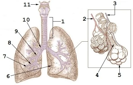
This file was derived from: Illu bronchi lungs.jpg The original PD image is modified by replacing the text labels with numerical labels, using the same numbering scheme as File:Diagrama de los pulmones.svg. This image ca

Figure 12: This file was derived from: Illu bronchi lungs.jpg The original PD image is modified by replacing the text labels with numerical labels, using the same numbering scheme as File:Diagrama de los pulmones.svg (this image ca).
The trachea appears as a vertical, air-filled column in the midline of the upper mediastinum. It bifurcates at the level of the sternal angle (T4/T5 vertebrae) into the right and left main bronchi. The right main bronchus is shorter, wider, and more vertical than the left, which explains the higher incidence of foreign body aspiration into the right lung. The left main bronchus passes under the aortic arch and anterior to the oesophagus. [2]
The lung fields are divided into lobes by fissures. The right lung has three lobes (upper, middle, and lower) separated by the horizontal and oblique fissures. The left lung has two lobes (upper and lower) separated by the oblique fissure. The cardiac notch on the left lung creates a concavity for the heart. The hilum, where vessels and bronchi enter, is visible as a dense shadow lateral to the heart border.
DefinitionLung Lobes
The right lung has three lobes; the left has two, separated by oblique fissures.
Computed tomography (CT) provides cross-sectional detail, revealing the bronchial tree's branching pattern and the vascular supply. The pulmonary arteries accompany the bronchi, while the pulmonary veins drain the lung tissue separately. The pleura lines the lungs and the thoracic cavity, with the visceral pleura adhering to the lung surface and the parietal pleura lining the chest wall.
| Structure | Origin | Course | Termination |
|---|---|---|---|
| Trachea | Larynx | Midline neck/thorax | Bifurcation at T4/T5 |
| Right Main Bronchus | Trachea | Short, wide, vertical | Lobar bronchi |
| Left Main Bronchus | Trachea | Long, narrow, horizontal | Lobar bronchi |
Understanding these radiological features is essential for interpreting chest imaging. Abnormalities such as consolidation, effusion, or masses can be localized based on their anatomical position relative to these landmarks. The clarity of the costophrenic angles and the sharpness of the diaphragm dome are also critical indicators of normal pleural space status.
The bronchial tree is the central conduit for air, branching from the trachea into the right and left main bronchi. The right main bronchus is shorter, wider, and more vertical than the left, making it the common site for aspirated foreign bodies. This anatomical arrangement dictates the distribution of pathology and the approach to surgical intervention.
The lungs are divided into bronchopulmonary segments, which are the functional and surgical units of the lung parenchyma. Each segment is a discrete pyramidal mass of lung tissue with its own segmental bronchus, artery, and lymphatic drainage, but shares a common venous drainage with adjacent segments. This independent blood supply allows for segmental resection without compromising the viability of surrounding lung tissue.
The right lung has ten segments: three in the upper lobe (apical, posterior, anterior), two in the middle lobe (lateral, medial), and five in the lower lobe (apical, anterior basal, medial basal, posterior basal, lateral basal). The left lung has eight or nine segments: four in the upper lobe (apicoposterior, anterior, superior lingular, inferior lingular) and four or five in the lower lobe (apical, anterior basal, medial basal, posterior basal, lateral basal). The superior segment of the lower lobe is often considered separately due to its distinct apical position.
The apical segment of the right upper lobe is located at the very top, while the basal segments of the lower lobe form the inferior base. Understanding this spatial arrangement is crucial for localising lesions on imaging and planning resections.
Lung resection is guided by the segmental anatomy. A lobectomy involves removing an entire lobe, while a segmentectomy removes a specific bronchopulmonary segment. The segmental arteries are ligated first to reduce bleeding, followed by the bronchus and veins.
| Lobe | Segment | Artery | Bronchus |
|---|---|---|---|
| Right Upper | Apical | Apical | Apical |
| Right Upper | Posterior | Posterior | Posterior |
| Right Upper | Anterior | Anterior | Anterior |
| Right Middle | Lateral | Lateral | Lateral |
| Right Middle | Medial | Medial | Medial |
| Right Lower | Apical | Apical | Apical |
| Right Lower | Anterior Basal | Anterior Basal | Anterior Basal |
| Right Lower | Medial Basal | Medial Basal | Medial Basal |
| Right Lower | Posterior Basal | Posterior Basal | Posterior Basal |
| Right Lower | Lateral Basal | Lateral Basal | Lateral Basal |
Clinical use of this anatomy includes localising infections or tumours. For example, tuberculosis often affects the apical segments due to higher oxygen tension. Pneumonia may be segmental, corresponding to the affected bronchopulmonary segment. Surgical resection for lung cancer aims to remove the segment containing the tumour with clear margins, preserving as much healthy lung tissue as possible. The vascular supply is critical; the segmental artery is the primary blood supply, and its ligation is key to safe resection.
An interlobular septum is a wall, composed of connective tissue, which separates lobules from one another. [1]
The bronchial tree begins with the trachea, which bifurcates at the carina, a ridge of cartilage that is more prominent on the right side. This anatomical asymmetry explains why aspirated foreign bodies are more likely to enter the right main bronchus. The right main bronchus is shorter, wider, and runs more vertically than the left. This orientation facilitates the passage of inhaled objects.
The conducting zone extends from the trachea to the terminal bronchioles. It serves to warm, humidify, and filter inspired air. The respiratory zone begins at the respiratory bronchioles and includes the alveolar ducts and sacs. Gas exchange occurs exclusively in the respiratory zone. The alveoli are lined by type I pneumocytes, which are thin squamous cells facilitating diffusion, and type II pneumocytes, which produce surfactant. Surfactant reduces surface tension, preventing alveolar collapse during expiration.
Clinical relevance often focuses on the vascular supply. The lungs have a dual blood supply. The pulmonary arteries carry deoxygenated blood from the right ventricle to the alveoli for gas exchange. The bronchial arteries arise from the aorta and supply the lung tissue itself, including the bronchi and connective tissue. This distinction is vital in haemoptysis; massive bleeding usually originates from the high-pressure bronchial circulation, not the pulmonary circulation.
Common traps include confusing the lobar anatomy. The right lung has three lobes: upper, middle, and lower. The left lung has two lobes: upper and lower. The lingula of the left upper lobe is homologous to the right middle lobe. Misidentifying these segments can lead to errors in localising pathology on chest X-rays or CT scans. Remember that the oblique fissures separate the lower lobes from the upper lobes on both sides, while the horizontal fissure on the right separates the upper and middle lobes.
The lymphatic drainage follows the bronchial tree. Lymph flows from the peripheral lung tissue to the hilar nodes, and then to the tracheobronchial nodes. This pathway is important for the spread of lung cancer. Metastasis to the mediastinal nodes indicates advanced disease. The thoracic duct drains lymph from the left side of the thorax, while the right lymphatic duct drains the right side.
Students frequently confuse the cellular origins of airway secretions, attributing all mucus production to a single cell type. It is essential to distinguish between the large airways and the smaller bronchioles. In the large airways, the submucosal gland harbours the mucus and serous cells that are responsible for the production of most airway mucus. Towards the bronchioles, goblet and club cells produce mucus, whereas in the alveoli AEC1 and AEC2 secrete pulmonary surfactant. Confusing these locations leads to errors in understanding regional pathology. [2]
Another frequent error involves the function of epithelial junctions. Students often view tight junctions merely as structural barriers without appreciating their regulatory role. Tight junctions are the main regulators of paracellular permeability, whereas the adherens junctions mechanically connect adjacent cells, and initiate the formation and maturation of cell−cell contacts. During disease, barrier function is impaired leading to increased epithelial permeability and pathogen entry. Recognising this distinction clarifies why permeability increases before structural collapse occurs. [2]
Misidentifying the specific ligands for Toll-like receptors (TLRs) is a common examination trap. TLRs are not generic sensors but have specific nucleic acid or cell wall targets. The main function of TLRs in mammals is to recognize distinct PAMPs, including components of microbial cell walls (TLR1, TLR2, TLR4 and TLR6), flagellin (TLR5), single‐stranded or double‐stranded viral RNA (TLR3, TLR7 and TLR8) or CpG‐containing DNA (TLR9). Furthermore, the TLR3, 7, 8 and 9 are specifically localized in endosomes a [2]
Students often overlook the specific triggers for asthma exacerbations in different age groups. In asthma, infection by respiratory viruses is a major trigger of wheezing in infants as well as exacerbations of asthma in older children. The infectious agents associated with these wheezing events include respiratory syncytial virus (RSV), human rhinovirus, parainfluenza, coronavirus and other viruses. Assuming bacterial causes are primary for wheezing in infants ignores this viral predominance. [2]
Confusion also arises regarding the role of E-cadherin in immune signalling. E‐cadherin, in addition to having a structural role at adherens junctions, is also the ligand for CD103, which is expressed on innate and adaptive immune cells and a subset of dendritic cells. It also binds to KLRG1, which is expressed by natural killer cells and regulatory T‐cells and group 2 innate lymphoid cells. Viewing it solely as a structural protein misses its immunomodulatory ligand function. [2]
Finally, students may misinterpret Wnt signalling pathways in lung disease. Wnt/β‐catenin signalling is also implicated in the pathogenesis of COPD, but in direct contrast to what is observed in IPF, signalling in the mesenchymal compartment is inactivated rather than activated. In IPF, a shift from canonical Wnt signalling (β‐catenin‐dependent) to non‐canonical pathways is suggested to drive pathology. Reversing these activation states leads to incorrect mechanistic explanations. [2]
This knowledge is applied at the bedside to interpret wheezing in infants. Clinicians recognise that respiratory viruses are the primary drivers, guiding supportive care rather than immediate antibiotic use. Understanding the viral triggers helps in counselling parents about the natural history of recurrent wheeze.
In the operating theatre, awareness of submucosal gland function is critical during airway management. Knowing that large airways produce most mucus via submucosal glands informs suction strategies and humidification requirements to prevent plugging. It also highlights why bronchiolar obstruction involves different cellular mechanisms, affecting drug delivery targets.
On imaging, recognising the distribution of surfactant-producing cells (AEC2) aids in interpreting diffuse alveolar damage. Pathology reports referencing surfactant dysfunction rely on the anatomical fact that AEC2 secrete pulmonary surfactant. This distinguishes alveolar pathology from bronchial mucus hypersecretion seen in chronic bronchitis.
In examinations, precise identification of TLR ligands is frequently tested. Students must match TLR4 to lipopolysaccharide (cell wall component) and TLR3 to double-stranded RNA. The localization of TLR3, 7, 8 and 9 in endosomes is a key detail for understanding intracellular pathogen detection. Misassigning these receptors results in lost marks.
Clinical reasoning also uses the concept of barrier dysfunction. When epithelial permeability increases, pathogen entry is facilitated. This explains the susceptibility to secondary infections in asthma. Recognising that defensins are the most abundant antimicrobial peptides helps in understanding innate immune responses in the airway. [2]
DefinitionAirway Mucus Production
In the large airways, the submucosal gland harbours the mucus and serous cells that are responsible for the production of most airway mucus.
Exam trapTLR Localization
The TLR3, 7, 8 and 9 are specifically localized in endosomes and detect pathogen nucleic acids following endocytosis. [2]
Innate Immune Recognition
- Pathogen EntryVirus or Bacteria
- Question: Location?
Surface
- TLR1,2,4,5,6Recognise cell walls/flagellin
Endosome
- TLR3,7,8,9Recognise nucleic acids
| TLR | Ligand Type |
|---|---|
| TLR1, 2, 4, 6 | Microbial cell wall components |
| TLR5 | Flagellin |
| TLR3, 7, 8 | Viral RNA |
| TLR9 | CpG-containing DNA |
In examinations, the distinction between canonical and non-canonical Wnt signalling is vital. In COPD, signalling in the mesenchymal compartment is inactivated rather than activated. This contrasts with IPF where canonical signalling is active. This specific detail is often tested in pathology questions.
Understanding E-cadherin's role in Th2 responses is also clinically relevant. This links barrier dysfunction directly to allergic inflammation, a key concept in asthma pathophysiology.
Clinical pearlE-cadherin Function
E‐cadherin, in addition to having a structural role at adherens junctions, is also the ligand for CD103.
Submucosal glands produce most large airway mucus. Goblet and club cells produce mucus in bronchioles. Tight junctions regulate paracellular permeability. TLR3, 7, 8, 9 are endosomal and detect nucleic acids. Defensins are the most abundant antimicrobial peptides. E-cadherin is a ligand for CD103. Wnt signalling is inactivated in COPD mesenchyme. Viruses are major triggers for infant wheezing.
Answer each one in your head first, then open the answer.
Which cells produce most airway mucus in large airways?
Show answerHide answer
Submucosal gland cellsWhere are TLR3, 7, 8 and 9 localized?
Show answerHide answer
EndosomesWhat is the main function of tight junctions?
Show answerHide answer
Regulate paracellular permeabilityWhich TLRs recognize cell wall components?
Show answerHide answer
TLR1, TLR2, TLR4 and TLR6What happens to Wnt signalling in COPD mesenchyme?
Show answerHide answer
It is inactivated
- Betts JG, Young KA, Wise JA, et al. The Lungs. OpenStax, Anatomy and Physiology 2e (CC BY-NC-SA 4.0). Accessed .
- Invernizzi R, Lloyd CM, Molyneaux PL. Respiratory microbiome and epithelial interactions shape immunity in the lungs. Immunology 2020;160(2):171-182. doi:10.1111/imm.13195. Accessed .
Figure credits
- Figure 1: Henry Vandyke Carter, via Wikimedia Commons, Public domain, Public domain.
- Figure 2: Henry Vandyke Carter, via Wikimedia Commons, Public domain, Public domain.
- Figure 3: Henry Vandyke Carter, via Wikimedia Commons, Public domain, Public domain.
- Figure 4: Unknown author (see the source page), via Wikimedia Commons, Public domain, Public domain.
- Figure 5: Henry Vandyke Carter, via Wikimedia Commons, Public domain, Public domain.
- Figure 6: Henry Vandyke Carter, via Wikimedia Commons, Public domain, Public domain.
- Figure 7: Henry Vandyke Carter, via Wikimedia Commons, Public domain, Public domain.
- Figure 8: Edelhart Kempeneers, via Wikimedia Commons, Public domain, Public domain.
- Figure 9: Henry Vandyke Carter, via Wikimedia Commons, Public domain, Public domain.
- Figure 10: Henry Vandyke Carter, via Wikimedia Commons, Public domain, Public domain.
- Figure 11: Henry Vandyke Carter, via Wikimedia Commons, Public domain, Public domain.
- Figure 12: An officer or employee of the United States Government, as described at File:Illu bronchi lungs.jpg, with modifications by User:Adrian J. Hunter, via Wikimedia Commons, Public domain, Public domain.
Educational content for medical students and doctors. It is not a substitute for clinical judgement or local guidelines.