Pleura
The pleura is a serous membrane enclosing the lungs and lining the thoracic cavity.
The lungs are pyramid-shaped, paired organs connected to the trachea by the right and left bronchi[1]. They are enclosed by the pleurae, which are attached to the mediastinum[1]. The right lung is shorter and wider than the left lung, and the left lung occupies a smaller volume than the right[1]. The cardiac notch is an indentation on the surface of the left lung, and it allows space for the heart[1]. The apex of the lung is the superior region, whereas th
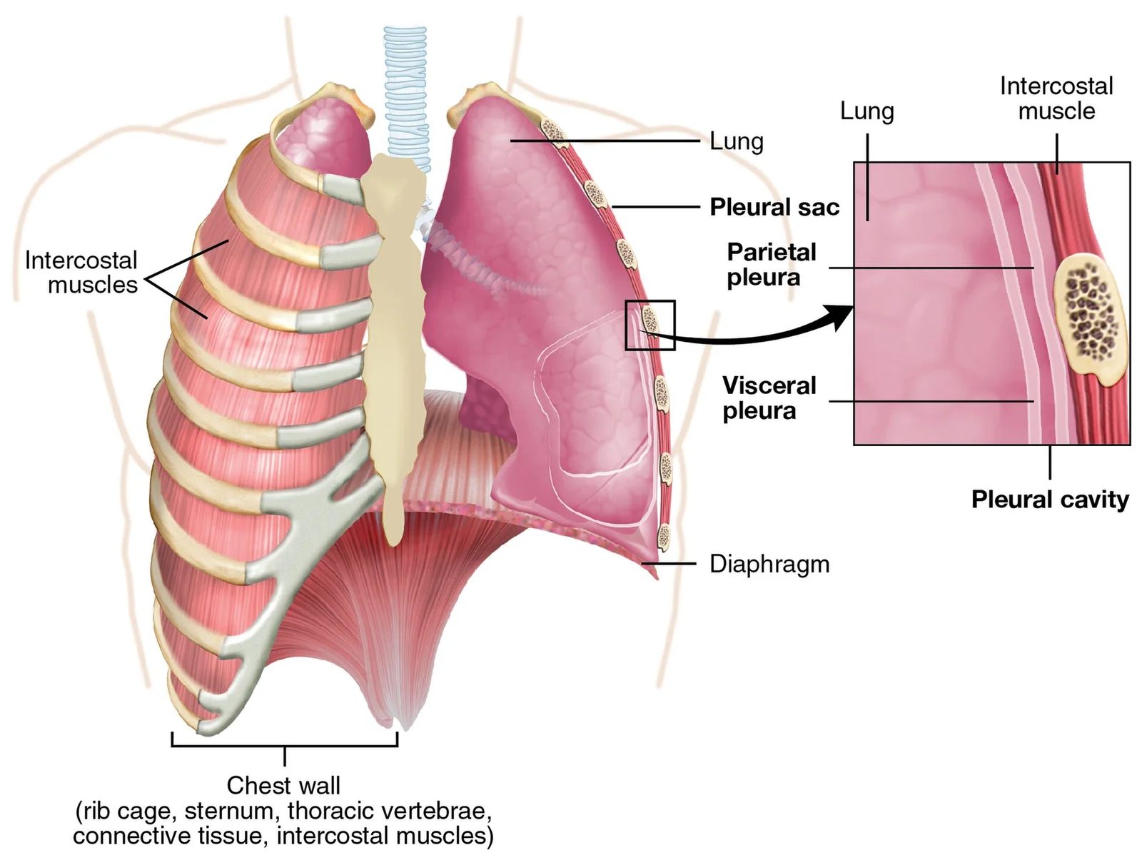
Each lung is enclosed within a cavity that is surrounded by the pleura[1]. The pleura (plural = pleurae) is a serous membrane that surrounds the lung[1]. The right and left pleurae, which enclose the right and left lungs, respectively, are separated by the mediastinum[1]. The pleurae consist of two layers[1]. The visceral pleura is the layer that is superficial to the lungs, and extends into and lines the lung fissures[1]. In contrast, the parietal pleura is the outer layer that connects to the thoracic wall, the mediastinum, and the diaphragm[1]. The visceral and parietal pleurae connect to each other at the hilum[1]. The pleural cavity is the space between the visceral and parietal layers[1].
This cavity is covered by the parietal pleura, which is attached to the chest wall and is covered by a continuous 'parietal' mesothelial cell layer. The lungs themselves are enclosed by the visceral pleura which is integral to the lung surface and which has a surface 'visceral' mesothelial layer. The tight fit of the lungs to the inside of the chest wall means that the two mesothelial layers are closely apposed and there is a thin space between them that contains the pleural fluid (Figure ) and also a population of pleural macrophages. [3]
DefinitionPleural Recesses
The pleural cavity contains recesses where the parietal pleura reflects upon itself, allowing lung expansion during deep inspiration.
The pleural fluid turns over rapidly , continuously exiting through stomata in the parietal (not the visceral) pleura via lymphatic capillaries. These stomatal openings on the parietal pleural surface are between 3 and 10 μm in diameter (Figure ). They are often found in association with 'milky spots', large accumulations of leukocytes present on the parietal pleura and presumed to be involved in immune activity in the pleural space. The pleural fluid outflow through these stomata drains to the [3]
NoteThoracentesis
Thoracentesis involves inserting a needle into the pleural space to remove fluid or air, typically entering through the intercostal space below the rib to avoid neurovascular bundles.
| Pathology | Description |
|---|---|
| Pleural Effusion | A build up of fluid within the pleural space. |
| Pneumothorax | Air in the pleural cavity, disrupting negative pressure. |
| Mesothelioma | Tumour originating in the parietal pleura. |
Figure 1Gray966
Long description
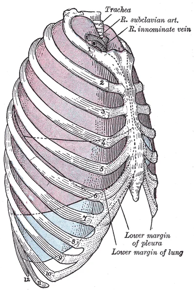
Lateral view of thorax, showing the relations of the pleuræ and lungs to the chest wall. Pleura in blue; lungs in purple.

Figure 1: Lateral view of thorax, showing the relations of the pleuræ and lungs to the chest wall.
The pleura forms a continuous serous membrane that encloses each lung within its own pleural cavity, the right and left cavities being completely separated by the mediastinum. The membrane has two distinct layers. The visceral pleura invests the lung surface directly, dipping into and lining the interlobar fissures so that it follows the contours of the lung parenchyma. The parietal pleura lines the inner aspect of the thoracic wall, covers the mediastinal surfaces and the superior surface of the diaphragm, and is continuous with the visceral layer at the hilum where structures enter and leave the lung. The potential space between these two layers is the pleural cavity, which normally contains only a thin film of lubricating fluid. [4]
DefinitionParietal pleural stomata
Microscopic openings (stomata) in the parietal pleura provide the normal clearance pathway for particles and fluid from the pleural space into the lymphatic system; long fibres are retained at these sites because their length exceeds the stomatal diameter.
The parietal pleura is functionally and pathologically distinct from the visceral layer. Experimental and human evidence shows that a fraction of all inhaled particles and fibres translocate to the pleural space, but only long fibres are retained in the parietal pleura, specifically at the stomata. This length‑dependent retention creates a focussed ‘dose’ of long fibres at the parietal mesothelium, driving sustained inflammation and pleural pathology including mesothelioma. Thoracoscopic studies confirm that mesothelioma originates on the parietal pleura; early disease confined to this layer carries a prognosis better by around 30 months than when the visceral pleura is involved. Staging systems therefore recognise parietal‑only disease as the earliest stage. [5]
| Feature | Visceral pleura | Parietal pleura |
|---|---|---|
| Anatomical relations | Covers lung surface, lines fissures | Lines thoracic wall, mediastinum, diaphragm |
| Continuity | Continuous with parietal layer at hilum | Continuous with visceral layer at hilum |
| Innervation | Autonomic (insensitive to pain) | Somatic (sensitive to pain) |
| Stomata | Absent | Present; clearance pathway for pleural fluid and particles |
| Fibre retention | Minimal; fibres recovered are often short | Major site of long fibre retention at stomata |
| Mesothelioma origin | Involved in advanced disease | Primary site of origin; early disease confined here |
Pleural fibre translocation and retention
- InhalationParticles and fibres deposit in lung parenchyma
- TranslocationA fraction of all deposited particles/fibres translocate to pleural space
- Clearance attemptNormal clearance via parietal pleural stomata into lymphatics
- Question: Fibre length vs stomatal diameter
Short fibres/particles
- Cleared through stomataLow‑grade parietal pleural 'black spots' may form transiently
Long fibres (e.g. asbestos, long nanotubes)
- Retained at parietal pleural stomataLength prevents passage; focal accumulation on parietal mesothelium
- Sustained mesothelial injury and inflammationDrives pleural pathology including mesothelioma
The practical implication is that assessment of mesothelioma risk should focus on whether fibres are retained in the parietal pleura rather than merely whether they reach the pleural space. Lung parenchymal or visceral pleural fibre burdens, which often contain only short fibres, do not reflect the effective parietal pleural dose.
The visceral pleura is the layer that is superficial to the lungs, and extends into and lines the lung fissures.
Figure 22313 The Lung Pleurea
Long description
Illustration from Anatomy & Physiology, Connexions Web site. http://cnx.org/content/col11496/1.6/, Jun 19, 2013.

Figure 2: Illustration from Anatomy & Physiology, Connexions Web site (http://cnx.org/content/col11496/1.6/, jun 19, 2013).
The peritoneal cavity is lined by a mesothelium similar to that of the pleura. This similarity has led to the use of the peritoneal cavity as a surrogate model for studying pleural mesothelium in fibre toxicity research. However, the clearance mechanisms differ. These stomata allow particles to be drawn cranially through the diaphragm to the parathymic lymph nodes. In contrast, the parietal pleura retains long fibres, which are implicated in the pathogenesis of mesothelioma. The parietal pleura is the primary site of fibre retention and subsequent pathology. The visceral pleura is less involved in fibre retention compared to the parietal layer. [3]
| Feature | Visceral Pleura | Parietal Pleura |
|---|---|---|
| Location | Covers lung surface | Lines thoracic cavity |
| Sensation | Insensitive to pain | Sensitive to pain |
| Fibre Retention | Low | High |
| Clearance | Limited | Stomata present in peritoneum, not pleura |
The relationship between the pleura and the peritoneum is anatomical but functionally distinct in terms of clearance and pathology. While both are lined by mesothelium, the pleural cavity is designed to facilitate lung movement within the rigid thoracic cage. The peritoneal cavity has its own clearance system via stomata. This retention is critical in the development of pleural diseases such as mesothelioma. The parietal pleura is the key site for this retention. The visceral pleura remains relatively free of retained fibres. [3]
Figure 3Gray968
Long description
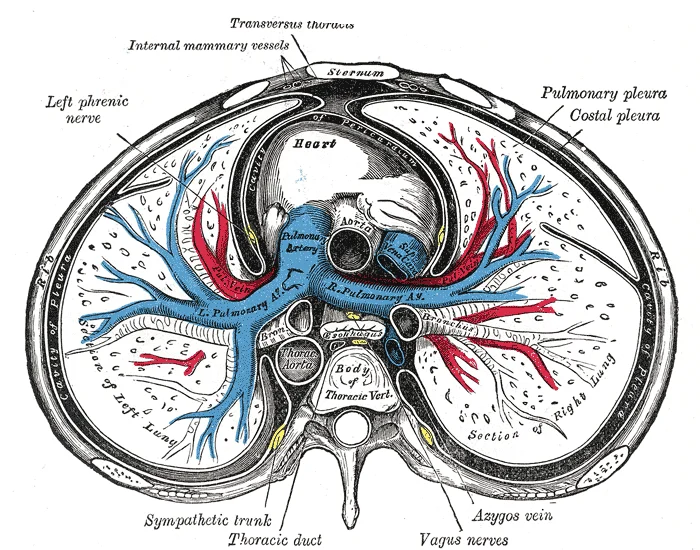
A transverse section of the thorax, showing the contents of the middle and the posterior mediastinum. The pleural and pericardial cavities are exaggerated since normally there is no space between parietal and visceral pleura and between pericardium and heart.

Figure 3: A transverse section of the thorax, showing the contents of the middle and the posterior mediastinum.
The parietal pleura is further subdivided based on its anatomical location.
The visceral pleura is shown enveloping the lung parenchyma. The narrow gap between the parietal and visceral layers represents the pleural cavity. The diagram highlights the continuity of the membrane as it reflects at the hilum, where the visceral pleura becomes continuous with the parietal pleura. This reflection creates the recesses, such as the costodiaphragmatic recess, which is the lowest point of the pleural cavity. Understanding this spatial relationship is crucial for interpreting imaging and performing procedures like thoracentesis.
The structural integrity of the pleura relies on its cellular composition. The mesothelial cells lining the pleural cavity form a protective barrier. Beneath the mesothelium lies a layer of connective tissue containing blood vessels, lymphatics, and nerves. The lymphatic drainage is particularly important for fluid balance and immune surveillance. Lymphatic stomata, primarily located in the parietal pleura, facilitate the removal of fluid and particles from the pleural space. These stomata act as a sieve, selectively retaining larger particles while allowing fluid to pass into the lymphatic system. [3]
DefinitionPleural Cavity
A potential space between the visceral and parietal pleura containing a thin layer of serous fluid that facilitates lung movement.
The vascular supply to the parietal pleura is derived from systemic arteries, including the intercostal arteries and the internal thoracic artery. This dual blood supply reflects the different embryological origins and functional roles of the two layers. The innervation also differs: the parietal pleura is sensitive to pain, temperature, and touch, supplied by somatic nerves, whereas the visceral pleura is insensitive to pain, being supplied by autonomic nerves. [3]
| Layer | Blood Supply | Innervation | Sensation |
|---|---|---|---|
| Parietal | Systemic (Intercostal, Internal Thoracic) | Somatic (Intercostal nerves) | Pain, Touch, Temperature |
| Visceral | Pulmonary | Autonomic | Insensitive to Pain |
The arterial supply to the pleura is distinct from the pulmonary circulation, reflecting its role in somatic sensation and structural support rather than gas exchange. This distinction is critical because the parietal pleura is innervated by somatic nerves and is sensitive to pain, temperature, and touch, while the visceral pleura is insensitive to these stimuli.
The parietal pleura is supplied by branches of the systemic circulation that correspond to the region of the thoracic cavity it lines. The costal and diaphragmatic parietal pleura are primarily supplied by the intercostal arteries and the musculophrenic artery, which are branches of the aorta. The mediastinal parietal pleura receives blood from the internal thoracic arteries and the pericardiophrenic arteries. The cervical parietal pleura (cupula) is supplied by branches from the subclavian artery, such as the supreme intercostal artery. [1]
The visceral pleura, which covers the lung surface and extends into the fissures, is supplied by the pulmonary arteries. These arteries arise from the pulmonary trunk and carry deoxygenated blood. As the pulmonary arteries branch and follow the bronchi, they become progressively smaller in diameter. One arteriole and an accompanying venule supply and drain one pulmonary lobule. As they near the alveoli, the pulmonary arteries become the pulmonary capillary network. This network consists of tiny vessels with very thin walls that lack smooth muscle fibers, facilitating gas exchange. The major function of the lungs is to perform gas exchange, which requires blood from the pulmonary circulation. [1]
Understanding the dual blood supply is essential for interpreting pleural pathology. Systemic hypertension can affect the parietal pleura, while pulmonary hypertension affects the visceral pleura. The parietal pleura's sensitivity to pain from systemic nerve supply explains why pleuritic pain is sharp and localized, whereas visceral pleural irritation causes diffuse, poorly localized discomfort.
The venous drainage of the pleura follows a pattern that mirrors its arterial supply, ensuring efficient return of blood from the thoracic cavity walls and the visceral surface of the lungs. Understanding this system requires looking at the distinct pathways for the parietal and visceral layers, as they do not merge immediately but serve different physiological roles before converging into the systemic circulation.
The parietal pleura, which lines the thoracic cavity, receives its venous drainage through a network that aligns with the regional anatomy of the chest wall. The blood from the costal, diaphragmatic, mediastinal, and cervical parts of the parietal pleura drains into the intercostal veins. These veins then empty into the azygos system on the right side and the hemiazygos or accessory hemiazygos systems on the left. This arrangement ensures that blood from the chest wall is returned to the superio
In contrast, the visceral pleura, which covers the lungs, has a venous drainage system that is intimately linked with the pulmonary circulation. This pathway is crucial because it integrates the pleural circulation with the gas exchange function of the lungs. Any pathology affecting the visceral pleura, such as inflammation or tumour, can therefore impact the pulmonary venous return.
The balance of forces within the thorax is maintained by the integrity of these vascular and lymphatic systems. Competing forces within the thorax cause the formation of the negative intrapleural pressure. The surface tension of alveolar fluid, which is mostly water, also creates an inward pull of the lung tissue. This inward tension from the lungs is countered by opposing forces from the pleural fluid and thoracic wall. Since the parietal pleura is attached to the thoracic wall, the natural ela [2]
The lymphatic drainage of the pleura is a critical anatomical pathway that facilitates the clearance of interstitial fluid and particulate matter. Understanding this system requires distinguishing between the two distinct layers of the pleura, as their drainage patterns differ significantly in direction and destination.
The parietal pleura, which lines the thoracic cavity, connects to the thoracic wall, the mediastinum, and the diaphragm.
In contrast, the visceral pleura, which covers the lung surface, has a more limited drainage route. Its lymphatics follow the bronchovascular bundles towards the hilum of the lung. At the hilum, these vessels enter the pulmonary nodes and subsequently the bronchopulmonary (hilar) nodes. From the hilar nodes, lymph flows into the tracheobronchial nodes and then into the bronchomediastinal trunks. This centralised drainage pattern means that pathological processes affecting the visceral pleura often manifest in the hilar and mediastinal lymph nodes.
The structural integrity of the lymphatic capillaries is vital for normal pleural physiology. Recent hypotheses suggest that pleural plaques, often associated with asbestos exposure, represent a unique pathological response. This mechanism highlights the role of the stomatal openings in the mesothelium, which allow particulate matter to access the underlying lymphatic system. [3]
DefinitionStomatal openings
Small pores in the mesothelium that allow particulate matter to access the sub-mesothelial interstitium and lymphatic capillaries.
| Pleural Layer | Primary Drainage Route | Destination Nodes |
|---|---|---|
| Parietal (Diaphragmatic) | Inferiorly via phrenic vessels | Anterior diaphragmatic, Retrosternal |
| Parietal (Costal) | Laterally via intercostal spaces | Intercostal, Posterior mediastinal |
| Visceral | Along bronchovascular bundles | Pulmonary, Bronchopulmonary (Hilar) |
The pleura receives its nerve supply from both autonomic and somatic sources, which dictates the nature of pain perception. The parasympathetic and sympathetic nervous systems provide autonomic control over the airways and associated structures. These fibers converge to form the pulmonary plexus, a region located on the lung root at the hilum. From this plexus, nerves follow the bronchi into the lungs, branching to innervate muscle fibers, glands, and blood vessels. [1]
Pain referral is a critical clinical concept derived from this innervation. The parietal pleura is innervated by somatic nerves, including the intercostal nerves and the phrenic nerve. Irritation of the diaphragmatic pleura, supplied by the phrenic nerve (C3-C5), often refers pain to the shoulder region. In contrast, the costal pleura is supplied by intercostal nerves, and irritation here causes pain that corresponds to the specific dermatome involved. This distinction is vital for diagnosing pleural pathology.
The pleura is a serous membrane that lines the thoracic cavity and covers the lungs. It consists of two layers: the parietal pleura, which lines the chest wall, diaphragm, and mediastinum, and the visceral pleura, which adheres directly to the lung surface. Between these layers lies the pleural cavity, a potential space containing a thin film of serous fluid. This fluid reduces friction during respiratory movements, allowing the lungs to expand and contract smoothly against the chest wall. [1]
Figure 4Gray965
Long description
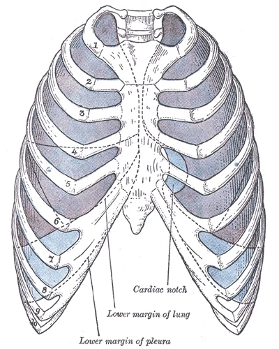
Front view of thorax, showing the relations of the pleuræ and lungs to the chest wall. Pleura in blue; lungs in purple.

Figure 4: Front view of thorax, showing the relations of the pleuræ and lungs to the chest wall.
The histological structure of the pleura is simple yet functional. Both layers are composed of a single layer of mesothelial cells resting on a thin basement membrane. These mesothelial cells are flat and squamous, providing a smooth, non-adherent surface. Underlying the mesothelium is a sparse connective tissue layer containing fibroblasts, collagen fibres, and elastic fibres. The presence of elastic fibres allows the pleura to stretch and recoil with lung movement. Blood vessels, lymphatics, and nerves are located in the submesothelial connective tissue, particularly in the parietal pleura, which is more vascular and sensitive to pain than the visceral pleura.
The parietal pleura is further subdivided based on its anatomical location.
The visceral pleura is tightly adherent to the lung parenchyma and extends into the interlobar fissures. It is insensitive to pain because it is innervated by the autonomic nervous system, which does not convey pain sensations. In contrast, the parietal pleura is innervated by somatic nerves, making it sensitive to pain, temperature, and touch. This difference in innervation explains why pleuritic pain is typically sharp and localized, arising from inflammation of the parietal pleura.
The pleural cavity normally contains a small volume of fluid, approximately 10 to 20 mL in adults. This fluid is produced by the parietal pleura and absorbed by the lymphatics. The balance between production and absorption maintains the negative intrapleural pressure, which is essential for lung expansion. Disruption of this balance can lead to pleural effusion, where excess fluid accumulates in the cavity, potentially compromising lung function. [3]
The pleura develops from the lateral plate mesoderm during the early stages of embryogenesis. This mesodermal layer splits into two distinct layers: the somatic (parietal) mesoderm and the splanchnic (visceral) mesoderm. The somatic mesoderm associates with the somatic ectoderm to form the body wall, while the splanchnic mesoderm associates with the endoderm to form the gut tube. Between these two layers lies the intraembryonic coelom, which eventually becomes the pleural, pericardial, and peritoneal cavities.
As the embryo grows, the lungs begin to develop as outgrowths of the foregut. These lung buds grow into the pericardioperitoneal canals, which are part of the intraembryonic coelom. The visceral pleura is derived from the splanchnic mesoderm that covers the developing lung buds. The parietal pleura is derived from the somatic mesoderm that lines the body wall. The two layers are continuous with each other at the hilum of the lung, where the visceral pleura reflects onto the parietal pleura.
The diaphragm also plays a crucial role in the development of the pleural cavities. The septum transversum, a thick mass of mesoderm, forms the central tendon of the diaphragm. The pleuroperitoneal membranes close the pericardioperitoneal canals, separating the pleural cavities from the peritoneal cavity. This separation is essential for the proper formation of the thoracic and abdominal cavities.
The embryological origin of the pleura is thus a complex interplay between mesodermal layers and the developing organs. Understanding this development is crucial for appreciating the anatomical relationships and potential pathological processes that can affect the pleura. The continuity between the visceral and parietal layers ensures that the pleural cavity remains a closed, potential space, allowing for the smooth movement of the lungs during respiration. Any disruption to this delicate balance, whether through developmental anomalies or external insults, can lead to significant clinical manifestations.
Understanding the pleura through imaging requires correlating surface landmarks with underlying anatomical structures to ensure safe clinical interventions. The pleura consists of two layers: the parietal pleura, which lines the thoracic cavity, and the visceral pleura, which covers the lungs. These layers are continuous at the hilum and enclose the pleural cavity, a potential space containing a small amount of lubricating fluid.
The caption states this is a sagittal ultrasound scan demonstrating the diaphragm and pleural interface. The costophrenic angle is clearly defined where the parietal pleura of the chest wall meets the parietal pleura of the diaphragm. This angle is crucial for thoracocentesis as it is the deepest part of the pleural cavity in the upright position, allowing for maximal fluid accumulation. The needle insertion site is typically marked just above the upper border of the rib to avoid the neurovascular bundle running along the inferior margin.
Thoracocentesis is a procedure to remove fluid or air from the pleural space. Ultrasound guidance has become standard practice to identify the optimal entry point. The sonographer identifies a safe window, usually in the mid-scapular or posterior axillary line, avoiding intercostal vessels and nerves. The needle is advanced slowly until the parietal pleura is penetrated, confirmed by a change in resistance and the appearance of fluid on the screen.
Clinical pearlSafe Zone
Always insert the needle above the upper border of the rib to avoid the intercostal neurovascular bundle located in the costal groove.
The anatomical relations are critical for safety. The intercostal muscles lie between the ribs, providing structural support and aiding in respiration. The parietal pleura is innervated by somatic nerves, making it sensitive to pain, unlike the visceral pleura, which is insensitive to pain but sensitive to stretch.
| Structure | Origin | Insertion | Action | Nerve Supply |
|---|---|---|---|---|
| External Intercostal | Inferior border of rib above | Superior border of rib below | Elevates ribs during inspiration | Intercostal nerve |
| Internal Intercostal | Rib above | Rib below | Depresses ribs during expiration | Intercostal nerve |
| Innermost Intercostal | Rib above | Rib below | Depresses ribs during expiration | Intercostal nerve |
Ultrasound anatomy also allows for the identification of pathological conditions. Pleural effusions appear as anechoic (black) spaces between the parietal and visceral pleura. Pneumothorax is identified by the absence of lung sliding and the presence of a 'lung point' where aerated and non-aerated lung meet. The clarity of the diaphragm and pleural interface in this view underscores the importance of understanding normal anatomy before identifying abnormalities.
The parietal pleura is the outer layer that connects to the thoracic wall, the mediastinum, and the diaphragm.
DefinitionPleural Cavity
A potential space between the visceral and parietal pleura containing a small amount of lubricating fluid, maintaining negative pressure relative to atmospheric pressure.
The reflections of the parietal pleura create recesses where the layers fold back on themselves. These recesses allow for lung expansion during deep inspiration. The costodiaphragmatic recess is the largest and most dependent part of the pleural cavity. It lies between the costal and diaphragmatic pleura. The costomediastinal recess is smaller and located anteriorly near the sternum. These recesses are clinically significant because fluid or air may accumulate here. [1]
The costal pleura is attached to the inner surfaces of the ribs. Understanding these relations is essential for interpreting chest radiographs and performing thoracentesis. The needle for thoracentesis is typically inserted just above the upper border of a rib to avoid the neurovascular bundle running along the inferior margin of the rib above. [1]
| Muscle | Origin | Insertion | Action | Nerve Supply |
|---|---|---|---|---|
| External Intercostal | Inferior border of rib | Superior border of rib below | Elevates ribs during inspiration | Intercostal nerves |
| Internal Intercostal | Rib angle | Costal cartilage of rib below | Depresses ribs during forced expiration | Intercostal nerves |
| Innermost Intercostal | Rib angle | Inner surface of rib below | Depresses ribs during forced expiration | Intercostal nerves |
The neurovascular bundle runs in the costal groove on the inferior border of each rib. This bundle contains the intercostal vein, artery, and nerve, arranged from superior to inferior. Protection of this bundle is critical during surgical procedures involving the chest wall. The pleura is innervated by somatic nerves. The parietal pleura is sensitive to pain, temperature, and touch. The visceral pleura is insensitive to pain but sensitive to stretch. This difference explains why pleuritic pain i [1]
The pleurae are serous membranes that perform two major functions: they produce pleural fluid and create cavities that separate the major organs. Pleural fluid is secreted by mesothelial cells from both pleural layers and acts to lubricate their surfaces. This lubrication reduces friction between the two layers to prevent trauma during breathing, and creates surface tension that helps maintain the position of the lungs against the thoracic wall. This adhesive characteristic of the pleural fluid [1]
Pulmonary ventilation is dependent on three types of pressure: atmospheric, intra-alveolar, and intrapleural[1]. Atmospheric pressure is the amount of force that is exerted by gases in the air surrounding any given surface, such as the body[1]. Atmospheric pressure can be expressed in terms of the unit atmosphere, abbreviated atm, or in millimeters of mercury (mm Hg)[1]. One atm is equal to 760 mm Hg, which is the atmospheric pressure at sea level[1]. Typically, for respiration, other pressure values are discussed in relation to atmospheric pressure[1]. Therefore, negative pressure is pressure lower than the atmospheric pressure, whereas positive pressure is pressure that it is greater than the atmospheric pressure[1]. A pressure that is equal to the atmospheric pressure is expressed as zero[1]. [2]
Pulmonary ventilation comprises two major steps: inspiration and expiration[1]. Inspiration is the process that causes air to enter the lungs, and expiration is the process that causes air to leave the lungs[1]. A respiratory cycle is one sequence of inspiration and expiration[1]. In general, two muscle groups are used during normal inspiration: the diaphragm and the external intercostal muscles[1]. Additional muscles can be used if a bigger breath is required[1]. When the diaphragm contracts, it moves inferiorly toward the abdominal cavity, creating a larger thoracic cavity and more space for the lungs[1]. Contraction of the external intercostal muscles moves the ribs upward and outward, causing the rib cage to expand, which increases the volume of the thoracic cavity[1]. Due to the adhesive force of the pleural fluid, the expansion of the thoracic cavity forces the lungs to stretch and expand as well[1]. [2]
Each lung is composed of smaller units called lobes. Fissures separate these lobes from each other. The right lung consists of three lobes: the superior, middle, and inferior lobes. The left lung consists of two lobes: the superior and inferior lobes. A bronchopulmonary segment is a division of a lobe, and each lobe houses multiple bronchopulmonary segments. Each segment receives air from its own tertiary bronchus and is supplied with blood by its own artery. Some diseases of the lungs typically affect one or more bronchopulmonary segments, and in some cases, the diseased segments can be surgically removed with little influence on neighboring segments. [1]
An interlobular septum is a wall, composed of connective tissue, which separates lobules from one another. [1]
Exam trapPressure Zero
A pressure that is equal to the atmospheric pressure is expressed as zero[1].
Clinical pearlPleural Adhesion
This adhesive characteristic of the pleural fluid causes the lungs to enlarge when the thoracic wall expands during ventilation, allowing the lungs to fill with air. [1]
DefinitionBronchopulmonary Segment
A bronchopulmonary segment is a division of a lobe, and each lobe houses multiple bronchopulmonary segments.
NoteSegmental Surgery
Some diseases of the lungs typically affect one or more bronchopulmonary segments, and in some cases, the diseased segments can be surgically removed with little influence on neighboring segments.
Exam trapRespiratory Stimulus
The major factor that stimulates the medulla oblongata and pons to produce respiration is surprisingly not oxygen concentration, but rather the concentration of carbon dioxide in the blood[1].
Clinical pearlInspiration Muscles
In general, two muscle groups are used during normal inspiration: the diaphragm and the external intercostal muscles[1].
DefinitionInterlobular Septum
An interlobular septum is a wall, composed of connective tissue, which separates lobules from one another.
NoteLobar Anatomy
The right lung consists of three lobes: the superior, middle, and inferior lobes.
Students frequently confuse the static volumes of the lung with the dynamic capacities that represent combinations of these volumes. A primary error is assuming that residual volume is exhaled during normal breathing. Residual volume (RV) is the air left in the lungs if you exhale as much air as possible. This volume prevents alveolar collapse and is never voluntarily expelled. Another frequent mistake involves the definition of functional residual capacity. Students often omit the residual volume co [2]
A third pitfall relates to pressure gradients. Learners often state that intrapleural pressure is positive during inspiration. In reality, intrapleural pressure is lower, or negative to, intra-alveolar pressure. This negative pressure is essential for keeping the lungs expanded against the chest wall. Confusing intra-alveolar pressure with atmospheric pressure is also common. Intrapleural pressure is the force exerted by gases in the pleural cavity. Air flows when a pressure gradient is created, from a space of higher pressure to a space of lower pressure. [2]
Students also struggle with dead space distinctions. Anatomical dead space is air that is present in the airway that never reaches the alveoli and therefore never participates in gas exchange. It is not merely "wasted" air but air that remains in conducting zones. Alveolar dead space involves air found within alveoli that are unable to function, such as those affected by disease or abnormal blood flow. Total dead space is the anatomical dead space and alveveolar dead space together.
Finally, students misinterpret the role of fibre length in pleural pathology. Short fibres are easily cleared through diaphragmatic stomata. Short fibres did not cause this effect, easily exiting through the stomata. Long fibres accumulate preferentially at the peritoneal face of the diaphragm around the stomata since they could not be cleared through them due to their length. Retention of long fibres at the diaphragmatic mesothelial surface initiated inflammation, proliferation and granuloma formation. Carcinogenicity was related to 'durable' fibres longer than 10 μm. [3]
Exam trapResidual Volume
Residual volume is never exhaled; it prevents alveolar collapse and is part of the functional residual capacity.
DefinitionTranspulmonary Pressure
The difference in pressure between intrapleural and intra-alveolar pressures is called transpulmonary pressure.
At the bedside, understanding respiratory volumes aids in interpreting spirometry. Tidal volume is the amount of air that normally enters the lungs during quiet breathing, which is about 500 milliliters. Expiratory reserve volume is the amount of air you can forcefully exhale past a normal tidal expiration, up to 1200 milliliters for males. This knowledge helps assess restrictive versus obstructive patterns. Inspiratory reserve volume is produced by a deep inhalation, past a tidal inspiration. This is the extra volume that can be brought into the lungs during a forced inspiration.
In the operating theatre, anaesthetists monitor pressures to prevent lung injury. The control of ventilation is a complex interplay of multiple regions in the brain that signal the muscles used in pulmonary ventilation to contract. The result is typically a rhythmic, consistent ventilation rate that provides the body with sufficient amounts of oxygen, while adequately removing carbon dioxide. Positive pressure ventilation alters natural pressure gradients. Pulmonary ventilation is the process of breathing, which is driven by pressure differences between the lungs and the atmosphere.
On imaging, particularly CT scans, radiologists assess for pneumothorax by evaluating the pleural space. Intrapleural pressure is normally negative. If air enters, the gradient collapses. The force exerted by gases within the alveoli is called intra-alveolar (intrapulmonary) pressure. Atmospheric pressure is the force exerted by gases present in the atmosphere. In addition, intra-alveolar pressure will equalize with the atmospheric pressure. Loss of negative intrapleural pressure allows the lung to recoil.
In examinations, students must distinguish between volumes and capacities. Respiratory capacity is the combination of two or more volumes. Tidal volume refers to the amount of air that enters the lungs during quiet breathing. Expiratory reserve volume is the extra amount of air that can leave with forceful expiration, following tidal expiration. Residual volume is the amount of air that is left in the lungs after expelling the expiratory reserve volume. Anatomical dead space refers to the air within the respiratory structures that never participates in gas exchange, because it does not reach functional alveoli.
Pleural Pressure Dynamics
- Start: Normal InspirationDiaphragm contracts
- Question: Is intrapleural pressure negative?
Yes
- Lung expands
- Transpulmonary pressure maintains expansion
No
- Pneumothorax occurs
- Lung collapses
NoteDead Space
Total dead space is the anatomical dead space and alveolar dead space together.
Residual volume is the air left in the lungs if you exhale as much air as possible. The functional residual capacity is the amount of air that remains in the lung after a normal tidal expiration. [2] It is the sum of expiratory reserve volume and residual volume. [2] Intrapleural pressure is lower, or negative to, intra-alveolar pressure. Air flows when a pressure gradient is created, from a space of higher pressure to a space of lower pressure. Short fibres did not cause this effect, easily exiting through the stomata. Retention of long fibres at the diaphragmatic mesothelial surface initiated inflammation, proliferation and granuloma formation. Carcinogenicity was related to 'durable' fibres longer than 10 μm.
Answer each one in your head first, then open the answer.
What is the approximate tidal volume during quiet breathing?
Show answerHide answer
500 millilitersWhat is the maximum expiratory reserve volume for males?
Show answerHide answer
1200 millilitersWhat two volumes make up the functional residual capacity?
Show answerHide answer
Expiratory reserve volume and residual volumeWhat is the minimum length for durable fibres associated with carcinogenicity?
Show answerHide answer
10 μmWhy do long fibres accumulate at the diaphragm?
Show answerHide answer
They could not be cleared through the stomata due to their length
- Betts JG, Young KA, Wise JA, et al. The Lungs. OpenStax, Anatomy and Physiology 2e (CC BY-NC-SA 4.0). Accessed .
- Betts JG, Young KA, Wise JA, et al. The Process of Breathing. OpenStax, Anatomy and Physiology 2e (CC BY-NC-SA 4.0). Accessed .
- Donaldson K, Murphy FA, Duffin R, Poland CA. Asbestos, carbon nanotubes and the pleural mesothelium: a review of the hypothesis regarding the role of long fibre retention in the parietal pleura, inflammation and mesothelioma. Particle and fibre toxicology 2010;7:5. doi:10.1186/1743-8977-7-5. Accessed .
- Betts JG, Young KA, Wise JA, et al. The Lungs. OpenStax, Anatomy and Physiology 2e (CC BY-NC-SA 4.0). Accessed .
- Donaldson K, Murphy FA, Duffin R, Poland CA. Asbestos, carbon nanotubes and the pleural mesothelium: a review of the hypothesis regarding the role of long fibre retention in the parietal pleura, inflammation and mesothelioma. Particle and fibre toxicology 2010;7:5. doi:10.1186/1743-8977-7-5. Accessed .
Figure credits
- Figure 1: Henry Vandyke Carter, via Wikimedia Commons, Public domain, Public domain.
- Figure 2: OpenStax College, via Wikimedia Commons, CC BY 3.0, CC BY 3.0. resized.
- Figure 3: Henry Vandyke Carter, via Wikimedia Commons, Public domain, Public domain.
- Figure 4: Henry Vandyke Carter, via Wikimedia Commons, Public domain, Public domain.
Educational content for medical students and doctors. It is not a substitute for clinical judgement or local guidelines.