Diaphragm
The diaphragm is the primary muscle of respiration, separating the thoracic and abdominal cavities.
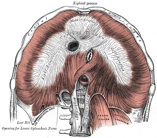
The diaphragm is a dome-shaped musculotendinous sheet that separates the thoracic cavity from the abdominal cavity. It serves as the primary muscle of inspiration, creating negative intrathoracic pressure to draw air into the lungs. The muscle fibres originate from three main sites: the sternum, the ribs, and the lumbar vertebrae. The sternal part arises from the xiphoid process. The costal part arises from the inner surfaces of the lower six ribs and their costal cartilages. The lumbar part arises from the lumbar vertebrae via two muscular crura and the arcuate ligaments.
Figure 1Gray391
Long description
The diaphragm. Under surface. Labelled illustration from Wikimedia Commons, used under its open licence.

Figure 1: The diaphragm (under surface).
The diaphragm contains three major openings that allow structures to pass between the thorax and abdomen. The caval opening is located at the level of the eighth thoracic vertebra. It transmits the inferior vena cava and branches of the right phrenic nerve. The oesophageal hiatus is located at the level of the tenth thoracic vertebra. It transmits the oesophagus, the vagus nerves, and gastric branches of the left gastric vessels. The aortic hiatus is located at the level of the twelft [3]
During quiet breathing, also known as eupnea, the diaphragm and external intercostal muscles must contract. Contraction of the diaphragm causes it to flatten and move inferiorly. This increases the vertical dimension of the thoracic cavity. Contraction and relaxation of the diaphragm and intercostals muscles cause most of the pressure changes that result in inspiration and expiration. The lungs themselves are passive during breathing, meaning they are not involved in creating the movement that helps inspiration and expiration. This is because of the adhesive nature of the pleural fluid, which allows the lungs to be pulled outward when the thoracic wall moves during inspiration. [2]
In forced breathing, also known as hyperpnea, inspiration and expiration both occur due to muscle contractions. During forced inspiration, muscles of the neck, including the scalenes, contract and lift the thoracic wall, increasing lung volume. During forced expiration, accessory muscles of the abdomen, including the obliques, contract, forcing abdominal organs upward against the diaphragm. This helps to push the diaphragm further into the thorax, pushing more air out. [2]
DefinitionDiaphragm
A dome-shaped musculotendinous sheet separating the thoracic and abdominal cavities, acting as the primary muscle of respiration.
| Opening | Vertebral Level | Structures Transmitted |
|---|---|---|
| Caval Opening | T8 | Inferior vena cava, right phrenic nerve branches |
| Oesophageal Hiatus | T10 | Oesophagus, vagus nerves, gastric vessels |
| Aortic Hiatus | T12 | Aorta, thoracic duct, azygos vein |
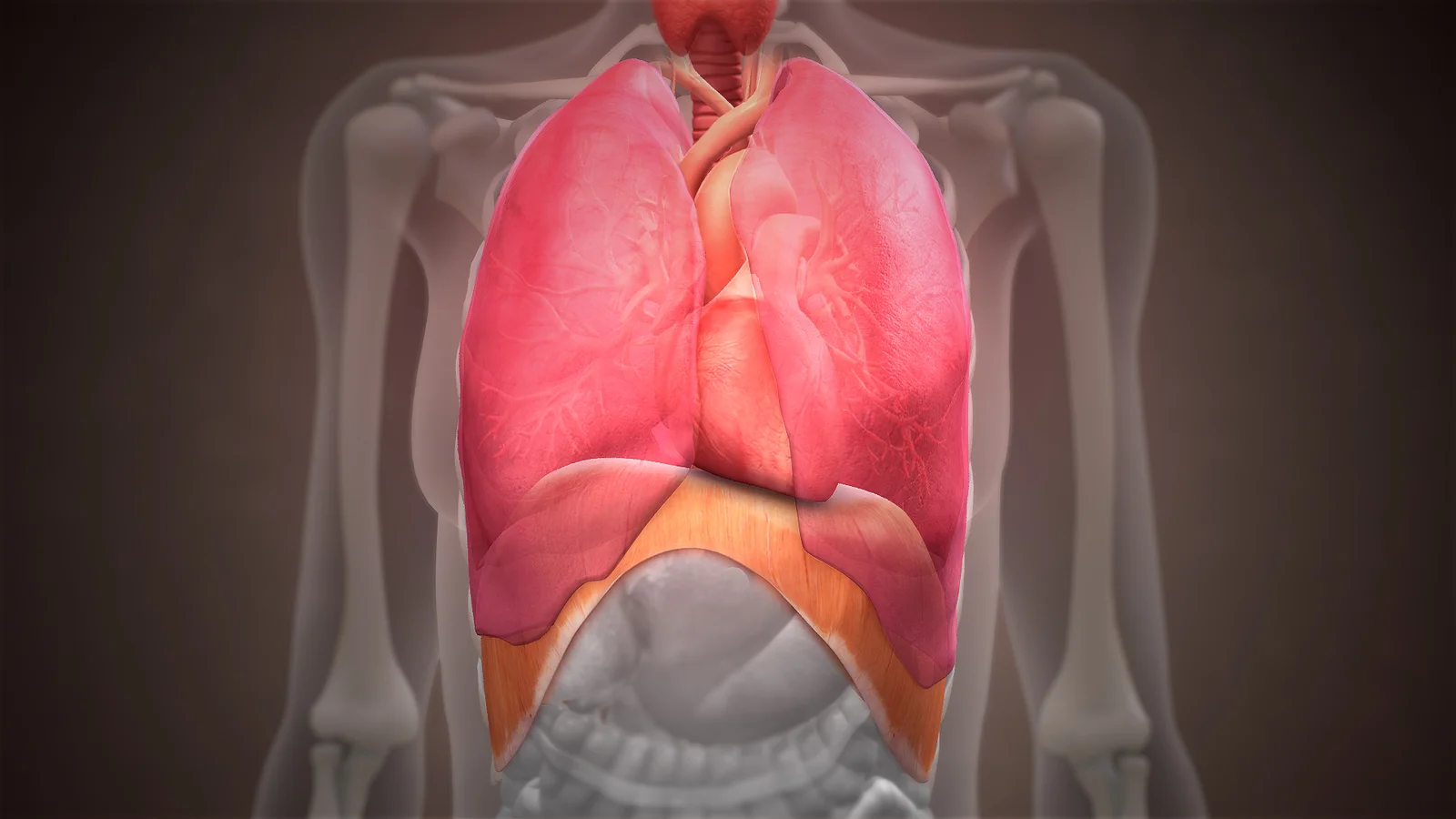
Figure 23D Medical Animation Diaphragm Structure
Long description
A 3D medical illustration showing structure of diaphragm and how it supports heart and lungs.

Figure 2: A 3D medical illustration showing structure of diaphragm and how it supports heart and lungs.
When the central tendon or the parietal peritoneum covering the diaphragm is irritated, the brain may interpret the signal as coming from the shoulder region. [1]
The diaphragm is a dome‑shaped musculotendinous partition separating the thoracic and abdominal cavities. Its motor drive originates in the ventral respiratory group (VRG) of the medulla, which maintains a constant breathing rhythm by stimulating the diaphragm and external intercostal muscles to contract, resulting in inspiration. During quiet inspiration, the diaphragm and external intercostal muscles contract, causing the rib cage to expand and move outward, and expanding the thoracic cavity and lung volume. When VRG activity ceases, these muscles relax, allowing elastic recoil to produce expiration. [4]
Forced breathing recruits additional musculature. During forced inspiration, muscles of the neck, including the scalenes, contract and lift the thoracic wall, increasing lung volume. During forced expiration, accessory muscles of the abdomen, including the obliques, contract, forcing abdominal organs upward against the diaphragm. In maximal exhalation, the internal intercostals and abdominal muscles may be involved in forcing air out of the lungs. [4]
DefinitionValsalva maneuver
Cooperation between a steady diaphragmatic contraction and abdominal muscle tightening raises intra‑abdominal pressure for defecation, urination, or childbirth.
The diaphragm serves as a key anatomical landmark. The descending aorta pierces the diaphragm at the aortic hiatus, passing through an opening in the diaphragm known as the aortic hiatus. Superior to the diaphragm the vessel is the thoracic aorta; inferior to the diaphragm it becomes the abdominal aorta. The abdominal aorta formally ends at approximately the level of vertebra L4, where it bifurcates into the common iliac arteries. The aortic arch itself ends at the level of the intervertebral disk between the fourth and fifth thoracic vertebrae. [5]
Venous drainage is also referenced to the diaphragm. If you draw an imaginary line at the level of the diaphragm, systemic venous circulation from above that line will generally flow into the superior vena cava; beneath the diaphragm, systemic venous flow enters the inferior vena cava.
| Structure | Vertebral level / description | Passage |
|---|---|---|
| Aortic arch termination | Intervertebral disk between T4 and T5 | P58 |
| Aortic hiatus (aortic opening) | Diaphragm, inferior to T4/T5 | P58 |
| Abdominal aorta termination | Approximately L4 vertebra | P72 |
| Diaphragm as venous landmark | Imaginary line separating SVC vs IVC drainage | P88 |
During the Valsalva maneuver, you hold your breath by a steady contraction of the diaphragm; this stabilizes the volume and pressure of the peritoneal cavity. When the abdominal muscles contract, the pressure cannot push the diaphragm up, so it increases pressure on the intestinal, urinary, or reproductive tracts. [6]
The diaphragm is a dome-shaped musculotendinous partition that separates the thoracic cavity from the abdominal cavity. It serves as the primary muscle of inspiration and establishes critical anatomical boundaries for major vascular and visceral structures. Understanding its relations requires examining the three major apertures through which structures pass, as well as the peritoneal coverings that define its superior and inferior surfaces.
The central tendon is the broad, aponeurotic centre. Anterior to the central tendon lies the sternocostal triangle. The aortic hiatus is located posteriorly.
The superior surface is covered by the mediastinal pleura and the pericardium. The inferior surface is covered by the peritoneum, specifically the diaphragmatic part of the peritoneum. This peritoneal covering reflects onto the liver, forming the coronary and triangular ligaments. The liver rests directly against the right dome of the diaphragm. The stomach and spleen lie against the left dome. The kidneys and adrenal glands are retroperitoneal and lie posterior to the diaphragm, separated only by the fascia.
Hiatus hernias occur when abdominal viscera, typically the stomach, protrude through the oesophageal hiatus into the thorax. This can compromise the lower oesophageal sphincter, leading to gastro-oesophageal reflux. Congenital diaphragmatic hernias, such as Bochdalek hernias, result from failure of the pleuroperitoneal membranes to close during embryological development. These defects usually occur posterolaterally, allowing abdominal contents to enter the thoracic cavity and compress the developing lungs. This compression can lead to pulmonary hypoplasia and persistent pulmonary hypertension of the newborn. The severity depends on the size of the defect and the degree of lung compression.
| Structure | Level | Side |
|---|---|---|
| Caval opening | T8 | Right |
| Oesophageal hiatus | T10 | Left |
| Aortic hiatus | T12 | Midline |
This occurs because the sensory fibres from the shoulder also [1]
Surrounding this tendon are the three main muscular components: the right crus, the left crus, and the costal part. The crura arise from the lumbar vertebrae and pass upwards to join the central tendon. The costal part arises from the lower six ribs and their costal cartilages, arching upwards to insert into the periphery of the central tendon. The caval opening is located within the central tendon at the level of the eighth thoracic vertebra, transmitting the inferior vena cava. The aortic hiatus lies posterior to the diaphragm, behind the median arcuate ligament, at the level of the twelfth thoracic vertebra, transmitting the aorta and thoracic duct.
The diaphragm is the primary muscle of inspiration. During normal inspiration, the diaphragm contracts and flattens, increasing the vertical diameter of the thoracic cavity. This action decreases intrapleural pressure, causing air to flow into the lungs. The process of normal expiration is passive, meaning that energy is not required to push air out of the lungs. Instead, the elasticity of the lung tissue causes the lung to recoil, as the diaphragm and intercostal muscles relax following inspiration. In turn, the thoracic cavity and lungs decrease in volume, causing an increase in intrapulmonary pressure. The intrapulmonary pressure rises above atmospheric pressure, creating a pressure gradient that causes air to leave the lungs.
DefinitionDiaphragm
The dome-shaped muscle separating the thoracic and abdominal cavities, serving as the primary driver of respiration.
The blood supply to the diaphragm is dual in nature. The superior surface receives blood from the pericardiophrenic and musculophrenic arteries, which are branches of the internal thoracic artery. The internal thoracic artery, or mammary artery, supplies blood to the thymus, the pericardium of the heart, and the anterior chest wall. The inferior surface is supplied by the inferior phrenic arteries, which arise directly from the abdominal aorta. The remaining thoracic aorta branches are collectively referred to as parietal branches or somatic branches, and include the intercostal and superior phrenic arteries. Each intercostal artery provides blood to the muscles of the thoracic cavity and vertebral column.
| Component | Origin | Insertion | Action | Nerve Supply |
|---|---|---|---|---|
| Right Crus | Lumbar vertebrae L1-L3 | Central tendon | Inspiration | Phrenic nerve |
| Left Crus | Lumbar vertebrae L1-L2 | Central tendon | Inspiration | Phrenic nerve |
| Costal Part | Lower six ribs | Central tendon | Inspiration | Phrenic nerve |
| Central Tendon | N/A | N/A | Tendon for muscle attachment | Phrenic nerve |
The innervation of the diaphragm is provided by the phrenic nerves, which arise from the cervical spinal nerves C3, C4, and C5. The right phrenic nerve descends on the right side of the superior vena cava and the right atrium. Both nerves pierce the diaphragm to supply the inferior surface. Sensory innervation to the central tendon is also provided by the phrenic nerves, which is why irritation of the diaphragm can refer pain to the shoulder region via the C3-C5 dermatomes.
Exam trapInnervation
Remember that the diaphragm is supplied by C3, C4, and C5. If C5 is damaged, the diaphragm may be affected.
The diaphragm also plays a role in increasing intra-abdominal pressure during activities such as coughing, sneezing, and defecation. This function is mediated by the contraction of the diaphragm against a closed glottis, known as the Valsalva manoeuvre. This action assists in forced expiration.
The diaphragm is vascularized by paired arteries from both the thoracic and abdominal aortae. The superior phrenic arteries arise from the thoracic aorta and descend to supply the superior (thoracic) surface of the muscle. The inferior phrenic arteries originate from the abdominal aorta or the celiac trunk, pass superolaterally across the crus and the inferior (abdominal) surface, and divide into medial and lateral branches that anastomose with the superior phrenic and musculophrenic vessels. The musculophrenic arteries, terminal branches of the internal thoracic arteries, course along the costal margin to supply the peripheral costal fibers. Additional contributions come from the pericardiacophrenic arteries accompanying the phrenic nerves and the lower intercostal arteries. This rich anastomotic network ensures robust perfusion of the central tendon and muscular dome during sustained contraction. [1]
The right inferior phrenic artery is typically larger and longer than the left, reflecting the asymmetry of the abdominal cavity. These arteries divide into superior and inferior branches. The superior branches anastomose with the musculophrenic and pericardiophrenic arteries, which are branches of the internal thoracic artery. The inferior branches supply the inferior surface of the diaphragm and anastomose with the superior phrenic arteries.
In addition to the inferior phrenic arteries, the superior phrenic arteries contribute to the blood supply. These are small vessels that arise from the posterior intercostal arteries. They descend to supply the superior surface of the diaphragm. The anastomosis between the superior and inferior phrenic arteries ensures a robust collateral circulation. This is clinically significant, as it provides redundancy in the event of obstruction. The blood supply is complemented by branches from the pericardiophrenic and musculophrenic arteries, which supply the peripheral parts of the diaphragm.
DefinitionInferior Phrenic Arteries
Paired arteries arising from the abdominal aorta that supply the inferior surface of the diaphragm.
The venous drainage parallels the arterial supply. The inferior phrenic veins drain into the inferior vena cava on the right and the renal vein on the left. This drainage pattern is distinct from the pulmonary circulation, which returns oxygenated blood to the left atrium via the four pulmonary veins. The systemic circulation, including the diaphragm, is served by the aorta and its branches. Understanding this vascular anatomy is essential for interpreting imaging and planning surgical interventions involving the diaphragm. [1]
The venous system operates on a complementary principle to the arterial supply, returning deoxygenated blood to the right atrium. While arteries distribute blood, veins collect it, often running parallel to their arterial counterparts, though with greater anatomical variability. Understanding this drainage is critical for interpreting pathology and surgical approaches.
The right phrenic vein empties directly into the inferior vena cava. [1]
DefinitionPhrenic Veins
The primary venous drainage of the diaphragm itself, with asymmetric termination points reflecting the position of the inferior vena cava.
Each side of the diaphragm drains into a phrenic vein. [1]
| Structure | Drainage Pathway | Termination |
|---|---|---|
| Right Phrenic Vein | Directly from diaphragm | Inferior Vena Cava |
| Left Phrenic Vein | From diaphragm to left renal vein | Left Renal Vein |
| Hepatic Veins | From liver parenchyma | Inferior Vena Cava |
| Inferior Vena Cava | Receives phrenic and hepatic veins | Right Atrium |
Other than the small amount of blood drained by the azygos and hemiazygos veins, most of the blood inferior to the diaphragm drains into the inferior vena cava before it is returned to the heart. [1]
Clinical pearlClinical Pearl
The asymmetry of the phrenic veins means that pathology on the left side may present with different venous congestion patterns compared to the right.
The inferior vena cava parallels the abdominal aorta, where it can receive blood from abdominal veins. Lying just beneath the parietal peritoneum in the abdominal cavity, it serves as the main conduit for lower body return. This arrangement allows for efficient collection of blood from the lower limbs, pelvis, and abdomen. The variability in venous circulation is significant, so keep this variation in mind when you move from the classroom to clinical practice. Understanding these landmarks is essential for safe surgical intervention and accurate diagnostic imaging. [1]
The lymphatic system plays a critical role in maintaining the homeostasis of the pleural cavity, a function that is intimately linked to the mechanics of breathing. To understand the drainage of the diaphragm, one must first appreciate the forces at play within the thorax. Competing forces within the thorax cause the formation of the negative intrapleural pressure. This pressure gradient is essential for lung expansion, but it also creates a dynamic environment where fluid balance is precarious. [2]
One of these forces relates to the elasticity of the lungs themselves—elastic tissue pulls the lungs inward, away from the thoracic wall. Simultaneously, surface tension of alveolar fluid, which is mostly water, also creates an inward pull of the lung tissue. This inward tension from the lungs is countered by opposing forces from the pleural fluid and thoracic wall. This delicate equilibrium means that the volume of pleural fluid must be strictly regulated. Too much or too little pleural fluid would hinder the creation of the negative intrapleural pressure. [2]
DefinitionPleural Homeostasis
The lymphatic drainage of the diaphragm is not merely an excretory pathway but a vital mechanism for preserving the negative intrapleural pressure required for respiration.
The superior surface drains primarily into the anterior and posterior mediastinal lymph nodes. These nodes lie adjacent to the pericardium and the lower mediastinum. The inferior surface, particularly the central tendon and the muscular parts near the crura, drains into the phrenic nodes. These nodes are located on the inferior surface of the diaphragm, near the aortic hiatus. The lymphatics from the peripheral parts of the diaphragm follow the intercostal spaces, draining into the posterior intercostal nodes. These nodes lie along the posterior thoracic wall, adjacent to the ribs.
| Region | Primary Drainage Nodes | Anatomical Relation |
|---|---|---|
| Superior Central | Anterior Mediastinal | Adjacent to pericardium |
| Superior Peripheral | Posterior Intercostal | Along posterior thoracic wall |
| Inferior Central | Phrenic | On inferior surface near crura |
| Inferior Peripheral | Lumbar / Cisterna Chyli | Near aortic hiatus |
The parietal pleura is attached to the thoracic wall, the natural elasticity of the chest wall opposes the inward pull of the lungs. This attachment ensures that the lymphatic vessels embedded in the parietal pleura and the diaphragmatic muscle are subjected to the same mechanical stresses as the pleural space itself. Consequently, efficient drainage is required to prevent fluid accumulation that could disrupt this opposition. The lymphatic capillaries in the diaphragm are highly permeable, allowing for the rapid uptake of proteins and cells that may leak from the capillaries during the pressure changes of inspiration. This process ensures that the pleural space remains sterile and free of excess fluid, thereby maintaining the integrity of the respiratory pump. [2]
The diaphragm receives its primary motor and sensory innervation from the phrenic nerves. These nerves are somatic in nature, arising from the cervical spinal cord segments. [1]
In addition to the phrenic nerves, the diaphragm receives autonomic innervation. These fibres regulate the smooth muscle components and vascular tone but do not provide voluntary motor control. The somatic innervation by the phrenic nerve is crucial for voluntary breathing. Damage to the phrenic nerve, for instance during cervical spine surgery or trauma, can lead to diaphragmatic paralysis [1]
On the right side, the nerve passes anterior to the root of the lung and then descends along the right side of the superior vena cava and the right atrium. On the left side, the nerve passes anterior to the root of the lung and then descends along the left side of the aortic arch and the left ventricle. Both nerves pierce the diaphragm separately. The right phrenic nerve supplies the right hemidiaphragm, while the left supplies the left.
Understanding this anatomy is vital for diagnosing abdominal pathologies. A hernia, such as a hiatal hernia, can irritate the diaphragm. The resulting pain is often misinterpreted by patients as cardiac or shoulder pain. The table below summarises the key aspects of diaphragmatic innervation.
| Structure | Origin | Function | Clinical Relevance |
|---|---|---|---|
| Phrenic Nerve | C3, C4, C5 | Motor to diaphragm; Sensory to central tendon | Referred shoulder pain |
| Autonomic Fibres | Vagus and Sympathetic | Vascular and smooth muscle control | Minor role in breathing |
The interplay between somatic and autonomic systems ensures that breathing is both voluntary and involuntary. The phrenic nerve is the main driver of inspiration. Its integrity is essential for life. Any lesion affecting C3-C5 can compromise respiratory function. This underscores the importance of protecting these spinal segments during surgical procedures.
The diaphragm is a dome-shaped musculotendinous partition separating the thoracic and abdominal cavities. Its structure is optimized for rapid, forceful contraction during respiration. The peripheral portion consists of skeletal muscle fibres, allowing voluntary control under cortical influence, while the central portion is a dense aponeurosis, the central tendon, which serves as a non-contractile anchor. This histological arrangement ensures efficient transmission of force to the thoracic walls while maintaining structural integrity against intra-abdominal pressure.
The muscle fibres are arranged in bundles that radiate from the central tendon to the attachments. These fibres are striated, typical of somatic skeletal muscle, and are innervated by the phrenic nerve. The central tendon is composed of dense irregular connective tissue, providing a robust surface for muscle attachment and a stable point for pressure transmission. This distinction between contractile muscle and static tendon is fundamental to understanding diaphragmatic mechanics.
DefinitionCentral Tendon
A fibrous, non-contractile central area of the diaphragm where muscle fibres converge, serving as the primary attachment point for the muscular bundles.
The histological organization supports the diaphragm's role as the primary muscle of inspiration. Contraction of the peripheral muscle fibres flattens the dome, increasing the vertical diameter of the thoracic cavity. The central tendon moves inferiorly, transmitting this force effectively. The dense connective tissue of the central tendon prevents over-distension and provides a secure anchor for the muscular fibres, ensuring that the generated force is directed towards expanding the thoracic volume rather than tearing the tissue.
| Component | Tissue Type | Function |
|---|---|---|
| Peripheral Muscle | Skeletal Striated Muscle | Contraction to flatten dome and increase thoracic volume |
| Central Tendon | Dense Irregular Connective Tissue | Anchor for muscle fibres; transmits force; non-contractile |
| Crura | Skeletal Striated Muscle | Origin for diaphragmatic fibres; stabilizes aortic hiatus |
Understanding this histological basis is crucial for interpreting clinical signs. Weakness in the muscle fibres leads to paradoxical movement, while damage to the central tendon can compromise the seal necessary for effective pressure gradients. The innervation by the phrenic nerve (C3-C5) ensures that these striated fibres respond to neural signals for breathing. The histological simplicity of the central tendon contrasts with the complex arrangement of the muscle fibres, highlighting the division of labour between force generation and structural support. This anatomical design allows for the high-frequency, rhythmic contractions required for life, balancing strength with endurance. The integration of these histological features enables the diaphragm to function as a powerful pump, driving ventilation through efficient mechanical advantage.
The diaphragm is a dome-shaped musculotendinous partition separating the thoracic and abdominal cavities. Its formation is a complex process involving the coalescence of four distinct embryological structures. Understanding its development requires tracing the origin of these components, as their fusion determines the final anatomical configuration and potential sites of congenital weakness.
The primary component arises from the septum transversum. This thick mass of mesoderm initially forms at the cranial end of the embryo. It separates the pericardial cavity from the primitive peritoneal cavity. As the heart descends, the septum transversum moves caudally. It eventually forms the central tendon of the diaphragm. This central part is tendinous and serves as the insertion point for the muscular fibres.
The pleuroperitoneal membranes develop later. These are folds of lateral body wall mesoderm. They grow medially to close the pericardio-peritoneal canals. These canals are the initial communications between the thoracic and abdominal cavities. The closure of these canals is essential for the separation of the two cavities. Failure of these membranes to fuse completely results in congenital diaphragmatic hernias, typically posterolateral.
The oesophageal mesoderm contributes the posterior muscular portion. The caudal part of the foregut is surrounded by mesoderm. This tissue forms the crura of the diaphragm. The right crus arises from the bodies of the first three lumbar vertebrae. The left crus arises from the first two lumbar vertebrae. These muscular pillars support the oesophageal hiatus. They also play a role in the anti-reflux mechanism.
The dorsal mesentery of the oesophagus contributes to the posterior aspect. It degenerates in most areas but persists to form the oesophageal hiatus. The nerves of the diaphragm reflect this embryological origin. The central tendon is innervated by the phrenic nerve, which arises from cervical roots C3 to C5. The peripheral muscular parts receive innervation from the lower intercostal nerves. This dual innervation pattern is a key anatomical feature.
The final structure is the adult diaphragm. It is a thin, dome-shaped muscle. It separates the thorax from the abdomen. It is the primary muscle of respiration. Contraction flattens the dome, increasing thoracic volume. This creates negative intrathoracic pressure, drawing air into the lungs. Relaxation allows the dome to return to its resting position. The diaphragm also assists in increasing intra-abdominal pressure during vomiting, defecation, and childbirth.
DefinitionDiaphragm
A dome-shaped musculotendinous partition separating the thoracic and abdominal cavities, formed by the septum transversum, pleuroperitoneal membranes, oesophageal mesoderm, and dorsal mesentery.
| Structure | Embryological Origin | Adult Component |
|---|---|---|
| Septum transversum | Mesoderm | Central tendon |
| Pleuroperitoneal membranes | Lateral body wall mesoderm | Posterolateral crura |
| Oesophageal mesoderm | Foregut mesoderm | Crura and posterior muscle |
| Dorsal mesentery | Mesoderm | Oesophageal hiatus |
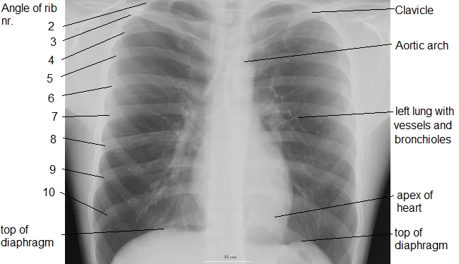
Figure 3Chest labeled
Long description
An X-ray of a human chest area, with some structures labeled. X-ray of chest, showing top of diaphragm.

Figure 3: An X-ray of a human chest area, with some structures labeled.
The diaphragm is the primary muscle of respiration, separating the thoracic and abdominal cavities. Its dome-shaped structure creates a pressure gradient essential for ventilation. Understanding its radiological appearance requires identifying its three major apertures, which transmit vital structures between the chest and abdomen.
The diaphragm contains three major openings that allow structures to pass between the thorax and abdomen. The caval opening is located at the level of the eighth thoracic vertebra. It transmits the inferior vena cava and branches of the right phrenic nerve. The oesophageal hiatus is located at the level of the tenth thoracic vertebra. It transmits the oesophagus, the vagus nerves, and gastric branches of the left gastric vessels. The aortic hiatus is located at the level of the twelft
The caval opening is the most superior aperture, located at the level of the eighth thoracic vertebra (T8). It transmits the inferior vena cava and branches of the right phrenic nerve. The opening is situated within the central tendon of the diaphragm. Because it is high and central, it is relatively fixed in position. Compression here can impede venous return, leading to haemodynamic compromise.
The oesophageal hiatus is located at the level of the tenth thoracic vertebra (T10). It is situated within the right crus of the diaphragm. It transmits the oesophagus, the anterior and posterior vagal trunks, and branches of the left gastric vessels. This hiatus is less fixed than the caval opening, allowing for the passage of the oesophagus during swallowing. It is a common site for hiatal hernias, where the stomach protrudes into the thoracic cavity. [3]
The aortic hiatus is the most posterior aperture, located at the level of the twelfth thoracic vertebra (T12). It is not an opening in the muscle itself but rather a space behind the median arcuate ligament, which connects the crura. It transmits the aorta, the thoracic duct, and the azygos vein. The aorta passes posterior to the diaphragm, meaning it is not enclosed by muscle fibres. This anatomical arrangement protects the aorta from respiratory movement but makes it susceptible to compression [1][3]
The diaphragm consists of three parts: the sternal, costal, and lumbar portions. The sternal part arises from the xiphoid process. The costal part arises from the inner surfaces of the lower six ribs and their cartilages. The lumbar part arises from the lumbar vertebrae via the crura and arcuate ligaments. All fibres converge to insert into the central tendon. The diaphragm is innervated by the phrenic nerve, which arises from cervical roots C3, C4, and C5. This innervation explains why irritation of the diaphragm can cause referred pain to the shoulder tip. [1]
| Structure | Origin | Insertion | Action | Nerve Supply |
|---|---|---|---|---|
| Sternal Part | Xiphoid process | Central tendon | Inspiration | Phrenic nerve |
| Costal Part | Lower 6 ribs | Central tendon | Inspiration | Phrenic nerve |
| Lumbar Part | Lumbar vertebrae | Central tendon | Inspiration | Phrenic nerve |
The diaphragm is the primary muscle of respiration, separating the thoracic and abdominal cavities. Its dome-shaped structure allows for the mechanical expansion of the thoracic volume during inspiration. Understanding its anatomy is critical for interpreting imaging and managing thoracoabdominal trauma.
The defect itself is located in the posterior-lateral aspect of the right hemidiaphragm, a common site for traumatic rupture due to the protective effect of the liver on the central tendon. Recognising the relationship between the liver and the diaphragm is essential, as the liver’s position can mask or mimic diaphragmatic injuries on plain radiographs. By examining the continuity of the diaphragmatic contour, one can identify the precise location of the tear. The surrounding mediastinal structures remain central, indicating no significant mediastinal shift, which helps differentiate this from a tension pneumothorax. This visualisation aids in planning surgical repair, as the posterior-lateral location requires a specific approach. The clarity of the soft tissue interfaces in this imaging modality allows for the assessment of associated organ injuries, such as hepatic lacerations, which frequently accompany diaphragmatic ruptures.
The diaphragm consists of three main muscular parts: the sternal, costal, and lumbar portions. The sternal part arises from the xiphoid process. The costal part originates from the inner surfaces of the lower six ribs and their cartilages. The lumbar part comprises two crura and the arcuate ligaments. The right crus arises from the upper three lumbar vertebrae, while the left crus arises from the upper two. The central tendon is the aponeurotic centre where all muscle fibres converge. [3]
| Part | Origin | Insertion | Action | Nerve Supply |
|---|---|---|---|---|
| Sternal | Xiphoid process | Central tendon | Inspiration | Phrenic nerve |
| Costal | Lower 6 ribs | Central tendon | Inspiration | Phrenic nerve |
| Right Crus | L1-L3 vertebrae | Central tendon | Inspiration | Phrenic nerve |
| Left Crus | L1-L2 vertebrae | Central tendon | Inspiration | Phrenic nerve |
The diaphragm has three major openings: the caval opening at T8, the oesophageal hiatus at T10, and the aortic hiatus at T12. The caval opening transmits the inferior vena cava and right phrenic nerve. The oesophageal hiatus transmits the oesophagus and vagus nerves. These openings are clinically significant as they are potential sites for hiatal hernias or traumatic injuries.
Clinical pearlClinical Pearl
The phrenic nerve arises from C3, C4, and C5, which explains why shoulder tip pain may occur with diaphragmatic irritation.
Injury to the diaphragm often results from blunt or penetrating trauma. Blunt trauma can cause rupture due to sudden increases in intra-abdominal pressure. Penetrating trauma directly lacerates the muscle. Diagnosis is challenging as symptoms may be non-specific. Imaging is crucial for confirmation.
Exam trapCommon Trap
Do not assume a normal chest X-ray rules out diaphragmatic rupture, as up to 50% of cases may have normal initial radiographs.
Management typically involves surgical repair, especially for traumatic ruptures. Non-operative management is rarely indicated except in very select cases. Early diagnosis improves outcomes and reduces morbidity associated with herniation and strangulation of abdominal organs.
The diaphragm is the primary muscle of respiration, separating the thoracic and abdominal cavities. Its dome shape allows for efficient pressure changes during breathing.
The diaphragm originates from three main structures. The sternal part arises from the xiphoid process. The costal part arises from the inner surfaces of the lower six ribs and their cartilages. The lumbar part arises from the crura and arcuate ligaments. The right crus arises from the upper three lumbar vertebrae. The left crus arises from the upper two lumbar vertebrae. The medial and lateral arcuate ligaments span across the psoas major and quadratus lumborum muscles respectively.
| Structure | Origin |
|---|---|
| Sternal part | Xiphoid process |
| Costal part | Lower six ribs |
| Right crus | L1-L3 vertebrae |
| Left crus | L1-L2 vertebrae |
| Medial arcuate ligament | Psoas major |
| Lateral arcuate ligament | Quadratus lumborum |
Three major apertures allow structures to pass between the thorax and abdomen. The caval opening is at T8. It transmits the inferior vena cava and right phrenic nerve. The oesophageal hiatus is at T10. It transmits the oesophagus and vagus nerves. The aortic hiatus is at T12. It transmits the aorta, thoracic duct, and azygos vein. These levels are mnemonic: T8, T10, T12.
Exam trapAortic Hiatus Level
The aortic hiatus is at T12, not T10. It is posterior to the diaphragm, not within it.
The diaphragm is innervated by the phrenic nerve. The right phrenic nerve arises from C3, C4, and C5. The left phrenic nerve arises from C3, C4, and C5. Contraction of the diaphragm flattens the dome, increasing thoracic volume. This decreases intrathoracic pressure, drawing air into the lungs. Relaxation allows the dome to return to its resting position. [3]
DefinitionPhrenic Nerve
The phrenic nerve provides motor innervation to the diaphragm and sensory innervation to the central tendon and mediastinal pleura.
Phrenic nerve injury leads to diaphragmatic paralysis. This can result from surgical damage or viral infection. Paralysis causes paradoxical movement of the diaphragm. The affected side moves up during inspiration. This reduces vital capacity and causes dyspnoea. Diaphragmatic hernias occur when abdominal contents protrude into the thorax. Bochdalek hernias are posterolateral and congenital. Morgagni hernias are anteromedial and congenital. Traumatic ruptures usually occur on the left side due t [3]
Clinical pearlDiaphragmatic Hernia
Bochdalek hernias are posterolateral. Morgagni hernias are anteromedial. Traumatic ruptures are more common on the left. [3]
The diaphragm develops from four structures. The septum transversum forms the central tendon. The pleuroperitoneal membranes close the pericardioperitoneal canals. The dorsal mesentery of the oesophagus forms the crura. The muscular walls of the body wall contribute to the peripheral parts. Failure of the pleuroperitoneal membranes to close results in a congenital diaphragmatic hernia. This is a life-threatening condition in newborns.
NoteDevelopment
The central tendon derives from the septum transversum. The crura derive from the dorsal mesentery of the oesophagus. [3]
Students frequently confuse the diaphragm with the surrounding thoracic structures, leading to errors in both anatomy and physiology. A primary error is assuming the diaphragm is a single, uniform muscle sheet. In reality, it is composed of three distinct parts: the sternal, costal, and lumbar portions. The sternal part arises from the xiphoid process, the costal part from the lower six ribs, and the lumbar part from the crura and arcuate ligaments. Failing to distinguish these origins obscures the mechanism of its contraction. The muscle fibres converge centrally to form the central tendon, a strong aponeurosis that does not contract but serves as the insertion point for the muscle fibres. This central tendon is the key to understanding how pressure changes are transmitted.
Another common mistake is misidentifying the innervation. Students often assume the diaphragm is innervated by intercostal nerves because of its location. This is incorrect. The diaphragm is innervated by the phrenic nerve, which arises from the cervical spinal cord segments C3, C4, and C5. This is why irritation of the diaphragm can refer pain to the shoulder tip, via the supraclavicular nerves (C3, C4). Forgetting this cervical origin leads to errors in clinical assessment of phrenic nerve injury.
A third pitfall involves the relationship between the diaphragm and the inferior vena cava. Students often place the inferior vena cava at the same level as the oesophagus. This is wrong. The oesophagus passes through the diaphragm at the oesophageal hiatus at the level of T10. The aorta passes posterior to the diaphragm at the aortic hiatus at the level of T12. The inferior vena cava passes through the caval opening at the level of T8. Memorising the mnemonic "I 8 10 Eggs At 12" helps, but understanding the anatomical reasoning is superior. The caval opening is located in the central tendon, whereas the oesophageal hiatus is in the muscular part.
A fourth error is confusing the diaphragm's role in pressure dynamics. Some students think the diaphragm creates positive pressure to push air out. While it relaxes during expiration, the primary driver of inspiration is the creation of negative pressure. As the diaphragm contracts, it flattens and descends, increasing the vertical dimension of the thoracic cavity. This increases the volume, which, according to Boyle's law, decreases the intra-alveolar pressure below atmospheric pressure, drawing air in. Pulmonary ventilation is dependent on three types of pressure: atmospheric, intra-alveolar, and intrapleural. Pulmonary ventilation is the process of breathing, which is driven by pressure differences between the lungs and the atmosphere. [2]
A fifth mistake is neglecting the embryological origin. The diaphragm is derived from the septum transversum, the pleuroperitoneal membranes, and the dorsal mesentery of the oesophagus. The central tendon is derived from the septum transversum. Understanding this helps explain congenital defects like congenital diaphragmatic hernias, which usually occur posterolaterally due to failure of the pleuroperitoneal membranes to close.
Exam trapInnervation Error
The diaphragm is innervated by the phrenic nerve (C3-C5), not the intercostal nerves. Injury here causes respiratory distress and shoulder tip pain.
Knowledge of the diaphragm is critical at the bedside for assessing respiratory effort. Clinicians observe the movement of the abdominal wall during inspiration. In healthy individuals, the descent of the diaphragm pushes the abdominal contents down, causing the abdomen to rise. In conditions such as diaphragmatic paralysis or severe chronic obstructive pulmonary disease, this movement may be absent or paradoxical (abdomen moves in during inspiration). This physical sign is a direct application
In the operating theatre, awareness of the diaphragm's anatomy is vital during abdominal and thoracic surgeries. Surgeons must identify the crura to avoid injury to the aorta and oesophagus. The aortic hiatus is located at T12, posterior to the diaphragm, between the crura. The oesophageal hiatus is at T10, within the muscular part. The caval opening is at T8, in the central tendon. The difference in pressure between intrapleural and intra-alveolar pressures is called transpulmonary pressure. Understanding these levels prevents accidental puncture of major vessels or the oesophagus. [2]
On imaging, particularly CT scans and X-rays, the diaphragm appears as a dome-shaped structure. Radiologists look for elevation or flattening. Flattening suggests hyperinflation, as seen in asthma or emphysema, where the diaphragm is pushed down. Elevation may indicate phrenic nerve palsy, subpulmonic effusion, or abdominal pathology pushing up. The costophrenic angles, where the diaphragm meets the chest wall, are checked for blunting, which indicates pleural effusion. Respiratory volume is the term used for various volumes of air moved by or associated with the lungs at a given point in the respiratory cycle. [2]
In examinations, students are often asked to identify the hiatuses and their contents. The mnemonic "I 8 10 Eggs At 12" is standard. T8: Vena cava, T10: Oesophagus, T12: Aorta. Additionally, students must know the nerve supply. The phrenic nerve (C3, C4, C5) supplies the motor innervation. The sensory innervation of the central tendon is also via the phrenic nerve, while the peripheral part is supplied by intercostal nerves. This distinction explains referred pain patterns.
Diaphragmatic Hiatus Anatomy
- Identify Vertebral LevelCheck T8, T10, or T12
- Question: Is it T8?
Yes
- Caval OpeningContains Inferior Vena Cava and right phrenic nerve
No
- Check T10...
- Question: Is it T10?
Yes
- Oesophageal HiatusContains Oesophagus, vagus nerves, and gastric vessels
No
- Check T12...
- Question: Is it T12?
Yes
- Aortic HiatusContains Aorta, thoracic duct, and azygos vein
No
- Error in LevelRe-evaluate anatomy
| Level | Opening | Contents |
|---|---|---|
| T8 | Caval Opening | Inferior Vena Cava, Right Phrenic Nerve |
| T10 | Oesophageal Hiatus | Oesophagus, Vagus Nerves, Gastric Vessels |
| T12 | Aortic Hiatus | Aorta, Thoracic Duct, Azygos Vein |
DefinitionTranspulmonary Pressure
The difference between intra-alveolar and intrapleural pressure, which keeps the lungs inflated.
The diaphragm is the primary muscle of inspiration, innervated by the phrenic nerve (C3-C5). It consists of sternal, costal, and lumbar parts converging on the central tendon. The caval opening is at T8, the oesophageal hiatus at T10, and the aortic hiatus at T12. Contraction of the diaphragm flattens it, increasing thoracic volume and decreasing intra-alveolar pressure. Referred pain to the shoulder indicates diaphragmatic irritation due to phrenic nerve supply. The central tendon is aponeurotic and does not contract, but transmits force. Paradoxical abdominal movement suggests diaphragmatic dysfunction. Imaging shows the diaphragm as a dome; flattening indicates hyperinflation.
Answer each one in your head first, then open the answer.
What is the vertebral level of the caval opening?
Show answerHide answer
T8Which nerve supplies the diaphragm?
Show answerHide answer
Phrenic nerveWhat is the origin of the sternal part of the diaphragm?
Show answerHide answer
Xiphoid processWhat happens to intra-alveolar pressure during inspiration?
Show answerHide answer
It decreases below atmospheric pressureWhat structure passes through the aortic hiatus?
Show answerHide answer
Aorta
- Betts JG, Young KA, Wise JA, et al. Circulatory Pathways. OpenStax, Anatomy and Physiology 2e (CC BY-NC-SA 4.0). Accessed .
- Betts JG, Young KA, Wise JA, et al. The Process of Breathing. OpenStax, Anatomy and Physiology 2e (CC BY-NC-SA 4.0). Accessed .
- Menshawi K, Mohr JP, Gutierrez J. A Functional Perspective on the Embryology and Anatomy of the Cerebral Blood Supply. Journal of stroke 2015;17(2):144-158. doi:10.5853/jos.2015.17.2.144. Accessed .
- Betts JG, Young KA, Wise JA, et al. The Process of Breathing. OpenStax, Anatomy and Physiology 2e (CC BY-NC-SA 4.0). Accessed .
- Betts JG, Young KA, Wise JA, et al. Circulatory Pathways. OpenStax, Anatomy and Physiology 2e (CC BY-NC-SA 4.0). Accessed .
- Betts JG, Young KA, Wise JA, et al. Axial Muscles of the Abdominal Wall, and Thorax. OpenStax, Anatomy and Physiology 2e (CC BY-NC-SA 4.0). Accessed .
Figure credits
- Figure 1: Henry Vandyke Carter, via Wikimedia Commons, Public domain, Public domain.
- Figure 2: In source box put https://www.scientificanimations.com, via Wikimedia Commons, CC BY-SA 4.0, CC BY-SA 4.0. resized.
- Figure 3: Chikumaya and Mikael Häggström (User:Mikael Häggström), via Wikimedia Commons, CC BY-SA 3.0, CC BY-SA 3.0.
Educational content for medical students and doctors. It is not a substitute for clinical judgement or local guidelines.