Intercostal spaces
Anatomical study of the spaces between ribs and their muscular, vascular and neural contents.
The intercostal spaces are the anatomical intervals situated between adjacent ribs, forming the lateral and anterior boundaries of the thoracic cavity. These spaces are not empty voids but are filled with layers of muscles, connective tissue, neurovascular bundles, and fat. The walls of these spaces are defined superiorly and inferiorly by the costal grooves of the ribs above and below. The internal thoracic artery runs vertically along the posterior aspect of the anterior chest wall, typically one to two centimetres lateral to the sternum, supplying blood to the anterior intercostal spaces. This vessel is a crucial landmark for surgical procedures and anatomical dissection.
The neurovascular bundle is a critical structure within each intercostal space. It contains the intercostal vein, artery, and nerve (VAN), arranged from superior to inferior. These structures run in the costal groove of the rib above, protected by the bony rim. For clinical safety, particularly during chest drain insertion, the safe site is along the upper border of the rib below. This avoids injury to the neurovascular bundle which lies immediately beneath the lower rib. Inserting a needle or tube too low risks damaging these vital structures, leading to haemorrhage or nerve injury.
Herpes zoster dermatomes provide a clinical correlation for intercostal anatomy. The varicella-zoster virus remains latent in the dorsal root ganglia of the intercostal nerves. Reactivation causes a painful, vesicular rash that follows the specific dermatomal distribution of the affected intercostal nerve. This presentation helps clinicians identify the specific spinal level involved, as the rash typically appears on one side of the trunk, respecting the midline.
The boundaries of the intercostal spaces extend from the vertebral column posteriorly to the sternum anteriorly. Laterally, the spaces are bounded by the ribs themselves. The muscles within these spaces change the dimensions of the thoracic cavity to facilitate breathing. The principal role of the intercostal muscles is to assist in breathing by changing the dimensions of the rib cage. This mechanical action is essential for pulmonary ventilation.
Clinical pearlSafe Zone
Always insert chest drains along the upper border of the rib to avoid the neurovascular bundle located in the costal groove.
The intercostal spaces are vital for both respiratory mechanics and clinical interventions. Understanding their anatomy ensures safe practice and accurate diagnosis of thoracic pathologies. The relationship between the ribs, muscles, and neurovascular structures is fundamental to thoracic surgery and emergency medicine.
| Muscle | Primary Action | Nerve Supply |
|---|---|---|
| External Intercostal | Elevates ribs (Inspiration) | Intercostal nerve |
| Internal Intercostal | Depresses ribs (Expiration) | Intercostal nerve |
| Innermost Intercostal | Synergist to internal | Intercostal nerve |
The vertebral column lies centrally, providing the midline reference. The ribs curve downwards and forwards from the vertebrae. The intercostal spaces are the gaps between adjacent ribs. The angle of the rib is a prominent posterior landmark. This anatomical relationship is critical for safe needle insertion.
DefinitionIntercostal Space
The anatomical gap between two adjacent ribs, containing muscles, nerves, and vessels.
Palpable landmarks include the sternal angle, which corresponds to the second rib and the second intercostal space anteriorly. This landmark is essential for counting ribs and identifying the correct space for procedures. The nipple in males typically lies at the fourth intercostal space, although this varies. In thoracentesis, the needle is inserted just above the superior border of the lower rib to avoid the neurovascular bundle located in the costal groove of the rib above. This "safe triangle" avoids damage to the intercostal vessels and nerves.
| Muscle | Action | Nerve Supply |
|---|---|---|
| External intercostal | Elevates ribs during inspiration | Intercostal nerves |
| Internal intercostal | Depresses ribs during expiration | Intercostal nerves |
| Innermost intercostal | Depresses ribs during expiration | Intercostal nerves |
Understanding the intercostal spaces requires correlating surface anatomy with cross-sectional imaging. The intercostal spaces are the gaps between the ribs, containing the neurovascular bundle and muscles essential for respiration. On a standard posterior-anterior chest X-ray, the ribs appear as curved, radiopaque structures. The intercostal spaces themselves are radiolucent, appearing as darker gaps between the bony ribs. The costophrenic angles, where the diaphragm meets the chest wall, shoul [1]
Computed tomography (CT) provides superior detail of the intercostal structures. In axial CT slices, the intercostal muscles are visible as layers of soft tissue density between the ribs. The neurovascular bundle, containing the intercostal vein, artery, and nerve, runs in the costal groove of the rib above. This bundle is protected by the overlying rib. On CT, the intercostal vessels appear as small, circular or oval structures adjacent to the inner surface of the rib. The intercostal nerve is [1]
The intercostal spaces are bounded superiorly and inferiorly by the ribs, forming distinct anatomical compartments that house neurovascular bundles and muscles. Understanding the layered arrangement of fascia and muscle is critical for procedures such as thoracentesis or intercostal nerve blocks, where inadvertent injury to these structures can lead to significant complications. The wall of the thorax is not merely a passive cage but an active system of compartments defined by fascial planes.
The outermost layer is the skin, followed by the superficial fascia. Its fibres run obliquely downwards and forwards. Deep to the internal intercostals lies the innermost intercostal layer, which is continuous with the transversus thoracis muscle anteriorly. The neurovascular bundle runs in the costal groove of the rib above, protected by the overlying internal intercostal muscle. The bundle contains the intercostal vein, artery, and nerve, arranged from superior to inferior as VAN. This protective positioning prevents injury during needle insertion if the approach is from below the rib.
The fascia enveloping these muscles provides structural integrity and serves as a pathway for the spread of infection or fluid. The endothoracic fascia lines the inner surface of the thoracic wall, separating the parietal pleura from the muscles and bones. This potential space is clinically significant in the context of empyema or haemothorax, where fluid may accumulate between the pleura and the chest wall.
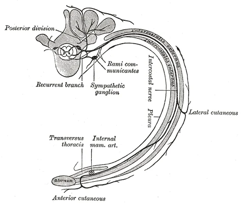
Although distinct from the thoracic wall, the fascial continuity between the abdominal and thoracic compartments allows for the spread of pathology, such as subcutaneous emphysema or abscesses, across these regions. The intercostal muscles themselves are innervated by the intercostal nerves, which are the anterior rami of the thoracic spinal nerves. These nerves run with the vessels in the costal groove, providing both motor supply to the muscles and sensory innervation to the overlying skin and parietal pleura. [3]
Figure 1Gray819
Long description
Diagram of the course and branches of a typical intercostal nerve.

Figure 1: Diagram of the course and branches of a typical intercostal nerve.
The intercostal spaces are defined by the skeletal framework of the thorax and the muscular layers that fill the gaps between the ribs. These muscles are essential for changing the volume of the thoracic cavity, thereby driving pulmonary ventilation. The lungs themselves are passive structures; they do not generate the movement required for breathing. Instead, the pressure changes that result in inspiration and expiration are caused by the contraction and relaxation of the diaphragm and the intercostal muscles. The adhesive nature of the pleural fluid ensures that when the thoracic wall moves, the lungs are pulled outward, allowing for expansion. [2]
The intercostal muscles are arranged in three layers within each intercostal space. The most superficial layer is the external intercostal muscle. The intermediate layer is the internal intercostal muscle. The deepest layer is the innermost intercostal muscle. These muscles work in concert with the diaphragm to control ventilation. Accessory muscles, primarily the internal intercostals, also help to compress the rib cage, which reduces the volume of the thoracic cavity during forced expiration. [2]
| Muscle | Origin | Insertion | Action | Nerve Supply |
|---|---|---|---|---|
| External Intercostal | Inferior border of the rib above | Superior border of the rib below | Elevates ribs; assists in inspiration | Intercostal nerve |
| Internal Intercostal | Superior border of the rib below | Inferior border of the rib above | Depresses ribs; assists in forced expiration | Intercostal nerve |
| Innermost Intercostal | Similar to internal intercostals | Similar to internal intercostals | Similar to internal intercostals | Intercostal nerve |
The neurovascular bundle runs along the inferior border of each rib, protected by the costal groove. This bundle contains the intercostal vein, artery, and nerve. It is crucial to remember this anatomical relationship when performing procedures such as chest drain insertion. The needle or catheter should be inserted just above the upper border of the rib to avoid damaging the neurovascular bundle.
The neurovascular bundle is located in the costal groove, just below the rib. The chest drain site is indicated above the rib.
The internal thoracic artery runs along the inner surface of the anterior chest wall. It provides blood supply to the anterior intercostal spaces. This artery is a key landmark for surgeons and anatomists. It runs parallel to the sternum, deep to the costal cartilages.
The muscles of the vertebral column, thorax, and abdominal wall extend, flex, and stabilize different parts of the body’s trunk. The intercostal muscles are part of this complex system. They help maintain posture and facilitate breathing.
The arterial supply to the intercostal spaces is derived from two distinct sources: posterior and anterior intercostal vessels. These vessels run within the intercostal spaces, providing blood to the intercostal muscles, parietal pleura, and overlying skin. Understanding the origin and course of these vessels is essential for procedures such as thoracentesis, where knowledge of the neurovascular bundle prevents iatrogenic injury.
The posterior intercostal arteries arise from the thoracic aorta. For the first two intercostal spaces, these arteries are branches of the supreme intercostal artery, which itself originates from the costocervical trunk. From the third intercostal space downwards, the posterior intercostal arteries branch directly from the thoracic aorta. Each posterior intercostal artery runs along the inferior border of its corresponding rib, within the intercostal space. It lies deep to the intercostal muscles and superficial to the parietal pleura. Accompanying the artery are the intercostal vein and nerve, forming the neurovascular bundle. This bundle is protected by the costal groove of the rib above. The posterior intercostal arteries give off collateral branches that run along the superior border of the rib below. These collateral branches supply the intercostal muscles and anastomose with the anterior intercostal arteries. [1]
The anterior intercostal arteries are smaller than their posterior counterparts. They arise from the internal thoracic artery for the first six intercostal spaces. The internal thoracic artery is a branch of the subclavian artery. It runs vertically down the posterior aspect of the anterior chest wall, just lateral to the sternum. The anterior intercostal arteries branch off the internal thoracic artery at each intercostal space. They run along the inferior border of the rib, deep to the internal intercostal muscle. For the last two intercostal spaces, the anterior intercostal arteries are derived from the musculophrenic artery, a terminal branch of the internal thoracic artery. These anterior vessels anastomose with the posterior intercostal arteries within the intercostal space, ensuring a continuous blood supply.
Lymphatic drainage from the intercostal spaces follows the venous drainage patterns. Superficial lymphatics from the skin and subcutaneous tissues of the chest wall drain into the axillary lymph nodes and the parasternal lymph nodes. The parasternal lymph nodes lie alongside the internal thoracic vessels. Deep lymphatics from the intercostal muscles and parietal pleura generally follow the posterior intercostal veins. They drain into the bronchomediastinal trunks. The right lymphatic duct receives lymph from the upper right side of the body, including the right intercostal spaces. The thoracic duct receives lymph from the rest of the body, including the left intercostal spaces. These ducts empty lymph into the venous system at the junction of the jugular and subclavian veins.
Clinical pearlClinical Pearl
Always insert needles above the superior border of the rib to avoid the neurovascular bundle located in the costal groove.
The venous drainage of the intercostal spaces follows a systematic pattern that mirrors the arterial supply, ensuring efficient return of deoxygenated blood from the thoracic wall to the central circulation. Understanding this pathway is critical for procedures involving the chest wall, as inadvertent injury to these vessels can lead to significant haemorrhage. The primary vessels involved are the posterior intercostal veins, which drain the majority of the intercostal spaces.
The posterior intercostal veins originate from the venous plexuses located within the intercostal spaces. These veins run alongside the posterior intercostal arteries and nerves. In the lower intercostal spaces, the veins typically drain into the azygos system. Specifically, the right posterior intercostal veins from the third to eleventh spaces drain directly into the azygos vein. The left posterior intercostal veins from the second to eleventh spaces drain into the hemiazygos and accessory hemiazygos veins, which then cross over to join the azygos vein. This arrangement ensures that blood from the left side of the thorax is eventually returned to the right side of the superior vena cava.
Clinical pearlClinical Pearl
The azygos vein serves as a crucial collateral pathway. If the inferior vena cava is obstructed, blood can flow retrograde through the azygos system to reach the superior vena cava.
The anterior intercostal veins drain the anterior aspects of the intercostal spaces. These veins accompany the anterior intercostal arteries. In the upper intercostal spaces, they drain into the internal thoracic vein. The internal thoracic vein then descends along the inner surface of the anterior chest wall, running parallel to the internal thoracic artery. It typically terminates by joining the brachiocephalic vein. In the lower intercostal spaces, the anterior intercostal veins may drain into the musculophrenic vein, which is a branch of the internal thoracic vein, or directly into the superior epigastric vein.
DefinitionAnatomical Definition
The internal thoracic vein is formed by the union of the anterior intercostal veins and drains into the brachiocephalic vein.
| Structure | Drainage Destination |
|---|---|
| Right posterior intercostal veins (3-11) | Azygos vein |
| Left posterior intercostal veins (2-11) | Hemiazygos/Accessory hemiazygos veins |
| Anterior intercostal veins (upper) | Internal thoracic vein |
| Anterior intercostal veins (lower) | Musculophrenic/Superior epigastric veins |
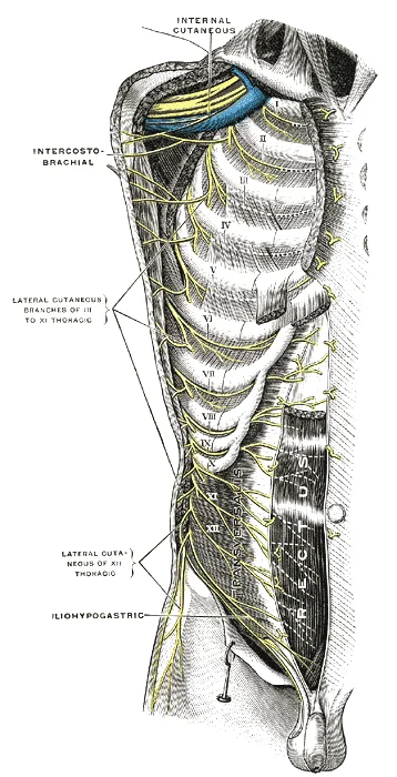
The intercostal spaces are innervated by the thoracic spinal nerves, which emerge from the spinal cord and pass through the intervertebral foramina. Each thoracic nerve divides into a dorsal ramus and a ventral ramus. The ventral ramus of the upper eleven thoracic nerves is known as the intercostal nerve, while the twelfth is the subcostal nerve. These nerves provide both motor supply to the muscles of the chest wall and cutaneous supply to the skin of the thorax and abdomen.
Figure 2Gray821
Long description
Intercostal nerves, the superficial muscles having been removed. (Testut).

Figure 2: Intercostal nerves, the superficial muscles having been removed (Testut).
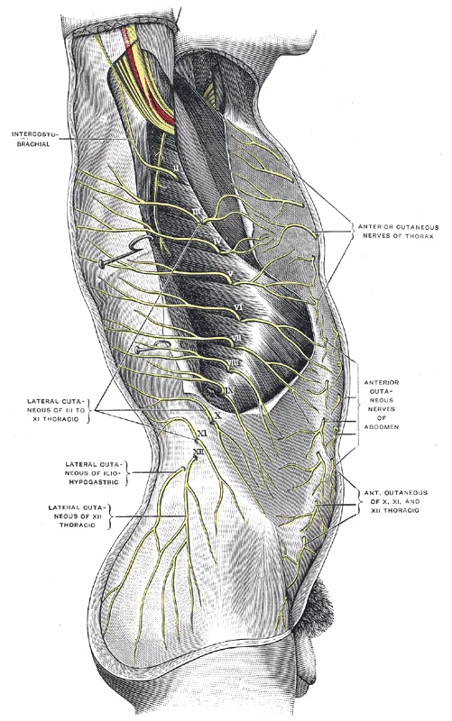
Figure 3Gray820
Long description
Cutaneous distribution of thoracic nerves. (Testut.)

Figure 3: Cutaneous distribution of thoracic nerves (Testut.).
Magnetic resonance imaging (MRI) offers excellent soft tissue contrast without ionising radiation. It is particularly useful for evaluating intercostal neuralgia or tumours involving the intercostal spaces. On T1-weighted images, the intercostal muscles have intermediate signal intensity, while fat appears bright. The neurovascular bundle is seen as a low-signal structure within the high-signal fat of the intercostal space. MRI can clearly delineate the relationship between the intercostal muscl [1]
The motor supply includes the external intercostal, internal intercostal, and innermost intercostal muscles. These muscles assist in respiration. The external intercostals elevate the ribs during inspiration. The internal and innermost intercostals depress the ribs during forced expiration. The subcostal muscles also aid in expiration. The transversus thoracis muscle is supplied by the intercostal nerves. These muscles stabilize the thoracic cage.
The cutaneous supply covers the skin of the anterior and lateral thorax. The intercostal nerves supply the skin overlying the intercostal spaces. The lateral cutaneous branches supply the skin of the lateral chest. The anterior cutaneous branches supply the skin of the anterior chest wall. The distribution of these nerves follows a dermatomal pattern. Each nerve supplies a specific band of skin. This pattern is important for localizing lesions.
| Muscle | Origin | Insertion | Action | Nerve Supply |
|---|---|---|---|---|
| External Intercostal | Lower border of rib above | Upper border of rib below | Elevates rib; inspiration | Intercostal nerve |
| Internal Intercostal | Upper border of rib below | Lower border of rib above | Depresses rib; expiration | Intercostal nerve |
| Innermost Intercostal | Upper border of rib below | Lower border of rib above | Depresses rib; expiration | Intercostal nerve |
| Subcostal | Inner surface of rib below | Inner surface of rib two below | Depresses rib; expiration | Intercostal nerve |
| Transversus Thoracis | Sternum | Inner surfaces of costal cartilages | Depresses ribs; expiration | Intercostal nerve |
Lesions of the intercostal nerves can cause pain and sensory loss. Intercostal neuralgia is a common condition. It presents with sharp, burning pain along the distribution of the nerve. The pain may be exacerbated by movement or coughing. Treatment involves analgesics and nerve blocks.
The lymphatic drainage of the intercostal spaces follows a pattern that mirrors the venous and arterial supply, ensuring efficient fluid return from the thoracic wall. Understanding this pathway is essential for appreciating how infections or malignancies in the chest wall might spread. The primary route involves drainage into the intercostal lymph nodes, which are situated along the posterior aspect of the intercostal spaces. These nodes receive lymph from the parietal pleura, the intercostal muscles, and the overlying skin. From these initial stations, the lymphatic vessels converge to drain into the posterior intercostal nodes. These nodes are located near the heads of the ribs and are closely associated with the posterior intercostal vessels. [1]
The efferent vessels from the posterior intercostal nodes do not drain directly into the venous system. Instead, they contribute to the formation of the thoracic duct on the left side and the right lymphatic duct on the right side, depending on the specific intercostal space involved. This bilateral drainage pattern highlights the importance of considering both sides when evaluating pathology. For instance, lesions in the upper intercostal spaces may drain into the apical nodes, while lower spaces drain towards the diaphragmatic nodes.
It is crucial to distinguish the lymphatic drainage of the intercostal spaces from that of the abdominal viscera. While the small intestine relies on lacteals for lipid transport, the thoracic wall utilizes a more direct nodal pathway. In the small intestine, lymphatic capillaries called lacteals are critical for the transport of dietary lipids and lipid-soluble vitamins to the bloodstream. This mechanism is specific to the gastrointestinal tract and does not apply to the intercostal spaces. The intercostal lymphatics primarily handle tissue fluid, immune cells, and potential pathogens from the chest wall structures. [1]
DefinitionIntercostal Lymph Nodes
Small lymph nodes located along the posterior intercostal spaces that drain the parietal pleura, intercostal muscles, and overlying skin.
| Structure | Primary Drainage Site | Secondary Drainage |
|---|---|---|
| Anterior Intercostal Spaces | Parasternal Nodes | Internal Thoracic Nodes |
| Posterior Intercostal Spaces | Posterior Intercostal Nodes | Thoracic/Right Lymphatic Duct |
| Lateral Intercostal Spaces | Axillary Nodes | Subclavian Trunk |
The clinical significance of this drainage pattern cannot be overstated. Knowledge of these pathways aids in the staging of thoracic malignancies and the management of infections. Surgeons must be aware of these routes to ensure complete excision of diseased tissue or effective drainage of abscesses. The proximity of these nodes to vital structures such as the aorta and vena cava also necessitates careful surgical planning.
The intercostal space is bounded superiorly and inferiorly by the costal grooves of adjacent ribs. Within this space, three primary structures run in a consistent vertical order from superior to inferior: the intercostal vein, the intercostal artery, and the intercostal nerve. This arrangement is often remembered by the mnemonic VAN. The intercostal vein lies most superiorly, closest to the upper border of the rib below. The intercostal artery is positioned immediately inferior to the vein. The intercostal nerve runs along the inferior border of the rib, protected within the costal groove. This protective positioning prevents injury to these structures during procedures such as thoracentesis, which should be performed along the superior border of the lower rib to avoid the neurovascular bundle. [1]
Clinical pearlSafe Zone
Always enter the intercostal space near the superior border of the rib to avoid the neurovascular bundle located in the costal groove.
The intercostal muscles occupy the space between the ribs. The external intercostal muscle originates from the inferior border of the rib above and inserts into the superior border of the rib below. Its fibres run obliquely downwards and forwards. The internal intercostal muscle originates from the costal groove of the rib above and inserts into the superior border of the rib below. Its fibres run obliquely downwards and backwards. The innermost intercostal layer lies deep to the internal intercostals and shares a similar fibre direction.
| Muscle | Origin | Insertion | Action | Nerve Supply |
|---|---|---|---|---|
| External Intercostal | Inferior border of rib above | Superior border of rib below | Elevates ribs during inspiration | Intercostal nerve |
| Internal Intercostal | Costal groove of rib above | Superior border of rib below | Depresses ribs during expiration | Intercostal nerve |
| Innermost Intercostal | Costal groove of rib above | Superior border of rib below | Depresses ribs during expiration | Intercostal nerve |
The breast tissue overlies the intercostal spaces and the pectoralis major muscle. The breast rests on the deep fascia covering the pectoralis major, serratus anterior, and external oblique muscles. It extends vertically from the second to the sixth rib and horizontally from the lateral border of the sternum to the midaxillary line. The retro-mammary space, a loose connective tissue plane, lies between the breast and the deep fascia, allowing for some mobility of the breast. This space is clinically significant in breast surgery and imaging.
The intercostal nerves provide sensory innervation to the skin of the thorax and abdomen. They also supply motor innervation to the intercostal muscles. The anterior rami of the thoracic spinal nerves form these intercostal nerves. The first two intercostal nerves do not follow the typical pattern; the first intercostal nerve joins the brachial plexus, and the second intercostal nerve provides the intercostobrachial nerve, which supplies sensation to the medial aspect of the arm.
Understanding the intercostal spaces requires correlating surface anatomy with cross-sectional imaging. The intercostal spaces are the gaps between the ribs, containing the neurovascular bundle and muscles essential for respiration. On a standard posterior-anterior chest X-ray, the ribs appear as curved, radiopaque structures. The costophrenic angles, where the diaphragm meets the chest wall, shoul
Computed tomography (CT) provides superior detail of the intercostal structures. In axial CT slices, the intercostal muscles are visible as layers of soft tissue density between the ribs. This bundle is protected by the overlying rib. On CT, the intercostal vessels appear as small, circular or oval structures adjacent to the inner surface of the rib. The intercostal nerve is
Magnetic resonance imaging (MRI) offers excellent soft tissue contrast without ionising radiation. It is particularly useful for evaluating intercostal neuralgia or tumours involving the intercostal spaces. On T1-weighted images, the intercostal muscles have intermediate signal intensity, while fat appears bright. MRI can clearly delineate the relationship between the intercostal muscl
Ultrasound is a dynamic, real-time modality often used for guided procedures. It is ideal for identifying the intercostal space for thoracentesis or intercostal nerve blocks. The ribs appear as hyperechoic, curved lines with posterior acoustic shadowing. The intercostal space between two ribs is visualised as a hypoechoic gap. Ultrasound allows for prec
The neurovascular bundle is located in the costal groove of the rib above, appearing as a small, dark spot adjacent to the inner rib surface. The lungs appear as large, dark areas filled with air. The mediastinum is central, containing the heart and great vessels.
| Structure | Location / Relation |
|---|---|
| External Intercostal Muscle | Outermost layer, fibres run obliquely downwards and forwards |
| Internal Intercostal Muscle | Middle layer, fibres run obliquely downwards and backwards |
| Innermost Intercostal Muscle | Deepest layer, shares innervation with internal intercostals |
| Intercostal Vein | Superior aspect of costal groove, most superficial |
| Intercostal Artery | Middle of costal groove, between vein and nerve |
| Intercostal Nerve | Inferior aspect of costal groove, most protected |
The intercostal spaces are critical landmarks for clinical procedures, serving as the primary route for thoracic access. The neurovascular bundle runs along the inferior border of each rib, protected by the costal groove. This anatomical arrangement dictates that needle insertion must occur just above the upper border of the lower rib to avoid damaging the intercostal nerve and vessels. Injury to these structures can result in significant haemothorax or pneumothorax, while nerve palsies may lead to intercostal neuralgia or muscle weakness affecting respiration.
Understanding this spatial relationship is vital for safe needle insertion during thoracentesis or chest drain placement.
| Muscle | Origin | Insertion | Action | Nerve Supply |
|---|---|---|---|---|
| External Intercostal | Inferior border of rib above | Superior border of rib below | Elevates ribs during inspiration | Intercostal nerve |
| Internal Intercostal | Superior border of rib below | Inferior border of rib above | Depresses ribs during forced expiration | Intercostal nerve |
| Innermost Intercostal | Similar to internal | Similar to internal | Assists internal intercostals | Intercostal nerve |
Surface landmarks are essential for locating these spaces. The angle of Louis corresponds to the second rib, providing a reliable reference point for counting ribs and identifying the correct intercostal space for procedures. The midclavicular line is often used as a vertical landmark for chest drain insertion, typically in the fourth or fifth intercostal space. Clinicians must palpate the rib borders carefully to ensure the needle is inserted in the safe zone, avoiding the neurovascular bundle.
Exam trapNeurovascular Bundle
Always insert needles just above the upper border of the rib to avoid the intercostal nerve and vessels which run in the costal groove of the rib above.
Injuries to the intercostal spaces can occur due to blunt trauma, leading to rib fractures and potential damage to the underlying lung. Nerve palsies may result from surgical trauma or compression, causing pain and sensory deficits in the corresponding dermatome. Procedures such as thoracentesis require precise anatomical knowledge to avoid complications. The clinical anatomy of the intercostal spaces is therefore fundamental for safe and effective medical practice.
The intercostal spaces are critical landmarks for clinical procedures, yet their anatomy is often misunderstood in examination settings. A common trap is assuming the neurovascular bundle is uniformly positioned; in reality, it lies in the costal groove of the rib above, protected by the rib itself. This arrangement means that when inserting a needle or tube, one must aim for the superior border of the lower rib to avoid vascular injury.
Exam trapNeurovascular Bundle
The intercostal vein, artery, and nerve (VAN) run in the costal groove of the rib above, not below. Always insert instruments along the superior border of the rib to avoid them.
Another high-yield fact involves the number of intercostal spaces. There are typically eleven pairs of intercostal spaces, located between the twelve pairs of ribs. The first space is narrow, while the lower spaces become progressively wider. This anatomical variation is crucial for determining safe zones for thoracentesis. [3]
The intercostal muscles are divided into three layers: external, internal, and innermost. The external intercostals run obliquely downwards and forwards, aiding in inspiration by elevating the ribs. In contrast, the internal intercostals run obliquely downwards and backwards, assisting in forced expiration. The innermost layer is often considered part of the internal layer but is separated by the neurovascular plane.
| Muscle | Fibre Direction | Action | Nerve Supply |
|---|---|---|---|
| External | Downwards and forwards | Elevates ribs (Inspiration) | Intercostal nerves |
| Internal | Downwards and backwards | Depresses ribs (Expiration) | Intercostal nerves |
| Innermost | Variable | Variable | Intercostal nerves |
A frequent exam question concerns the blood supply. The posterior intercostal arteries arise from the aorta for the lower nine spaces, while the upper two spaces are supplied by the supreme intercostal artery from the costocervical trunk. The anterior intercostal arteries are branches of the internal thoracic artery. This dual supply ensures robust perfusion but also creates potential sites for collateral circulation. [1]
Clinical pearlBlood Supply
Posterior intercostal arteries (spaces 3-11) come from the aorta. Spaces 1-2 come from the supreme intercostal artery. Anterior supply is from the internal thoracic artery.
The intercostal nerves are anterior rami of the thoracic spinal nerves. They provide motor innervation to the intercostal muscles and sensory innervation to the parietal pleura and skin. The first two nerves have unique distributions, with the first nerve supplying the skin of the axilla.
A common trap is confusing the intercostal space with the rib number. The space is named after the rib above it. For example, the first intercostal space is below the first rib. This nomenclature is essential for accurate documentation and communication.
The clinical significance of the intercostal spaces extends to chest drain insertion. The safe triangle for chest drain insertion is bounded by the lateral border of pectoralis major, the lateral border of latissimus dorsi, and a line superior to the horizontal level of the nipple. This area avoids major neurovascular structures and ensures effective drainage.
DefinitionSafe Triangle
The area for chest drain insertion, bounded by pectoralis major, latissimus dorsi, and the nipple line. It minimizes risk to neurovascular bundles.
Another key fact is the presence of the intercostal vein, which drains into the azygos system on the right and the hemiazygos system on the left. This drainage pattern is important in understanding the spread of infection or malignancy.
The intercostal spaces also contain lymphatic vessels that drain into the parasternal and posterior intercostal lymph nodes. These nodes are critical in staging lung cancer and other thoracic malignancies.
Finally, remember that the intercostal muscles are innervated by the intercostal nerves, which are segmental. This segmental nature means that pain from intercostal neuralgia follows a dermatomal pattern, aiding in diagnosis.
Students frequently confuse the anatomical location of the intercostal neurovascular bundle with the safe zone for needle insertion. A critical error is inserting a needle too close to the superior border of a rib. The intercostal vein, artery, and nerve run in the costal groove along the inferior margin of the rib above. Consequently, a needle inserted near the superior edge risks injuring these structures, leading to haemothorax or pneumothorax. The correct technique requires inserting the needle just above the superior border of the lower rib to avoid the bundle located below the upper rib. [1]
Ultrasound is a dynamic, real-time modality often used for guided procedures. It is ideal for identifying the intercostal space for thoracentesis or intercostal nerve blocks. The ribs appear as hyperechoic, curved lines with posterior acoustic shadowing. The intercostal space between two ribs is visualised as a hypoechoic gap. Within this gap, the neurovascular bundle can be identified as a small, hypoechoic structure lying against the inferior border of the upper rib. Ultrasound allows for prec [1]
A third pitfall is misunderstanding the relationship between the intercostal muscles and breathing mechanics. Some students believe all intercostal muscles are involved in quiet inspiration. In reality, quiet breathing, also known as eupnea, primarily involves the diaphragm and external intercostals. The internal intercostals are generally inactive during quiet breathing and are recruited during forced expiration. Confusing these roles leads to incorrect physiological explanations of respiratory mechanics. [2]
Fourth, students often overlook the segmental nature of the intercostal nerves. Each intercostal nerve supplies a specific dermatome and muscle group. Damage to a single intercostal nerve results in sensory loss and motor weakness in a narrow band, not a widespread deficit. Assuming a single nerve injury causes global chest wall paralysis is incorrect. The segmental distribution means that multiple nerves must be affected to cause significant respiratory compromise. [1]
Fifth, there is confusion regarding the blood supply to the intercostal spaces. The posterior intercostal arteries arise from the thoracic aorta, while the anterior intercostal arteries arise from the internal thoracic artery. Students may assume a single source supplies the entire space. Understanding this dual supply is crucial for interpreting angiograms and planning surgical approaches, as collateral circulation exists between anterior and posterior systems.
Sixth, students frequently misinterpret the pressure changes in the intercostal space during respiration. Intrapleural pressure is always negative relative to atmospheric pressure, approximately –4 mm Hg. This negative pressure maintains lung expansion. A common error is assuming intrapleural pressure becomes positive during normal expiration. It remains negative throughout the breathing cycle, fluctuating slightly but never reaching zero or positive values in healthy lungs. [2]
Seventh, the relationship between the intercostal muscles and rib movement is often reversed. External intercostals elevate the ribs during inspiration, increasing thoracic volume. Internal intercostals depress the ribs during forced expiration. Students sometimes think external intercostals depress the ribs. This error contradicts the basic mechanics of the pump-handle and bucket-handle movements that increase thoracic capacity. [2]
Eighth, students may ignore the anatomical variations in the number of intercostal spaces. Typically, there are eleven pairs of intercostal spaces. However, anatomical variations can occur, such as a cervical rib or an extra lumbar rib. Assuming a fixed number without palpating landmarks can lead to errors in clinical examinations and procedural planning. Always verify anatomical landmarks before proceeding with invasive procedures.
Knowledge of intercostal anatomy is vital at the bedside for performing thoracocentesis. This procedure involves inserting a needle into the pleural space to remove fluid or air. The needle must be inserted just above the superior border of the rib to avoid the neurovascular bundle. Correct identification of the intercostal space ensures safe access to the pleural cavity. This skill is fundamental for managing pleural effusions and pneumothorax.
In the operating theatre, intercostal anatomy guides chest drain insertion. A chest drain is used to evacuate air or fluid from the pleural space. The surgeon must identify the correct intercostal space, typically the fourth or fifth intercostal space in the mid-axillary line. The drain is inserted over the superior border of the rib to prevent injury to the intercostal vessels and nerve. Precise anatomical knowledge prevents complications such as haemothorax or nerve damage.
On imaging, intercostal anatomy helps interpret chest X-rays and CT scans. Radiologists identify the intercostal spaces to locate lesions or foreign bodies. The ribs serve as landmarks for measuring the size of pleural effusions or pneumothoraces. Understanding the three-dimensional arrangement of the intercostal muscles and nerves aids in distinguishing normal anatomical structures from pathological findings. This knowledge is essential for accurate diagnosis and treatment planning.
In examinations, students are tested on the structure and function of the intercostal muscles. Questions may ask about the origin, insertion, and action of the external and internal intercostals. Students must describe how these muscles contribute to respiration. They must also identify the neurovascular bundle and its position relative to the ribs. Mastery of these facts is required for passing anatomy and physiology exams.
DefinitionIntercostal Space
The space between two adjacent ribs, containing muscles, nerves, and vessels.
| Muscle | Origin | Insertion | Action | Nerve Supply |
|---|---|---|---|---|
| External Intercostal | Inferior border of rib above | Superior border of rib below | Elevates ribs during inspiration | Intercostal nerve |
| Internal Intercostal | Superior border of rib below | Inferior border of rib above | Depresses ribs during forced expiration | Intercostal nerve |
Safe Needle Insertion
- Identify RibPalpate ribs to locate the target intercostal space
- Question: Needle Position?
Above Superior Border
- SafeAvoids neurovascular bundle
Below Inferior Border
- UnsafeRisk of injury to vessels and nerve
The intercostal neurovascular bundle runs along the inferior margin of each rib. External intercostals elevate the ribs during inspiration. Internal intercostals depress the ribs during forced expiration. Intrapleural pressure is approximately –4 mm Hg and remains negative. Thoracocentesis requires needle insertion above the superior rib border. There are typically eleven pairs of intercostal spaces. The intercostal nerves provide segmental innervation to the chest wall. Quiet breathing primarily involves the diaphragm and external intercostals.
Answer each one in your head first, then open the answer.
Where does the intercostal neurovascular bundle run?
Show answerHide answer
Along the inferior margin of the ribWhich muscle elevates the ribs during inspiration?
Show answerHide answer
External intercostalWhat is the typical intrapleural pressure?
Show answerHide answer
Approximately –4 mm HgHow many pairs of intercostal spaces are there?
Show answerHide answer
ElevenWhich nerve supplies the intercostal muscles?
Show answerHide answer
Intercostal nerve
- Betts JG, Young KA, Wise JA, et al. Anatomy of the Lymphatic and Immune Systems. OpenStax, Anatomy and Physiology 2e (CC BY-NC-SA 4.0). Accessed .
- Betts JG, Young KA, Wise JA, et al. The Process of Breathing. OpenStax, Anatomy and Physiology 2e (CC BY-NC-SA 4.0). Accessed .
- Betts JG, Young KA, Wise JA, et al. Axial Muscles of the Abdominal Wall, and Thorax. OpenStax, Anatomy and Physiology 2e (CC BY-NC-SA 4.0). Accessed .
Figure credits
Educational content for medical students and doctors. It is not a substitute for clinical judgement or local guidelines.