Thoracic wall
The thoracic cage is a bony structure protecting the heart and lungs consisting of the sternum and twelve pairs of ribs.
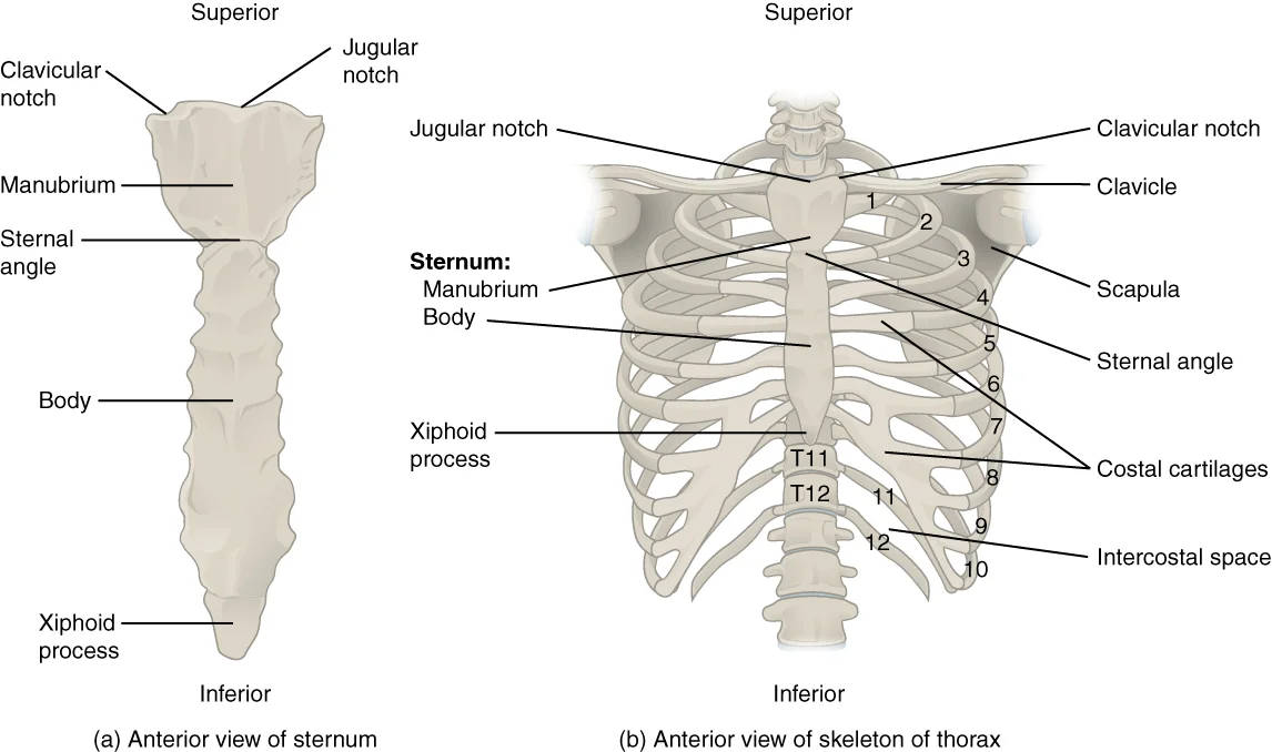
The thoracic cage forms the thorax portion of the body. It is composed of 12 pairs of ribs with their costal cartilages and the sternum. The cage is anchored posteriorly to the 12 thoracic vertebrae (T1–T12). The sternum consists of the manubrium, the body, and the xiphoid process. The manubrium and body join at the sternal angle, which forms a slight bend and serves as the attachment site for the second ribs. [1]
Figure 1Gray120
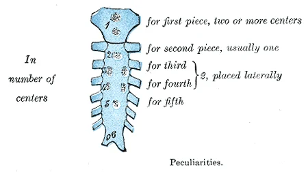
Long description
Peculiarities. Labelled illustration from Wikimedia Commons, used under its open licence.

Figure 1: Peculiarities.
Figure 2Gray114
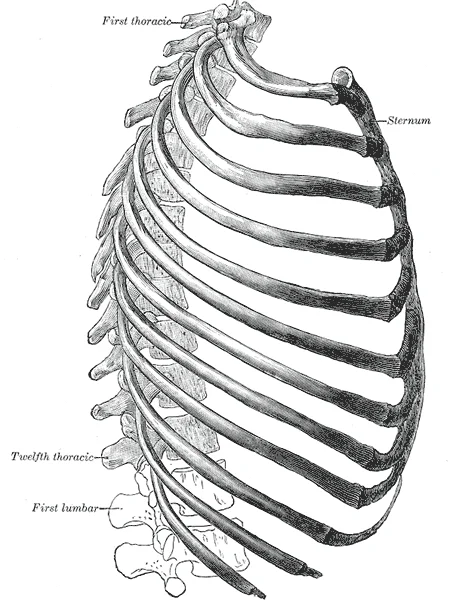
Long description
The thorax from the right. (Spalteholz.)

Figure 2: The thorax from the right (Spalteholz.).
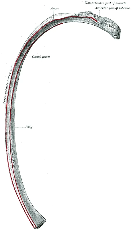
Each rib is a curved, flattened bone. The posterior head articulates with the costal facets of the thoracic vertebrae, while the tubercle articulates with the facet on the vertebral transverse process. The angle of the rib, located lateral to the tubercle, represents the point of greatest curvature and the most posterior extent of the thoracic cage. The inferior margin of each rib contains a costal groove that carries blood vessels and a nerve.
Ribs are classified based on their anterior attachment:
- True ribs (1–7): These vertebrosternal ribs attach directly to the sternum via their costal cartilages. [1]
- False ribs (8–12): These vertebrochondral ribs do not attach directly to the sternum. Ribs 8–10 attach to the cartilage of the next higher rib. [1]
- Floating ribs (11–12): These vertebral ribs are short and do not attach to the sternum or other ribs, terminating within the lateral abdominal wall musculature. [1]
Figure 3Gray318
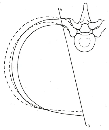
Long description
Diagram showing the axis of movement ( A B ) of a vertebrochondral rib. The interrupted lines indicate the position of the rib in inspiration.

Figure 3: Diagram showing the axis of movement ( A B ) of a vertebrochondral rib.
Figure 4Gray317
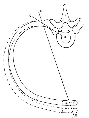
Long description
Diagram showing the axes of movement ( A B and C D ) of a vertebrosternal rib. The interrupted lines indicate the position of the rib in inspiration.

Figure 4: Diagram showing the axes of movement ( A B and C D ) of a vertebrosternal rib.
Figure 5Gray128
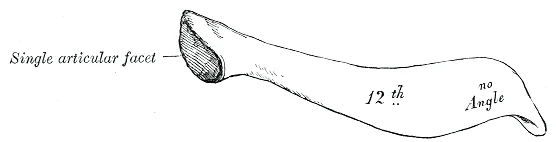
Long description
Peculiar ribs. Labelled illustration from Wikimedia Commons, used under its open licence.

Figure 5: Peculiar ribs.
Figure 6Gray127
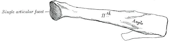
Long description
Peculiar ribs. Labelled illustration from Wikimedia Commons, used under its open licence.

Figure 6: Peculiar ribs.
Figure 7Gray126
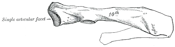
Long description
Peculiar ribs. Labelled illustration from Wikimedia Commons, used under its open licence.

Figure 7: Peculiar ribs.
Figure 8Gray123
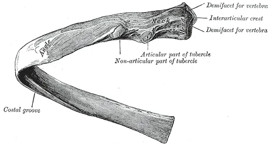
Long description
A central rib of the left side, viewed from behind.

Figure 8: A central rib of the left side, viewed from behind.
Figure 9Gray115
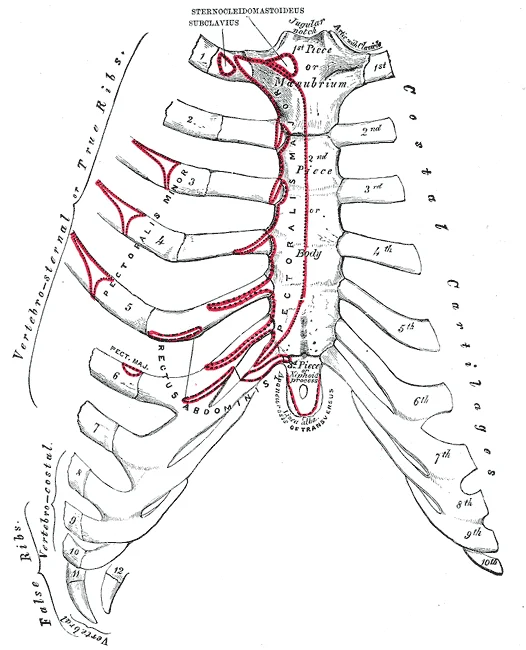
Long description
Anterior surface of sternum and costa cartilages.

Figure 9: Anterior surface of sternum and costa cartilages.
DefinitionThoracic boundaries
The thoracic inlet is the superior opening of the cage, while the thoracic outlet is the inferior boundary closed by the diaphragm.
Ultrasound can verify endotracheal tube positioning in emergency medicine. It differentiates unclear mass lesions of the chest wall accurately and with low risk. Localized fluid collections are rare but include hematomas, seromas, and abscesses. CEUS shows no enhancement in fluid collections. Hematomas result from trauma or surgery. Fresh hematomas are inhomogeneously hyperechoic. Organized hematomas are inhomogeneously hypoechoic or anechoic. Primary tumors of the chest wall are rare and have an incidence of less than 2% of th [3]
| Method | Utility |
|---|---|
| Chest X-ray | Standard initial imaging |
| Ultrasound | Superior to X-ray for detecting fractures |
| Oblique rib view | Limited sensitivity compared to ultrasound |
Rib identification
- Locate sternal anglePalpate the junction of manubrium and body
- Identify second ribThe second rib attaches at the sternal angle
- Count ribsUse the second rib as the landmark to count inferiorly
The sternum lies centrally, providing attachment for the costal cartilages. The clavicles articulate with the manubrium superiorly. The ribs curve downwards and forwards, becoming palpable as they extend laterally. The intercostal spaces are the gaps between adjacent ribs, containing the neurovascular bundle.
Palpation of the chest wall allows assessment of superficial and deep pathology. A small-part linear transducer with a frequency range of approximately 7–12 MHz is typically used for ultrasound evaluation. This tool acts as an extension of physical palpation for visible or dolent findings. The ribs run primarily horizontally in the dorsal arch part. In the lateral arch part, they run steeply caudally. Near the sternum, ribs 1–6 run mostly horizontally again. Ribs 7–10 run steeply cranially. The assessment is most favorable in the expiratory position. [3]
Figure 10Gray316
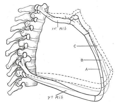
Long description
Lateral view of first and seventh ribs in position, showing the movements of the sternum and ribs in A, ordinary expiration; B, quiet inspiration; C, deep inspiration.

Figure 10: Lateral view of first and seventh ribs in position, showing the movements of the sternum and ribs in A, ordinary expiration; B, quiet inspiration; C, deep inspiration.
DefinitionThoracic Wall
The thoracic wall comprises skin, fascia, muscles, and bones that protect intrathoracic organs and facilitate respiration by altering cavity volume. [2]
| Muscle | Origin | Insertion | Action | Nerve Supply |
|---|---|---|---|---|
| Rectus abdominis | Pubic symphysis | Xiphoid process and costal cartilages | Flexes vertebral column | Intercostal nerves |
| External oblique | Lower eight ribs | Linea alba and pubic tubercle | Compresses abdomen | Intercostal nerves |
| Internal oblique | Inguinal ligament | Lower three ribs and linea alba | Compresses abdomen | Intercostal nerves |
| Transversus abdominis | Inguinal ligament and ribs | Linea alba and pubic crest | Compresses abdomen | Intercostal nerves |
Ultrasound can verify endotracheal tube positioning in emergency medicine. It differentiates unclear mass lesions with low risk. Localized fluid collections are rare but include hematomas, seromas, and abscesses. CEUS shows no enhancement in fluid collections. Hematomas result from trauma or surgery. Fresh hematomas are inhomogeneously hyperechoic. Organized hematomas are inhomogeneously hypoechoic or anechoic. Primary chest wall tumors are rare, with an incidence of less than 2%. Benign tumors usually grow slowly and expansively and are asymptomatic. [3]
Figure 11Gray113
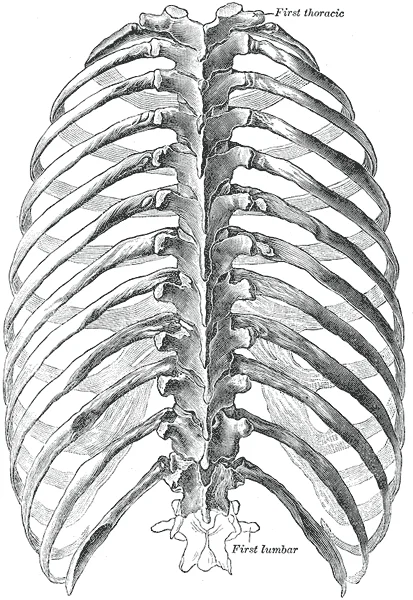
Long description
The thorax from behind. (Spalteholz.). Labelled illustration from Wikimedia Commons, used under its open licence.

Figure 11: The thorax from behind (Spalteholz.).
Figure 12Gray315
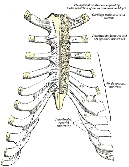
Long description
Sternocostal and interchondral articulations. Anterior view.

Figure 12: Sternocostal and interchondral articulations (anterior view).
The thoracic cage is formed by the thoracic vertebrae posteriorly, the sternum anteriorly, and the ribs laterally. These structures protect the heart and lungs while providing attachment points for respiratory muscles.
Figure 13Gray117
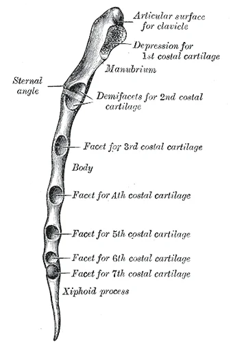
Long description
Lateral border of sternum. Labelled illustration from Wikimedia Commons, used under its open licence.

Figure 13: Lateral border of sternum.
Figure 14Gray116
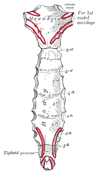
Long description
Posterior surface of sternum. Labelled illustration from Wikimedia Commons, used under its open licence.

Figure 14: Posterior surface of sternum.
The sternum is the elongated bony structure that anchors the anterior thoracic cage[1]. It consists of three parts: the manubrium, body, and xiphoid process[1]. The manubrium is the wider, superior portion of the sternum[1]. The top of the manubrium has a shallow, U-shaped border called the jugular (suprasternal) notch[1]. This can be easily felt at the anterior base of the neck, between the medial ends of the clavicles[1]. The clavicular notch is the shallow depression located on either side at the superior-lateral margins of the manubrium[1]. This is the site of the sternoclavicular joint, between the sternum and clavicle[1]. The first ribs also attach to the manubrium[1].
Figure 15Gray118
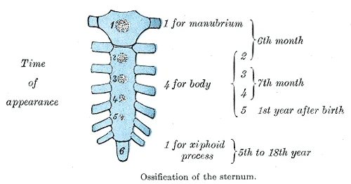
Long description
Ossification of the sternum. Labelled illustration from Wikimedia Commons, used under its open licence.

Figure 15: Ossification of the sternum.
The bony ribs do not extend anteriorly completely around to the sternum[1]. Instead, each rib ends in a costal cartilage[1]. These cartilages are made of hyaline cartilage and can extend for several inches[1]. Most ribs are then attached, either directly or indirectly, to the sternum via their costal cartilage[1]. The ribs are classified into three groups based on their relationship to the sternum[1]. [3]
The clavicular notches are lateral to the manubrium, articulating with the clavicles. The costal cartilages connect the ribs to the sternum, forming the anterior thoracic cage. The xiphoid process is the small, inferior tip of the sternum. [1]
Figure 16Gray312
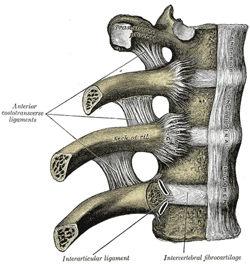
Long description
Costovertebral articulations. Anterior view.

Figure 16: Costovertebral articulations (anterior view).
The posterior thoracic wall is formed by the thoracic vertebrae. These vertebrae articulate with the ribs posteriorly. The ribs curve around the thorax, attaching to the vertebrae posteriorly and the sternum or its cartilages anteriorly. This arrangement creates a rigid yet flexible cage.
The diaphragm attaches to the lower thoracic wall. The skeletal muscle portions of the diaphragm insert into the central tendon. Origins include the xiphoid process of the sternum anteriorly, the inferior six ribs and their cartilages laterally, and the lumbar vertebrae and 12th ribs posteriorly. [2]
Figure 17Gray390
Long description
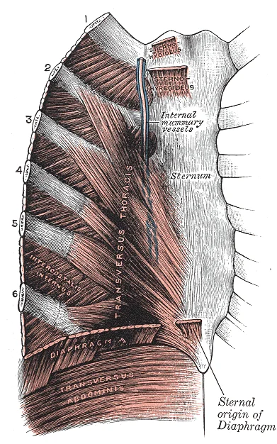
Posterior surface of sternum and costal cartilages, showing Transversus thoracis.

Figure 17: Posterior surface of sternum and costal cartilages, showing Transversus thoracis.
Figure 18Gray122
Long description
A central rib of the left side. Inferior aspect.

Figure 18: A central rib of the left side (inferior aspect).
The posterior abdominal wall is formed by the lumbar vertebrae, parts of the ilia of the hip bones, psoas major and iliacus muscles, and quadratus lumborum muscle. [2]
DefinitionSternum
The elongated bony structure that anchors the anterior thoracic cage, consisting of the manubrium, body, and xiphoid process.
DefinitionCostal Cartilage
Hyaline cartilage connecting the ribs to the sternum, allowing for thoracic expansion.
| Structure | Location | Function |
|---|---|---|
| Sternum | Anterior midline | Anchors anterior thoracic cage |
| Ribs | Lateral | Protect organs, aid respiration |
| Thoracic Vertebrae | Posterior | Support posterior thoracic wall |
| Diaphragm | Inferior | Primary muscle of respiration |
The thoracic wall comprises a complex arrangement of bones, cartilages, and joints that protect the thoracic viscera while permitting the mechanical movements required for respiration. The primary bony elements are the sternum, the twelve pairs of ribs, and the costal cartilages. The sternum consists of the manubrium, body, and xiphoid process. The ribs articulate posteriorly with the vertebral column and anteriorly with the sternum or costal cartilages. The thoracic inlet is bounded by the first rib, the superior border of the manubrium, and the first thoracic vertebra. The thoracic outlet is defined by the twelfth rib, the costal margin, and the twelfth thoracic vertebra.
Figure 19721 Rib Cage
Long description
The thoracic cage is formed by the (a) sternum and (b) 12 pairs of ribs with their costal cartilages. The ribs are anchored posteriorly to the 12 thoracic vertebrae. The sternum consists of the manubrium, body, and xiphoid process. The ribs are classified as true ribs (1–7) and false ribs (8–12). Th

Figure 19: The thoracic cage is formed by the (a) sternum and (b) 12 pairs of ribs with their costal cartilages.
The costal cartilages connect the anterior ends of the true ribs to the sternum. The costovertebral joints are located where the rib heads articulate with the vertebral bodies. The sternocostal joints are situated where the costal cartilages meet the sternum.
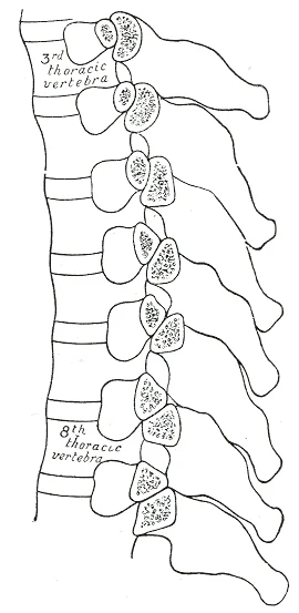
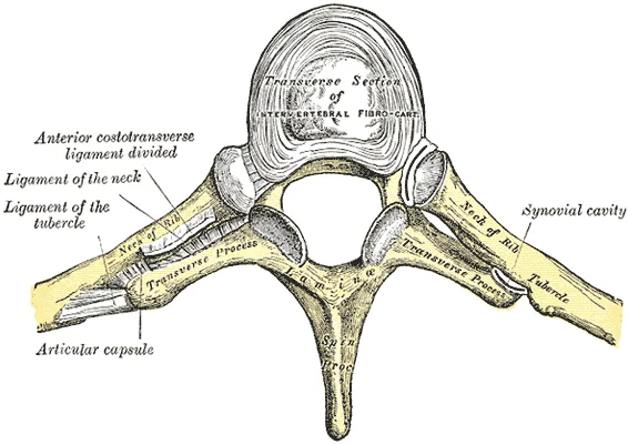
The costovertebral joints include the joints of the rib heads with the vertebral bodies and the costotransverse joints. The joints of the rib heads involve articulation with the demifacets on the vertebral bodies and the intervertebral disc. The costotransverse joints connect the tubercle of the rib to the transverse process of the vertebra. These joints allow for rotation and slight gliding movements. The sternocostal joints are synovial joints between the costal cartilages and the sternum. The first sternocostal joint is a synchondrosis, a cartilaginous joint that permits little movement. The second to seventh sternocostal joints are synovial plane joints.
Figure 20Gray314
Long description
Section of the costotransverse joints from the third to the ninth inclusive. Constrast the concave facets on the upper with the flattened facets on the lower transverse processes.

Figure 20: Section of the costotransverse joints from the third to the ninth inclusive.
Figure 21Gray313
Long description
Costotransverse articulation. Seen from above.

Figure 21: Costotransverse articulation (seen from above).
The intercostal muscles lie between the ribs. The external intercostal muscles run obliquely downwards and forwards. They aid in inspiration by raising the rib cage, which expands the thoracic cavity. The internal intercostal muscles lie deep to the externals and run obliquely downwards and backwards. They are used for expiration because they draw the ribs together to constrict the rib cage. The innermost intercostal muscles are the deepest layer and act as synergists for the action of the internal intercostals. [2]
Fractures of the ribs or sternum involve an interruption of the cortical bone contour. Frequently, a step formation with a surrounding hematoma is found, which is visualized as a blurred-bounded, inhomogeneous, hypoechoic structure. Traumatic fractures cannot be differentiated from pathological fractures unless a tumor soft tissue mantle can be visualized. Caution is required when diagnosing rib fractures near the costochondral joint, as a small step is often found here that can mimic a fracture [3]
Sternal fractures are difficult to diagnose on ultrasound due to synchondroses between the corpus and manubrium sterni and incomplete fusions of sternal bone spurs, which can mimic fractures. The diagnosis of sternal fractures is difficult on US, as cortical interruptions in the area of synchondrosis. Knowledge of these synchondroses allows for easier diagnosis of sternal fractures with ultrasound compared to normal X-ray. The idiopathic chondropathia of the costal cartilages at the base of the sternum presents as pain and swelling. Sonographically, it appears as a hypoechoic swelling of the sternoclavic [3]
In flail chest, paradoxical movement occurs with decreased lung compliancy and increased lung resistance, resulting in increased breathing effort. Impaired venous return due to pressure changes within the chest is also described. Moreover, the impaired venous return due to pressure changes within the chest is described. [4]
The thoracic wall is a complex structural unit designed to balance the body and stabilize the trunk. The muscles of the vertebral column, thorax, and abdominal wall extend, flex, and stabilize different parts of the body’s trunk. The deep muscles of the core of the body help maintain posture as well as carry out other functions. If any one group fails to function, body posture will be compromised. [2]
The most superficial layer is the skin, under which lies the superficial fascia containing subcutaneous fat and cutaneous nerves. Deep to this is the deep fascia, which invests the external intercostal muscles. The external intercostal muscles occupy the intercostal spaces, with their fibres running obliquely downwards and forwards. Deep to the external intercostals are the internal intercostal muscles, whose fibres run perpendicular to the external layer, downwards and backwards. The deepest muscular layer is the innermost intercostal muscles, separated from the internal intercostals by the intercostal neurovascular bundle. This bundle lies in the costal groove of the rib above, protected by the rib's inferior margin. The transversus thoracis muscle lies on the inner surface of the anterior thoracic wall, attaching to the sternum and costal cartilages. The parietal pleura lines the inner aspect of the thoracic cavity, separated from the endothoracic fascia by a potential space.
DefinitionThoracic Wall Layers
The thoracic wall consists of skin, superficial fascia, deep fascia, muscular layers (external, internal, and innermost intercostals), endothoracic fascia, and parietal pleura.
The arrangement of three bands of muscles in different orientations allows various movements and rotations of the trunk. The three layers of muscle also help to protect the internal abdominal organs in an area where there is no bone. [2]
| Layer | Fibre Direction | Primary Action |
|---|---|---|
| External Intercostals | Downwards and forwards | Elevate ribs during inspiration |
| Internal Intercostals | Downwards and backwards | Depress ribs during forced expiration |
| Innermost Intercostals | Variable | Assist in rib movement |
Sonographic examination of the prevascular mediastinum is limited by the ribs and sternum. Sonographic access to the mediastinum is via the supra- and parasternal, and occasionally also via the infrasternal, routes. The great vessels serve as guide structures because of their positional relationship to the heart at various levels. Suprasternal examination is performed with the patient in the supine position. The view into the upper mediastinum is facilitated by reclining the head, preferably wit Malignant tumors of the chest wall are characterized by infiltration of tumor tissue into the surrounding structures. [3]
The thoracic wall muscles are primarily responsible for respiration and structural integrity. They are grouped into compartments based on their location relative to the ribs.
The sternum lies centrally, providing attachment for the costal cartilages. The clavicles articulate with the manubrium superiorly. The ribs curve downwards and forwards, becoming palpable as they extend laterally. The intercostal spaces are the gaps between adjacent ribs, containing the neurovascular bundle.
The intercostal muscles assist breathing by changing rib cage dimensions. There are three sets of intercostal muscles spanning each intercostal space. The principal role of the intercostal muscles is to assist in breathing by changing the dimensions of the rib cage. The external intercostals elevate the ribs during inspiration. The internal intercostals depress the ribs during forced expiration. The innermost intercostals act similarly to the internals. They help change the shape of the rib cage and the volume-pressure [2]
The diaphragm is the principal muscle of respiration. It originates from the lower sternum, lower six ribs, and lumbar vertebrae. It inserts into the central tendon. It is innervated by the phrenic nerve. Contraction increases the vertical diameter of the thorax.
Each muscle is segmented by three transverse bands of collagen fibers called the tendinous intersections. They are enclosed by the rectus sheaths. These join at the anterior midline to form the linea alba. The linea alba is a white, fibrous band that is made of the bilateral rectus sheaths that join at the anterior midline of the body. [2]
The sternum lies centrally, providing attachment for the costal cartilages. The clavicles articulate with the manubrium superiorly. The ribs curve downwards and forwards, becoming palpable as they extend laterally. The intercostal spaces are the gaps between adjacent ribs, containing the neurovascular bundle.
| Muscle | Origin | Insertion | Action | Nerve Supply |
|---|---|---|---|---|
| External Intercostal | Inferior margin of rib above | Superior margin of rib below | Elevates ribs; aids inspiration | Intercostal nerves |
| Internal Intercostal | Superior margin of rib below | Inferior margin of rib above | Depresses ribs; aids expiration | Intercostal nerves |
| Innermost Intercostal | Similar to internals | Similar to internals | Depresses ribs; aids expiration | Intercostal nerves |
| Transversus Thoracis | Posterior surface of sternum | Costal cartilages 2-6 | Depresses ribs; aids expiration | Intercostal nerves |
| Diaphragm | Lower sternum, ribs, vertebrae | Central tendon | Principal muscle of inspiration | Phrenic nerve |
| Rectus Abdominis | Pubic crest and symphysis | Costal cartilages 5-7 and xiphoid | Compresses abdomen; flexes trunk | Intercostal nerves |
A shallow costal groove for the passage of blood vessels and a nerve is found along the inferior margin of each rib. This protects the neurovascular bundle. [1]
The arterial supply to the thoracic wall is segmental, derived from paired sources that ensure robust perfusion through anastomotic networks. Understanding this vascular architecture is fundamental for surgical approaches and interpreting imaging.
The posterior intercostal arteries originate from the thoracic aorta. They course along the inferior border of each rib, protected by the costal groove. Each artery gives off a dorsal ramus and a ventral ramus. The ventral ramus runs anteriorly along the intercostal space. It anastomoses with the anterior intercostal arteries. This connection ensures collateral circulation. [3]
The anterior intercostal arteries arise from the internal thoracic artery. The internal thoracic artery originates from the first part of the subclavian artery. It descends posterior to the costal cartilages, approximately 1 cm lateral to the sternum. It gives off perforating branches that supply the anterior chest wall. The anterior intercostal arteries run between the internal and transversus thoracis muscles. They anastomose with the posterior intercostal arteries.
The internal thoracic artery also gives off the musculophrenic artery. This branch follows the costal margin. It supplies the diaphragm and lower intercostal spaces. The superior epigastric artery is the terminal branch of the internal thoracic artery. It enters the rectus sheath and supplies the rectus abdominis muscle.
Vascular integrity is crucial during thoracic procedures. Injury to these vessels can lead to significant haemorrhage. The lymph node region along the parasternal mammary vessels must be explored conscientiously. This area is critical in oncological surgery to ensure complete clearance of metastatic disease. Surgeons must be aware of the proximity of these vessels to the sternum and costal cartilages. [3]
The musculophrenic artery can be traced along the costal margin.
The summary of invasive techniques provided here highlights the ingenuity of the treating surgeons. While these techniques vary, the underlying vascular anatomy remains constant. Understanding the origin, course, and branches of these arteries prevents iatrogenic injury. The internal thoracic artery is often used as a graft in coronary artery bypass surgery due to its reliable flow and size. Its branches provide essential collateral flow to the chest wall. [4]
The venous system of the thoracic wall is complex, featuring both superficial and deep networks that communicate extensively to ensure collateral circulation. This redundancy is clinically significant, particularly in cases of superior vena cava obstruction, where blood can bypass the blockage via alternative pathways.
It originates from the superior epigastric vein and ascends alongside the sternum, receiving intercostal veins from the upper six or seven intercostal spaces. These veins enter the internal thoracic vein at right angles. This pathway is crucial for draining the anterior mediastinum and the anterior chest wall. [1]
The azygos venous system provides the primary drainage for the posterior thoracic wall and serves as a vital collateral pathway. It consists of the azygos vein, hemiazygos vein, and accessory hemiazygos vein.
The azygos vein begins in the abdomen as the continuation of the right ascending lumbar vein. It ascends along the right side of the vertebral column, receiving the right superior intercostal veins and often the right bronchial veins. At the level of the fourth thoracic vertebra, it arches anteriorly over the root of the right lung to drain into the superior vena cava. [1]
The hemiazygos vein drains the left lower posterior intercostal spaces. It crosses the midline at the level of the ninth or tenth thoracic vertebra to join the azygos vein. The accessory hemiazygos vein drains the left middle intercostal spaces and may drain directly into the azygos vein or join the hemiazygos vein. [1]
| Vein | Origin | Termination | Key Relations |
|---|---|---|---|
| Internal Thoracic | Superior Epigastric | Brachiocephalic | Along sternum |
| Azygos | Right Ascending Lumbar | Superior Vena Cava | Right vertebral column |
| Hemiazygos | Left Lower Intercostals | Azygos | Left vertebral column |
Exam trapCommon Misconception
Do not confuse the internal thoracic vein with the internal jugular vein; they are distinct structures with different drainage territories.
The thoracic wall relies on a precise arrangement of nerves for both movement and sensation. Understanding this supply is fundamental before considering any surgical intervention or trauma assessment.
The primary motor and cutaneous supply comes from the anterior rami of the thoracic spinal nerves T1 to T11. These are known as intercostal nerves. The twelfth thoracic nerve (T12) is termed the subcostal nerve as it lies below the twelfth rib.
Each intercostal nerve runs within the neurovascular bundle of the intercostal space. This bundle is located between the internal intercostal muscle and the innermost intercostal muscle. The order from superior to inferior is Vein, Artery, Nerve (VAN). This anatomical relationship dictates that safe needle insertion for procedures must occur at the superior border of the rib to avoid vascular and neural injury.
The intercostal nerves provide motor innervation to the intercostal muscles, the transversus thoracis, and the subcostal muscles. They also supply the levatores costarum. Cutaneously, they provide sensation to the skin overlying the thoracic wall. The anterior rami of T2 to T6 supply the skin of the anterior and lateral thorax. The T7 to T11 nerves supply the lower anterior and lateral abdominal wall, extending their sensory field below the rib cage.
The viewer should note the nerve's position deep to the internal intercostals.
Damage to these nerves can result in significant sensory loss and motor weakness. Intercostal neuralgia presents as sharp, burning pain along the distribution of the affected nerve. This can occur post-thoracotomy or due to herpes zoster infection.
| Nerve | Motor Supply | Cutaneous Supply |
|---|---|---|
| T1-T11 | Intercostals, transversus thoracis, subcostals | Skin of thoracic wall and upper abdomen |
| T12 (Subcostal) | Abdominal muscles | Skin of lower abdomen |
Understanding the precise anatomical course allows clinicians to predict sensory deficits and plan analgesic techniques such as intercostal nerve blocks.
The lymphatic drainage of the thoracic wall follows a predictable pattern based on the anatomical location of the tissues, ensuring that fluid from the chest wall and underlying structures returns to the systemic circulation via specific nodal groups. Understanding this pathway is critical for assessing the spread of malignancies or infections originating in the thoracic region.
Deep lymphatics from the anterior intercostal spaces and the medial breast follow the internal thoracic vessels. [1]
DefinitionParasternal Nodes
Lymph nodes located along the internal thoracic vessels, draining the medial breast and anterior intercostal spaces.
Clinical pearlClinical Relevance
Malignancy in the medial breast quadrant often metastasizes to parasternal nodes, whereas lateral quadrant cancers typically spread to axillary nodes.
| Region | Primary Drainage Site | Key Vessels/Nerves |
|---|---|---|
| Anterior Thoracic Wall | Axillary Nodes (Lateral) | Lateral Thoracic Vessels |
| Medial Breast/Anterior Intercostals | Parasternal Nodes | Internal Thoracic Vessels |
| Posterior Thoracic Wall | Posterior Intercostal Nodes | Posterior Intercostal Vessels |
The thoracic wall forms the protective boundary of the thoracic cavity, separating the internal viscera from the external environment. Its structural integrity relies on the precise arrangement of bones, muscles, and fascial layers. Understanding the spatial relationships between these components is essential for interpreting clinical signs and imaging findings.
The sternum is centrally located, articulating with the costal cartilages of the true ribs. Lateral to the sternum, the intercostal spaces contain the neurovascular bundle. The diaphragm forms the inferior boundary, curving upwards into the thoracic cavity. The posterior third of the right diaphragm is visualised approximately 5 cm lateral to the inferior vena cava foramen. The transducer is positioned between the middle clavicular line and the anterior axillary line, directed medially, cranially, and dorsally. [3]
The intercostal muscles occupy the spaces between the ribs. The external intercostal muscles run obliquely downwards and forwards, originating from the inferior border of the rib above and inserting into the superior border of the rib below. They are responsible for elevating the ribs during inspiration. They assist in forced expiration.
The neurovascular bundle runs in the costal groove of each rib, protected by the overlying bone. It contains the intercostal vein, artery, and nerve, arranged from superior to inferior as vein, artery, nerve (VAN). This arrangement is critical to avoid injury during thoracentesis or intercostal nerve blocks. The pleura and peritoneum line the internal surfaces, with the parietal pleura adhering to the inner thoracic wall and the diaphragmatic pleura covering the superior surface of the diaphragm. The potential space between the visceral and parietal pleura is the pleural cavity, which contains a small amount of lubricating fluid. [2]
DefinitionDiaphragmatic Excursion
The amplitude of diaphragmatic movement, measured as the distance between the highest and lowest points during breathing, ranges from 1–2 cm in resting breathing to 7–9 cm in forced breathing. [3]
The diaphragm’s function is closely linked to the thoracic wall’s mechanics. Diaphragmatic excursion can be determined only in spontaneously breathing patients. The amplitude and speed are measured in both quiet breathing and forced inspiration, such as the sniff maneuver. This measurement provides insight into respiratory muscle strength and diaphragmatic function. [3]
| Muscle | Origin | Insertion | Action | Nerve Supply |
|---|---|---|---|---|
| External Intercostal | Inferior border of rib above | Superior border of rib below | Elevates ribs (inspiration) | Intercostal nerve |
| Internal Intercostal | Costal groove of rib above | Superior border of rib below | Depresses ribs (expiration) | Intercostal nerve |
| Innermost Intercostal | Costal groove of rib above | Superior border of rib below | Depresses ribs (expiration) | Intercostal nerve |
The thoracic wall is assessed using a multimodal imaging approach, with each technique offering distinct advantages for visualising bony and soft tissue structures. Plain X-ray remains the initial screening tool for gross bony abnormalities, though it lacks the resolution for detailed soft tissue characterisation. Computed tomography (CT) provides high-resolution cross-sectional anatomy, serving as the gold standard for evaluating cortical bone integrity and mediastinal relationships. Magnetic resonance imaging (MRI) offers superior soft tissue contrast, particularly useful for distinguishing between benign and malignant mediastinal lymphadenopathy, where CT may be ambiguous. Ultrasound (US) has emerged as a dynamic, point-of-care modality, particularly for traumatic pathologies and superficial structures, offering diagnostic accuracy comparable to CT in specific contexts. [3]
The sternum is centrally located, articulating with the costal cartilages of ribs 1–7. Laterally, the ribs curve posteriorly, forming the thoracic cage. The intercostal spaces contain the neurovascular bundle, lying in the costal groove of the rib above. The diaphragm forms the inferior boundary, appearing as a dome-shaped structure. The mediastinum lies centrally, containing the heart and great vessels. The lungs flank the mediastinum, appearing radiolucent.
Ultrasound appearance varies by tissue type. Osteolytic metastases present as an irregular interruption of the cortical bone contour, accompanied by inhomogeneously hypoechoic soft tissue. This destruction leads to pathological sound transmission. For diaphragmatic assessment, real-time US is the first imaging modality for diagnosing paresis. Diaphragmatic excursion is evaluated using M-mode with a low-frequency probe (2.5–5 MHz). Diaphragm thickness is measured with a linear transducer (≥7 MHz) in B- or M-mode. However, TUS is not suitable for acute diaphragmatic injuries due to low sensitivity. The diaphragm is not completely visible sonographically, especially on the left side. [3]
Contrast-enhanced US (CEUS) can evaluate malignancy in lesions visualised on B-mode US.
Clinical pearlUltrasound Utility
US is dynamic and point-of-care, ideal for trauma and diaphragmatic function, but limited for acute injuries and deep mediastinal structures.
| Modality | Primary Use | Key Advantage |
|---|---|---|
| X-ray | Initial screening | Availability, low cost |
| CT | Bone detail, mediastinum | High resolution, gold standard |
| MRI | Soft tissue contrast | Differentiation of lymph nodes |
| Ultrasound | Trauma, diaphragm, biopsy | Dynamic, point-of-care, real-time |
The thoracic wall serves as the protective framework for the mediastinal contents, yet its clinical utility extends far beyond passive protection. Understanding the surface landmarks and underlying structures is vital for interpreting imaging and performing procedures. In this anterior view, the sternum is central, flanked by the costal cartilages which articulate with the ribs. The intercostal spaces contain the neurovascular bundle, lying in the costal groove of the rib above. This anatomical arrangement dictates that safe needle insertion for thoracentesis must occur just above the upper border of the rib to avoid the intercostal vessels and nerve. The figure also demonstrates the diaphragm’s insertion on the lower ribs, a key landmark for assessing diaphragmatic excursion.
Nerve palsies of the thoracic wall typically involve the intercostal nerves. Injury to these nerves can result in sensory deficits or motor weakness of the intercostal muscles. Clinically, this may present as pain or difficulty in coughing. The intercostal nerves run along the inferior border of each rib, making them vulnerable during rib fractures or surgical procedures.
Procedures such as diaphragmatic ultrasound are increasingly important. Diaphragmatic atrophy is defined as a thickness of <1.5 mm in men and <1.1 mm in women. Furthermore, a DTF of <20% is compatible with paresis. Diaphragmatic paresis is evident on real-time clinical orienting examination and M-mode US as the absence of excursion during quiet and deep breathing and as the absence of or paradoxical movement during the sniff maneuver. In patients with sonographic follow-up, an increase in thickness during inspiration over time was associated with an improvement in inspiratory function and an increase in vital capacity. [3]
Clinical pearlIntercostal Neurovascular Bundle
The intercostal vein, artery, and nerve run in the costal groove of the rib above, in that order from superior to inferior. Always insert needles above the rib to avoid injury.
| Structure | Location/Relation | Clinical Significance |
|---|---|---|
| Intercostal Nerve | Inferior to rib | Vulnerable in fractures; causes pain/numbness |
| Diaphragm | Lower thoracic wall | Ultrasound assesses function; atrophy <1.5mm (men) |
| Sternum | Anterior midline | Attachment for flail chest fixation |
| Costal Cartilage | Anterior rib ends | Connects ribs to sternum; palpable landmark |
DefinitionThoracic Wall Anatomy
The thoracic wall comprises the bony cage, muscles, and neurovascular structures that protect the thoracic viscera and facilitate respiration. Understanding the mechanics of rib fixation and tracheal anatomy is critical for both surgical intervention and emergency airway management.
| Feature | Description | Clinical Relevance |
|---|---|---|
| Rib Material | Relatively soft bone | Screws prone to breakout |
| Labitzke Plate | Grasping mechanism, titanium | Prevents screw failure |
| RibLoc System | Slides over rib, locking screws | No bending required |
| Correct Intubation | One hyperechoic interface | Confirms tracheal placement |
| Esophageal Intubation | Two hyperechoic interfaces | Indicates misplacement |
Tracheal Ultrasound Interpretation
- Perform UltrasoundScan anterior neck
- Question: Visualize interfaces?
One interface
- Correct tracheal intubation
Two interfaces
- Esophageal intubation
Students frequently confuse the anatomical boundaries of the thoracic wall with adjacent structures, leading to errors in clinical localisation. A primary error involves misidentifying the inferior limit of the sternum. The inferior tip is the xiphoid process, a small structure that is cartilaginous early in life but gradually becomes ossified starting during middle age. Assuming it is fully bone in young adults can lead to incorrect landmark identification during resuscitation or examination. [1]
Another significant pitfall relates to the interpretation of ultrasound imaging in the anterior mediastinum. Students often mistake normal thymic tissue for pathology. In childhood, specifically between 2 and 8 years of age, thymic tissue is detectable in more than 90% of cases[1]. On ultrasound, this tissue predominantly shows similar homogeneous echogenicity to that of the liver and spleen, with multiple hypoechoic strands[1]. These strands appear as a “starry sky” pattern, which helps to identify thymic tissue as normal rather than a mass[1]. Failing to recognise this pattern may lead to unnecessary invasive procedures. Conversely, in adults, the involved thymus is mostly replaced by adipose tissue and usually cannot be visualised[1]. Assuming a visible anterior mediastinal mass is always pathological without considering age-related involution is a common diagnostic error. [3]
Misunderstanding the role of imaging in thoracic trauma is another frequent mistake. While ultrasound is of high importance in the detection of malignant lesions due to its location in the near-field region, its utility in acute trauma is specific[1]. It is considered a reliable, cost-effective, and safe method for use at the bedside in the neonatal unit, but its application in adult rib fractures requires careful technique[1]. Students often overestimate its ability to visua The primary importance of this benign pathology is the differentiation from malignant infiltration. [3]
Finally, students may confuse internal and external support mechanisms for chest wall instability. External support for a flail chest was first reported by Jones, appearing 25 years before mechanical ventilation was presented. However, internal support or splinting by mechanical positive pressure ventilation is a relatively modern technique compared to external splinting. Confusing the historical development or current indications for these two distinct approaches can lead to inappropriate manag. The flail chest as we know it today was described as early as 1955 by Cohen. [4]
Knowledge of the thoracic wall is essential at the bedside for physical examination and procedural safety. Clinicians must identify the xiphoid process to avoid injury during cardiopulmonary resuscitation, recognising that its cartilaginous nature in younger patients makes it prone to fracture if pressure is applied incorrectly. In neonatal units, lung ultrasound is used as a point-of-care tool for diagnosing pulmonary diseases, leveraging its safety and lack of radiation exposure[1].
In the operating theatre, understanding thoracic anatomy guides surgical approaches for chest wall stabilisation. Various techniques have been developed to stabilise rib fractures, ranging from early methods using K wires to modern dedicated systems. For instance, the Matrix® rib fixation system by Synthes™ uses titanium plates designed to mimic the biodynamic characteristics of ribs, fixed with locking screws. Surgeons must be aware that ribs are relatively soft compared to other bones, which may make them prone to breakage when attempting to bend clamps around them. This knowledge prevents iatrogenic injury during internal fixation. [4]
On imaging, particularly ultrasound, the thoracic wall is assessed for lesions and normal variants. Contrast-enhanced ultrasound (CEUS) requires careful probe selection; linear probes with high frequencies are preferred for superficial lesions, while curved arrays may be used for deeper targets[1]. The contrast agent, typically 2.4 mL of SonoVue, is administered as a bolus, and the lesion is observed continuously for at least the first 30 seconds[1]. This protocol helps distinguish between benign and malignant lesions, as US is highly important for detecting malignancies in the near-field region[1]. [3]
In examinations, students are tested on their ability to differentiate normal anatomical variants from pathology. Recognising that reactive thymus enlargement can occur after stressful situations such as surgery or burns is crucial[1]. Additionally, understanding that the thymus may have normal anatomic variants, such as cervical or retrocaval extensions, prevents misdiagnosis on imaging[1]. These facts are standard textbook knowledge required for clinical competence. [3]
The xiphoid process is the inferior tip of the sternum and ossifies from middle age onwards. Thymic tissue is detectable in over 90% of children aged 2–8 years. Normal thymus shows homogeneous echogenicity with hypoechoic “starry sky” strands on ultrasound. In adults, the thymus is mostly replaced by adipose tissue and is rarely visualised. Ultrasound is a safe, radiation-free bedside tool for neonatal pulmonary assessment. Linear probes are preferred for superficial thoracic lesions in contrast-enhanced ultrasound. Rib fractures may be stabilised using internal fixation systems like the Matrix® plate. Ribs are soft bones, requiring careful handling to avoid breakage during fixation.
Answer each one in your head first, then open the answer.
What is the inferior tip of the sternum called?
Show answerHide answer
Xiphoid processAt what age does the xiphoid process typically begin to ossify?
Show answerHide answer
Middle ageWhat percentage of children aged 2–8 years have detectable thymic tissue?
Show answerHide answer
More than 90%What ultrasound pattern helps identify normal thymic tissue?
Show answerHide answer
Starry skyWhat is the typical dose of SonoVue for contrast ultrasound?
Show answerHide answer
2.4 mL
- Betts JG, Young KA, Wise JA, et al. The Thoracic Cage. OpenStax, Anatomy and Physiology 2e (CC BY-NC-SA 4.0). Accessed .
- Betts JG, Young KA, Wise JA, et al. Axial Muscles of the Abdominal Wall, and Thorax. OpenStax, Anatomy and Physiology 2e (CC BY-NC-SA 4.0). Accessed .
- Safai Zadeh E, Görg C, Prosch H, et al. The Role of Thoracic Ultrasound for Diagnosis of Diseases of the Chest Wall, the Mediastinum, and the Diaphragm-Narrative Review and Pictorial Essay. Diagnostics (Basel, Switzerland) 2023;13(4):767. doi:10.3390/diagnostics13040767. Accessed .
- Bemelman M, Poeze M, Blokhuis TJ, Leenen LP. Historic overview of treatment techniques for rib fractures and flail chest. European journal of trauma and emergency surgery : official publication of the European Trauma Society 2010;36(5):407-415. doi:10.1007/s00068-010-0046-5. Accessed .
Figure credits
- Figure 1: Henry Vandyke Carter, via Wikimedia Commons, Public domain, Public domain.
- Figure 2: Henry Vandyke Carter, via Wikimedia Commons, Public domain, Public domain.
- Figure 3: Henry Vandyke Carter, via Wikimedia Commons, Public domain, Public domain.
- Figure 4: Henry Vandyke Carter, via Wikimedia Commons, Public domain, Public domain.
- Figure 5: Henry Vandyke Carter, via Wikimedia Commons, Public domain, Public domain.
- Figure 6: Henry Vandyke Carter, via Wikimedia Commons, Public domain, Public domain.
- Figure 7: Henry Vandyke Carter, via Wikimedia Commons, Public domain, Public domain.
- Figure 8: Henry Vandyke Carter, via Wikimedia Commons, Public domain, Public domain.
- Figure 9: Henry Vandyke Carter, via Wikimedia Commons, Public domain, Public domain.
- Figure 10: Henry Vandyke Carter, via Wikimedia Commons, Public domain, Public domain.
- Figure 11: Henry Vandyke Carter, via Wikimedia Commons, Public domain, Public domain.
- Figure 12: Henry Vandyke Carter, via Wikimedia Commons, Public domain, Public domain.
- Figure 13: Henry Vandyke Carter, via Wikimedia Commons, Public domain, Public domain.
- Figure 14: Henry Vandyke Carter, via Wikimedia Commons, Public domain, Public domain.
- Figure 15: Henry Vandyke Carter, via Wikimedia Commons, Public domain, Public domain.
- Figure 16: Henry Vandyke Carter, via Wikimedia Commons, Public domain, Public domain.
- Figure 17: Henry Vandyke Carter, via Wikimedia Commons, Public domain, Public domain.
- Figure 18: Henry Vandyke Carter, via Wikimedia Commons, Public domain, Public domain.
- Figure 19: OpenStax College, via Wikimedia Commons, CC BY 3.0, CC BY 3.0.
- Figure 20: Henry Vandyke Carter, via Wikimedia Commons, Public domain, Public domain.
- Figure 21: Henry Vandyke Carter, via Wikimedia Commons, Public domain, Public domain.
Educational content for medical students and doctors. It is not a substitute for clinical judgement or local guidelines.