Vertebral canal, spinal meninges and lumbar puncture
The vertebral canal is the bony passage protecting the spinal cord and meninges within the vertebral column.
Figure 1Gray301
Long description
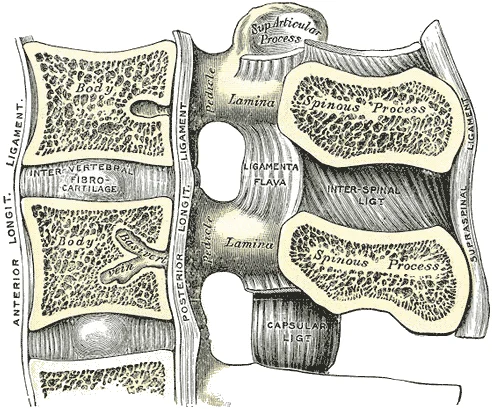
Median sagittal section of two lumbar vertebræ and their ligaments.

Figure 1: Median sagittal section of two lumbar vertebræ and their ligaments.
The vertebral canal is the continuous bony space formed by the alignment of the vertebral foramina of all vertebrae[1]. It serves as the primary protective passageway for the spinal cord and its associated meningeal coverings[1]. The canal is bounded anteriorly by the posterior surfaces of the vertebral bodies and the posterior longitudinal ligament[1]. The lateral and posterior walls are formed by the vertebral arches, specifically the pedicles and the laminae[1]. The ligamentum flavum, which contains elastic fibres, connects the laminae of adjacent vertebrae to complete the posterior wall of the canal[1].
DefinitionVertebral Canal
The vertebral canal is the bony space formed by the alignment of the vertebral foramina, housing the spinal cord and meninges.
The spinal cord typically terminates at the level of the L1 or L2 vertebrae. Below this point, the canal contains the cauda equina, a collection of nerve roots that descend from the end of the spinal cord to exit through the lower lumbar and sacral foramina.
Lumbar puncture is performed to access the subarachnoid space, typically between the L3 and L4 or L4 and L5 vertebrae. The needle traverses several layers to reach the cerebrospinal fluid:
- Skin and subcutaneous tissue
- Supraspinous ligament
- Interspinous ligament
- Ligamentum flavum
- Epidural space (containing fat and internal vertebral venous plexus)
- Dura mater
- Arachnoid mater
In epidural anaesthesia, the needle is advanced through the same posterior structures but is stopped within the epidural space, anterior to the dura mater. The ligamentum flavum provides a distinct tactile resistance that clinicians use to identify the depth of the needle before entering the epidural space.
| Ligament | Location | Function |
|---|---|---|
| Anterior longitudinal | Anterior vertebral bodies | Prevents hyperextension |
| Posterior longitudinal | Posterior vertebral bodies | Prevents hyperflexion |
| Ligamentum flavum | Between adjacent laminae | Supports column during bending |
| Supraspinous | Tips of spinous processes | Limits flexion |
Lumbar Puncture Access
- Skin and Subcutaneous TissueInitial entry point
- Supraspinous and Interspinous LigamentsDense connective tissue layers
- Ligamentum FlavumElastic ligament providing resistance
- Epidural SpaceFat and venous plexus
- Dura and Arachnoid MaterFinal barrier to subarachnoid space
Figure 2Gray661
Long description
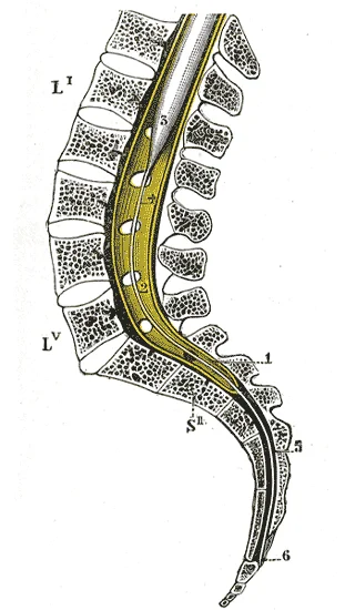
Sagittal section of vertebral canal to show the lower end of the medulla spinalis and the filum terminale. Li, Lv. First and fifth lumbar vertebræ. Sii. Second sacral vertebra. 1. Dura mater. 2. Lower part of tube of dura mater. 3. Lower extremity of medulla spinalis. 4. Intradural, and 5, Extradura

Figure 2: Sagittal section of vertebral canal to show the lower end of the medulla spinalis and the filum terminale (li, lv).
The vertebral column consists of vertebrae that vary in size and shape but follow a similar structural pattern. A typical vertebra includes a body, a vertebral arch, and seven processes. When aligned, notches in the pedicle margins form an intervertebral foramen, the opening for spinal nerve exit. Seven processes arise from the vertebral arch. Each paired transverse process projects laterally from the junction of the pedicle and lamina. The single spinous process projects posteriorly at the midline. These vertebral spines are easily felt as a series of bumps just under the skin down the middle of the back. [1]
Palpating these spinous processes allows clinicians to identify specific vertebral levels. The supraspinous ligament interconnects the spinous processes of thoracic and lumbar vertebrae. This strong ligament supports the column during forward bending. In the posterior neck, where cervical spinous processes are short, this ligament expands into the nuchal ligament. The nuchal ligament attaches to cervical spinous processes and extends to the external occipital protuberance. [1]
Adjacent vertebrae are united by ligaments running along the posterior and anterior aspects. These resist excess forward or backward bending. Intervertebral discs fill the gap between adjacent vertebral bodies. These fibrocartilaginous pads are anchored to adjacent vertebrae, uniting them strongly. Discs provide padding during weight bearing. They are thin in the cervical region and thickest in the lumbar region, which carries the most body weight. In total, intervertebral discs account for approximately 25 percent of body height between the top of the pelvis and the base of the skull. [1]
The sacral promontory is the anterior lip of the superior base of the sacrum. Lateral to this is the roughened auricular surface, joining the ilium to form the immobile sacroiliac joints. Passing inferiorly through the sacrum is the sacral canal, terminating at the sacral hiatus near the inferior tip. The anterior and posterior surfaces of the sacrum have paired openings called sacral foramina. These openings allow anterior and posterior branches of sacral spinal nerves to exit. [1]
Clinical pearlLandmark Identification
The intercristal line, passing between the highest points of the iliac crests, typically intersects the spinous process of L4 or the L4-L5 interspace, providing a reliable surface landmark for lumbar puncture.
| Landmark | Anatomical Relation | Clinical Utility |
|---|---|---|
| Iliac Crests | Highest point of hipbone | Intersects L4 spinous process |
| Spinous Processes | Posterior midline projections | Palpable bumps for level identification |
| Sacral Hiatus | Inferior termination of sacral canal | Landmark for caudal anaesthesia |
Figure 3Gray83
Long description
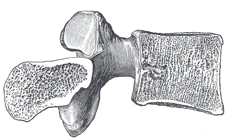
Sagittal section of a lumbar vertebra. Labelled illustration from Wikimedia Commons, used under its open licence.

Figure 3: Sagittal section of a lumbar vertebra.
The vertebral column comprises vertebrae separated by intervertebral discs. Each vertebra has a body anteriorly, which supports weight and increases in size inferiorly. Adjacent bodies are united by discs that provide padding and allow movement. The anterior longitudinal ligament runs along the anterior vertebral bodies, resisting backward bending. [1]
Cervical vertebrae are smaller than lumbar vertebrae. The atlas (C1) lacks a body, consisting of anterior and posterior arches. The axis (C2) features the dens, a bony projection articulating with the atlas. Typical cervical vertebrae have bifid spinous processes and transverse foramina. [1]
Thoracic vertebrae have long, downward-projecting spinous processes and facets for rib attachment. They form the thoracic curve, a primary curvature. [1]
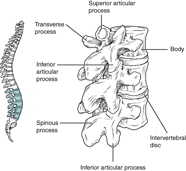
Lumbar vertebrae have large, thick bodies and short, blunt spinous processes. The superior articular processes face medially, and the inferior face laterally. These features allow significant weight bearing while permitting flexion and extension. [1]
Figure 4725 Lumbar Vertebrae
Long description
Lumbar vertebrae are characterized by having a large, thick body and a short, rounded spinous process.

Figure 4: Lumbar vertebrae are characterized by having a large, thick body and a short, rounded spinous process.
The coccyx is small, derived from four fused vertebrae. [1]
Disorders of curvature include kyphosis (excessive posterior thoracic curvature), lordosis (excessive anterior lumbar curvature), and scoliosis (abnormal lateral curvature with twisting). [1]
The bony end plate is a thickened, porous layer of fused trabecular bone, generally 0.2 to 0.8 mm thick. It is thinner centrally than peripherally at a given lumbar level. [3]
The bone marrow compartment adjacent to the end plate contains hematopoietic cells, fat cells, sinusoids, and nerves. Sinusoids form an arterial grid, terminating adjacent to cartilage end plates. [3]
Approximately two-thirds of spinal metastases arise from breast (16.5%), prostate (20.7%), and lung carcinoma (24.6%). [2]
The joints of the vertebral column are primarily cartilaginous joints located between the vertebral bodies, supported by synovial joints between the articular processes. The shape and orientation of these articular processes vary by region, which plays a major role in determining the type and range of motion available. [1]
In the thoracic region, the superior articular facets face posteriorly, while the inferior facets face anteriorly. These orientations restrict the range of movement compared to other regions. The vertebral bodies are separated by intervertebral discs, which contain a gel-like nucleus pulposus. This nucleus allows the disc to change shape as vertebrae rock during movement. Bending forward compresses the anterior portion of the disc and expands the posterior disc. [1]
DefinitionIntervertebral disc function
The nucleus pulposus acts as a shock absorber and pivot point, allowing vertebral bodies to tilt and rotate relative to their neighbours.
The typical marrow contact area, or effective exchange area, is between 10 and 40%. These plates are vulnerable to damage, which can lead to communication between the nucleus and the marrow, potentially causing nerve proliferation and pain. If the posterior anulus fibrosus is weakened, the nucleus pulposus may protrude posteriorly, potentially compressing a spinal nerve. [3]
| Feature | Description |
|---|---|
| Nucleus pulposus | Gel-like core allowing vertebral rocking |
| Thoracic facets | Superior face posteriorly; inferior face anteriorly |
| End plate contact | Effective exchange area is 10 to 40% |
| Disc herniation | Posterior protrusion of nucleus pulposus |
Vertebral bone marrow lesions, or Modic changes, are associated with discogenic pain. Type I changes involve fibrovascular replacement, while Type II changes correlate with fatty marrow replacement. These changes are dynamic and can convert between types or return to normal. Type III changes represent dense woven bone or sclerosis. [3]
The adult vertebral column does not form a straight line, but instead has four curvatures along its length. These curves increase the vertebral column’s strength, flexibility, and ability to absorb shock. When the load on the spine is increased, by carrying a heavy backpack for example, the curvatures increase in depth (become more curved) to accommodate the extra weight. They then spring back when the weight is removed. The four adult curvatures are classified as either primary or secondary curvatures. Primary curves are retained from the original fetal curvature, while secondary curvatures develop after birth. [1]
The vertebral bodies are anterior, with intervertebral discs between them. Posterior to the bodies lies the vertebral canal. Inside the canal, the spinal cord (or cauda equina in the lumbar region) is surrounded by meninges. The epidural space lies between the dura mater and the vertebral canal walls. The ligamentum flavum connects the laminae. The spinous processes project posteriorly. The subarachnoid space contains cerebrospinal fluid. The dura mater is the tough outer layer. The arachnoid mater is the middle layer. The pia mater adheres to the spinal cord. The transverse processes extend laterally. The spinal nerves exit through the intervertebral foramina. The epidural fat fills the space around the dural sac. The ligamentum nuchae is present in the cervical region, not lumbar. The supraspinous ligament connects the tips of the spinous processes. The interspinous ligaments connect adjacent spinous processes. The intertransverse ligaments connect transverse processes. The anterior longitudinal ligament runs along the anterior vertebral bodies. The posterior longitudinal ligament runs along the posterior vertebral bodies within the canal. [1]
Figure 5Gray770
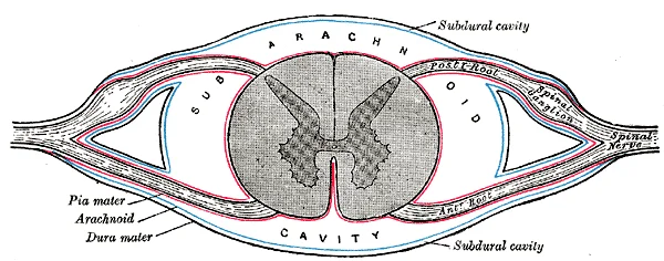
Long description
Diagrammatic transverse section of the medulla spinalis and its membranes.

Figure 5: Diagrammatic transverse section of the medulla spinalis and its membranes.
Typical cervical vertebrae, such as C4 or C5, have several characteristic features that differentiate them from thoracic or lumbar vertebrae. Cervical vertebrae have a small body, reflecting the fact that they carry the least amount of body weight. Cervical vertebrae usually have a bifid (Y-shaped) spinous process. The spinous processes of the C3–C6 vertebrae are short, but the spine of C7 is much longer. You can find these vertebrae by running your finger down the midline of the posterior neck until you encounter the prominent C7 spine located at the base of the neck. The transverse processes of the cervical vertebrae are sharply curved (U-shaped) to allow for passage of the cervical spinal nerves. Each transverse process also has an opening called the transverse foramen. An important artery that supplies the brain ascends up the neck by passing through these openings. [1]
The superior articular process of the sacrum, one of which is found on either side of the superior opening of the sacral canal, articulates with the inferior articular processes from the L5 vertebra. [1]
By age 18, the epiphyseal cartilage has thinned and a subchondral bone plate has formed, thus creating the adult end plate bilayer. Simultaneously, the ring apophysis fuses to the vertebral body. The structure of the end plate facilitates important biomechanical and nutritional functions. Biomechanically, the end plate is subjected to significant loads during activities of daily living as the trunk muscles contract to stabilize posture. Lumbar compression forces can be in the range of 800 N while standing upright to over 3,000 N during active lifting. The nucleus becomes pressurized in response to these forces, the values of which have been measured to vary from 0.4 MPa while lying, to 1.5 MPa while standing and sitting, to 2.3 MPa while lifting. The end plate distributes these intradiscal pressures onto the adjacent vertebrae and prevents the pressurized disk nucleus from bulging into the underlying trabecular bone. [3]
During spinal compression, the pressurized nucleus causes the end plate to be stretched, like a drumhead. Consequently, the end plate is most prone to fail in tension. Ultimately, thickness, porosity, and curvature are important structural determinants of end plate biomechanical function: thick, dense end plates with a high degree of curvature are stronger than thin, porous, and flat end plates. Consequently, the end plate must balance conflicting biophysical demands. It must be strong to resist mechanical failure but must also be porous to facilitate chemical transport. Thin, porous end plates may favor disk health and thick, impermeable end plates favor vertebral integrity. Recent data indicate that double-layer end plates may provide a more optimal balance between end plate strength and porosity, thereby protecting against damage while supporting improved transport to and from adjacent disks. [3]
When the spine is compressed, the bony end plate is subjected to high tensile strains as it deforms into the underlying trabecular bone. Several factors influence the susceptibility of the end plate to damage, including the nature of the mechanical loading, the local morphology of the end plate structure, the tissue material properties, and the condition of the intervertebral disk. End plates at the cranial vertebral aspect may be more susceptible to damage than caudal end plates because they ar [3]
Crock first proposed that vertebral BML result from inflammatory constituents that diffuse from the adjacent disks, because disk tissue can trigger an autoimmune response due to secretion of proinflammatory and neurogenic factors such as interleukin-1, -6, and -8; tumor necrosis factor-α, prostaglandin E2, monocyte chemotactic protein-1, and NGF. These nucleus-derived chemicals can both sensitize existing nerves and promote new nerve growth. Additionally, end plate nerves can be irritated via accumulated by-products of disk cell anaerobic metabolism, such as lactic acid. [3]
The muscles of the vertebral column are grouped into posterior and anterior compartments, though the anterior group is often considered part of the abdominal wall. The posterior compartment contains the intrinsic back muscles, which are further divided into superficial, intermediate, and deep layers. These muscles are primarily responsible for extending, laterally flexing, and rotating the vertebral column. They are innervated by the dorsal rami of the spinal nerves. [1]
The trapezius and latissimus dorsi have been resected to reveal the deeper erector spinae and transversospinalis groups. Deep to these lie the semispinalis, multifidus, and rotatores muscles, which attach between adjacent vertebrae.
The erector spinae group originates from the sacrum, iliac crest, and spinous processes of lumbar and lower thoracic vertebrae. They insert onto the ribs, transverse processes, and spinous processes of higher vertebrae. Their action is bilateral extension of the vertebral column and unilateral lateral flexion. They are supplied by the dorsal rami of spinal nerves. [1]
The transversospinalis group, including the multifidus and rotatores, originates from the transverse processes and inserts onto the spinous processes of superior vertebrae. These muscles rotate the vertebral column to the opposite side and stabilize the spine. They are also innervated by the dorsal rami of spinal nerves.
The epidural space lies between the dura mater and the periosteum of the vertebral canal. It contains loose connective tissue, fat, and the internal vertebral venous plexus. This plexus is a valveless network of veins that drains blood from the spinal cord and vertebral bones. It communicates with the dural venous sinuses and the systemic venous system. [1]
| Muscle Group | Origin | Insertion | Action | Nerve Supply |
|---|---|---|---|---|
| Erector Spinae | Sacrum, Iliac Crest, Spinous Processes | Ribs, Transverse Processes, Spinous Processes | Extension, Lateral Flexion | Dorsal Rami |
| Transversospinalis | Transverse Processes | Spinous Processes | Rotation, Stabilization | Dorsal Rami |
The internal vertebral venous plexus is located within the epidural space. It consists of anterior and posterior longitudinal venous plexuses. These plexuses are connected by intervertebral veins that pass through the intervertebral foramina. The plexus provides a route for the spread of infection or tumour cells from the pelvic and abdominal organs to the vertebral column. [1]
The arterial supply to the vertebral canal and spinal meninges is derived from segmental arteries that arise from the aorta and its major branches. These vessels enter the vertebral column through the intervertebral foramina. Once inside, they divide into anterior and posterior spinal branches. The anterior spinal branches anastomose to form the anterior spinal artery, which runs along the anterior median fissure of the spinal cord. The posterior spinal branches form the paired posterior spinal [3]
You can see the anterior spinal artery running longitudinally on the front of the cord. The meninges themselves receive blood from these segmental branches and from the vertebral arteries in the cervical region.
The vertebral arteries, arising from the subclavian arteries, contribute to the supply of the upper cervical spinal cord and meninges. They give off spinal branches that enter the vertebral canal. These branches join with the segmental arteries from the thoracic and lumbar regions. The confluence of these vessels ensures a robust blood supply to the neural elements.
DefinitionSpinal Arterial Supply
The spinal cord is supplied by the anterior and posterior spinal arteries, reinforced by radicular arteries arising from segmental vessels.
The arterial network is critical for the health of the spinal cord and meninges. Ischemia in this region can lead to significant neurological deficits. Understanding the origin and course of these vessels is essential for interpreting imaging and planning surgical interventions. The anterior spinal artery supplies the anterior two-thirds of the cord, while the posterior spinal arteries supply the posterior third. This vascular arrangement highlights the importance of collateral circulation in maintaining spinal cord viability.
The venous drainage of the vertebral canal and spinal meninges relies on a complex network that bypasses the typical caval system, ensuring rapid distribution of substances and providing a route for metastatic spread. This system is divided into internal and external plexuses, which are interconnected and valveless, allowing bidirectional flow.
The internal vertebral venous plexus, also known as the Batson plexus, lies within the epidural space. It consists of a network of valveless veins that surround the dural sac. These veins are arranged in longitudinal channels that run along the anterior and posterior aspects of the vertebral bodies and the posterior elements of the vertebrae. The plexus receives blood from the basivertebral veins of the vertebral bodies and from the emissary veins of the spinal cord. Because it lacks valves, pre [1]
Figure 6Gray579
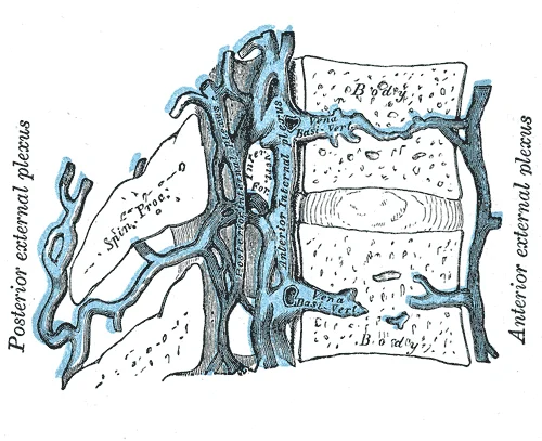
Long description
Median sagittal section of two thoracic vertebræ, showing the vertebral venous plexuses.

Figure 6: Median sagittal section of two thoracic vertebræ, showing the vertebral venous plexuses.
The external vertebral venous plexus is located outside the vertebral column, covering the posterior surface of the vertebrae and the ligaments. It communicates with the internal plexus through intervertebral foramina. This external network drains into the segmental veins, which accompany the spinal nerves. The segmental veins then connect to larger systemic veins such as the azygos system in the thorax and the iliac veins in the pelvis. This connection allows blood from the spinal cord and meninges to enter the general circulation. The valveless nature of these connections is clinically significant, as it permits retrograde flow during increased intra-abdominal pressure, such as during coughing or straining.
Understanding this drainage is crucial for interpreting the spread of disease. The lack of valves means that pathogens or tumour cells can travel from the pelvic organs to the vertebral column and subsequently to the brain. This pathway is particularly relevant in cases of prostate cancer or pelvic infections. The proximity of the veins to the dura means that vascular engorgement can occur, potentially complicating the procedure if not managed correctly.
Figure 7723 Cervical Vertebrae
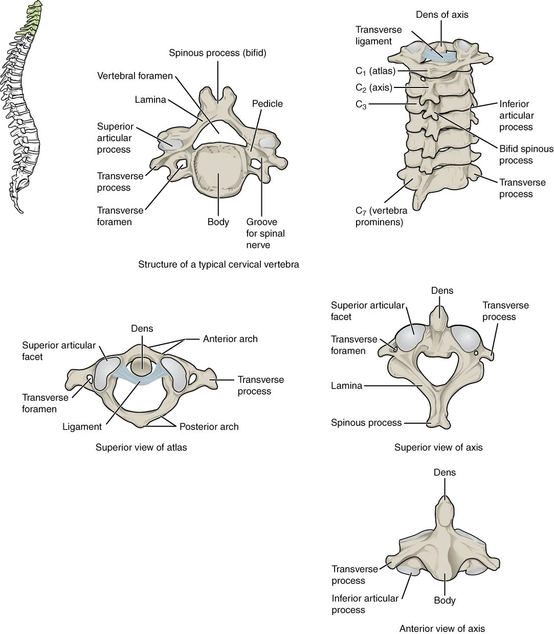
Long description
A typical cervical vertebra has a small body, a bifid spinous process, transverse processes that have a transverse foramen and are curved for spinal nerve passage. The atlas (C1 vertebra) does not have a body or spinous process. It consists of an anterior and a posterior arch and elongated transvers

Figure 7: A typical cervical vertebra has a small body, a bifid spinous process, transverse processes that have a transverse foramen and are curved for spinal nerve passage.
Figure 8718 Vertebra
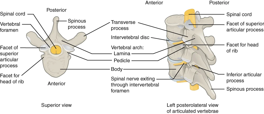
Long description
A typical vertebra consists of a body and a vertebral arch. The arch is formed by the paired pedicles and paired laminae. Arising from the vertebral arch are the transverse, spinous, superior articular, and inferior articular processes. The vertebral foramen provides for passage of the spinal cord.

Figure 8: A typical vertebra consists of a body and a vertebral arch.
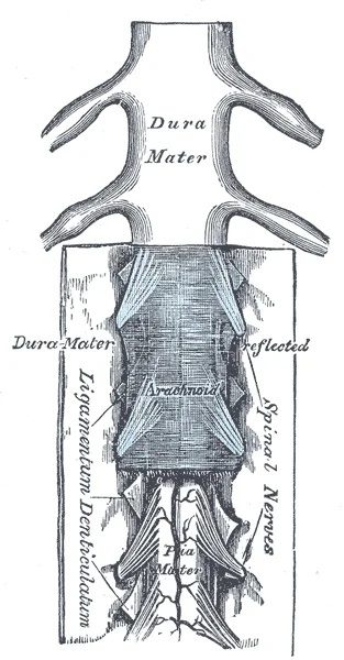
The vertebral canal houses the spinal cord and its associated nerve roots, which provide essential motor and cutaneous supply to the body. Understanding the anatomy of these nerves is critical for interpreting clinical signs and performing procedures such as lumbar puncture safely. This diagram shows the spinal cord within the dural sac, with nerve roots exiting through the intervertebral foramina. The cauda equina is visible as a bundle of nerve roots descending below the conus medullaris. The lumbar puncture site is indicated at the L3/L4 or L4/L5 interspace, below the termination of the spinal cord to avoid injury. The image provides a posterior view, allowing clear visualization of the meningeal layers and the path of the needle.
Figure 9Gray767
Long description
The medulla spinalis and its membranes. Labelled illustration from Wikimedia Commons, used under its open licence.

Figure 9: The medulla spinalis and its membranes.
The spinal nerves emerge from the spinal cord and divide into dorsal and ventral rami. The dorsal rami supply the intrinsic muscles of the back and the overlying skin. The ventral rami form the major plexuses, including the lumbar and sacral plexuses, which innervate the lower limbs and pelvic structures. It gives rise to the femoral nerve, which supplies the anterior compartment of the thigh, and the obturator nerve, which supplies the medial compartment.
Clinical assessment of these nerves involves testing motor strength and sensory distribution. Motor deficits may indicate compression of the nerve roots or the spinal cord itself. Sensory disturbances can help localize the level of lesion. For instance, radicular pain often follows a specific dermatomal pattern. Bladder dysfunction may suggest involvement of the sacral roots or the conus medullaris.
| Nerve | Muscles Supplied | Skin Area Supplied |
|---|---|---|
| Femoral | Quadriceps, Sartorius | Anterior thigh, medial leg |
| Obturator | Adductors | Medial thigh |
| Sciatic | Hamstrings, Calf muscles | Posterior thigh, leg, foot |
| Dorsal Ramus | Erector Spinae | Overlying skin of back |
The relationship between the nerves and the vertebral column is key to understanding pain patterns. Radicular pain occurs when nerve roots are compressed or irritated. This pain often radiates along the path of the nerve. MESCC may lead to more specific symptoms including radicular pain (50% as initial symptom and close to 80% at diagnosis). Ataxia (67%) is a more frequently presenting symptom compared to motor weakness (40% as initial symptom and close to 90% at diagnosis). Sensory disturbances (30% to 75%) and/or bladder dysfunction (5 to 60% at diagnosis) may be one of the presenting symptoms of MESCC. [2]
In oncologic patients with back pain (suggesting SM), radicular pain, motor weakness, sensory complaints and/or bladder dysfunction (suggesting MESCC), a careful and timely diagnostic work up is mandatory in order to avoid delayed diagnosis, that would increases the odds of a persisting neurological deficit and resulting loss of ambulation, which in turn would reduce QOL and LOL.
The lymphatic drainage of the vertebral canal and spinal meninges is a specialized system that parallels the venous plexuses, ensuring efficient removal of interstitial fluid and immune surveillance within the central nervous system's protective layers. Understanding this pathway is critical for comprehending how infections or malignancies may spread from the spinal cord to systemic circulation.
The epidural space, located between the dura mater and the vertebral periosteum, contains a rich network of valveless veins known as the internal vertebral venous plexuses. While primarily venous, this space also harbours initial lymphatic capillaries that drain the outer layers of the dura mater. These lymphatic vessels follow the course of the epidural veins, moving towards the intervertebral foramina. Here, they communicate with the paravertebral lymph nodes. This arrangement allows for the drainage of cerebrospinal fluid (CSF) components that may leak into the epidural space, although the primary route for CSF absorption remains the arachnoid villi projecting into the dural venous sinuses. The proximity of these lymphatic channels to the epidural veins means that pathological processes affecting the venous system can potentially influence lymphatic flow, although direct anatomical connections between the two systems are complex and not fully delineated in standard undergraduate texts.
The arachnoid mater itself is avascular and lacks direct lymphatic vessels. However, the subarachnoid space, which contains the CSF, is in close contact with the arachnoid villi. These villi act as one-way valves, allowing CSF to pass into the dural venous sinuses. Recent evidence suggests that a glymphatic system, involving perivascular spaces, facilitates the clearance of waste products from the brain and spinal cord interstitial fluid into the cervical lymph nodes via the cribriform plate of the ethmoid bone and along the optic nerve sheath. For the spinal cord specifically, lymphatic drainage is less direct. It is proposed that fluid from the spinal subarachnoid space drains into the perineural lymphatic spaces surrounding the spinal nerve roots. These perineural spaces extend through the intervertebral foramina and connect with the paravertebral lymph nodes. This pathway is particularly relevant for the drainage of inflammatory mediators and cells from the spinal meninges.
The spinal nerve roots are enveloped by extensions of the meninges. The dura mater extends along the nerve roots to form the dural sac, which terminates at the level of the second sacral vertebra. The arachnoid and pia mater also follow the nerve roots. Lymphatic vessels are present in the connective tissue surrounding the nerve roots, particularly in the perineural space. These vessels drain into the paravertebral lymph nodes located alongside the vertebral column. The drainage from the lumbar region, therefore, flows superiorly and laterally towards the para-aortic and lumbar lymph nodes. This anatomical arrangement explains why infections or tumours involving the lower spinal meninges can metastasize to the retroperitoneal lymph nodes. The valveless nature of the epidural venous plexuses also facilitates bidirectional flow, potentially allowing for retrograde spread of pathogens or malignant cells, although lymphatic drainage is generally unidirectional towards the central lymphatic ducts.
The vertebral canal is the bony enclosure formed by the vertebral foramina of stacked vertebrae. It contains the spinal cord, spinal nerve roots, meninges, and epidural fat. The spinal cord terminates in most adults at the level of the L1 or L2 vertebral body. Below this level, the subarachnoid space contains the cauda equina, a bundle of lumbar and sacral nerve roots floating in cerebrospinal fluid.
The spinal meninges consist of three layers. The dura mater is the tough, outermost fibrous layer. It lines the vertebral canal and forms the dural sac. The arachnoid mater is the delicate, avascular middle layer. It lies beneath the dura and is separated from it by the potential subdural space. The pia mater is the thin, vascular inner layer that adheres directly to the surface of the spinal cord and nerve roots.
Between the dura mater and the periosteum of the vertebral canal lies the epidural space. This space contains loose connective tissue, fat, and the internal vertebral venous plexus (Batson’s plexus). It does not contain cerebrospinal fluid.
The subarachnoid space lies between the arachnoid mater and the pia mater. It is filled with cerebrospinal fluid (CSF). This space surrounds the spinal cord and the cauda equina. It extends inferiorly to the level of S2.
Lumbar puncture is performed to access the subarachnoid space for diagnostic or therapeutic purposes. The needle passes through the skin, subcutaneous tissue, supraspinous ligament, interspinous ligament, ligamentum flavum, and epidural space before entering the subarachnoid space. The procedure is typically performed at the L3–L4 or L4–L5 interspace. This level is chosen because the spinal cord has usually terminated, reducing the risk of cord injury. The needle traverses the dura and arachnoid mater to enter the subarachnoid space.
The contents of the vertebral canal include the spinal cord (or cauda equina below L2), meninges, CSF, and epidural fat. The epidural space contains fat and veins. The meninges comprise dura, arachnoid, and pia. The lumbar puncture targets the subarachnoid space below L2.
The vertebral canal houses the spinal cord and its protective meninges. Radiological assessment is essential for identifying structural integrity and potential compression.
Plain radiographs provide an initial overview of the bony vertebral canal. The pedicles, laminae, and spinous processes are clearly defined. Computed Tomography (CT) offers superior detail of these osseous structures. The pedicles form the lateral walls, while the laminae and spinous process complete the posterior arch. CT is the gold standard for identifying fractures or bony encroachment into the canal.
Magnetic Resonance Imaging (MRI) is the primary modality for soft tissue evaluation, including the spinal cord, cerebrospinal fluid (CSF), and meninges. On T2-weighted sequences, CSF appears hyperintense (bright), providing a natural contrast that outlines the spinal cord and nerve roots. The dural sac is visible as a tubular structure containing the cauda equina nerve roots within the hyperintense CSF. Modic type I and II changes, which are specific observations for predicting pain, appear as distinct signal intensity alterations in the vertebral end plates. While standard MRI is effective, the end plate has a short T2, which may limit visualization of certain pathologies. [3]
Ultrasound is primarily utilised for procedural guidance, such as lumbar puncture. It allows for the real-time identification of bony landmarks, specifically the spinous processes and the interlaminar space. By visualising the ligamentum flavum and the posterior dura, clinicians can estimate the depth required for needle insertion. This reduces the reliance on anatomical landmarks alone, which can be challenging in patients with high body mass indices.
DefinitionModic changes
Modic changes are MRI-observed signal intensity alterations in the vertebral end plates, often associated with disc-related pain.
| Modality | Primary Use | Key Feature |
|---|---|---|
| X-ray | Bony alignment | Initial screening |
| CT | Bony detail | Fracture identification |
| MRI | Soft tissue | Cord and meninges |
| Ultrasound | Guidance | Real-time needle placement |
The spinal cord terminates at the conus medullaris, typically at the L1/L2 level in adults. Below this, the dural sac continues as the thecal sac, containing the cauda equina. The needle trajectory passes through the skin, supraspinous ligament, interspinous ligament, ligamentum flavum, and epidural space before entering the subarachnoid space.
Injuries to the intervertebral discs are a primary cause of nerve root compression. The most common sites for disc herniation are the L4/L5 or L5/S1 intervertebral discs. These herniations can cause sciatica, a widespread pain that radiates from the lower back down the thigh and into the leg. Similar injuries of the C5/C6 or C6/C7 intervertebral discs, following forcible hyperflexion of the neck from a collision accident or football injury, can produce pain in the neck, shoulder, and upper limb. [1]
Nerve palsies often result from metastatic compression. The most frequently cited incidence of spinal metastasis (SM) of 30% is based on autopsy studies. Approximately 1 out of 10 patients with SM will develop malignant epidural spinal cord compression (MESCC). Ataxia was more common compared to weakness as an initial symptom, affecting 67% versus 27%. [2]
Procedures such as lumbar puncture rely on precise surface landmarks. The intercristal line, connecting the highest points of the iliac crests, typically crosses the L4 spinous process or the L4/L5 interspace. This landmark guides the needle insertion to avoid the spinal cord. Understanding these epidemiologic data should increase awareness for first symptoms, allowing early diagnosis and subsequent treatment. However, studies show an important delay between onset of symptoms and diagnosis. This delay is occasionally or partially explained by late presentation of patients. [2]
Surface landmarks are critical for safe access. The spinous processes of L3, L4, and L5 are easily palpable. The subarachnoid space extends down to the S2 level. Insertion above L3 risks cord injury. Insertion below L4 is often technically difficult due to body habitus. Accurate identification of these structures prevents complications and ensures diagnostic yield.
The vertebral canal is the bony enclosure protecting the spinal cord and its coverings. The spinal meninges consist of three layers: the dura mater, arachnoid mater, and pia mater. The dura mater is the tough outer layer, attached to the periosteum of the vertebral canal at the foramen magnum and the second sacral vertebra. The arachnoid mater is the middle, web-like layer, separated from the dura by the subdural space. The pia mater is the delicate inner layer, adhering directly to the spinal cord surface.
Lumbar puncture is performed to obtain cerebrospinal fluid (CSF) for diagnostic analysis. The procedure is typically carried out at the L3-L4 or L4-L5 interspace to avoid injury to the spinal cord, which usually terminates at the L1-L2 level in adults. The needle passes through the skin, subcutaneous tissue, supraspinous ligament, interspinous ligament, and ligamentum flavum to reach the subarachnoid space. The ligamentum flavum provides a distinct 'pop' as the needle penetrates it.
Common traps include inserting the needle too high, risking cord injury, or too low, hitting the sacrum. Incorrect needle angle can lead to a 'dry tap' or post-dural puncture headache. Always ensure the patient is in the lateral decubitus position with the back flexed to open the interspinous spaces.
Clinical pearlSafe Zone
The spinal cord ends at L1-L2; lumbar puncture is safe below L3.
Exam trapLigamentum Flavum
Feel a distinct 'pop' when penetrating the ligamentum flavum.
DefinitionSubarachnoid Space
The space between the arachnoid and pia mater containing CSF.
NoteCSF Volume
Normal CSF volume is approximately 150 ml in adults.
Clinical pearlNeedle Depth
Needle depth varies by patient habitus; advance slowly after ligamentum flavum penetration.
Exam trapContraindication
Do not perform LP if signs of raised intracranial pressure are present.
DefinitionDura Mater
The tough, outermost meningeal layer attached to the foramen magnum and S2.
NotePost-LP Care
Keep the patient supine for 1-2 hours to reduce headache risk.
Clinical pearlCSF Appearance
Normal CSF is clear and colourless; xanthochromia suggests subarachnoid haemorrhage.
Exam trapInfection Risk
Strict aseptic technique is mandatory to prevent iatrogenic meningitis.
DefinitionPia Mater
The delicate inner meningeal layer adhering directly to the spinal cord.
Students often struggle with the precise anatomical landmarks and procedural nuances of the vertebral canal and lumbar puncture. Avoiding these common errors is essential for clinical safety and diagnostic accuracy.
- Misidentifying the interspace: A common mistake is assuming the iliac crests always align with the L4 vertebra. In practice, the L4-L5 interspace is often identified by drawing a line between the highest points of the iliac crests, but anatomical variation is common.
- Incorrect needle angulation: Students frequently angle the needle too steeply or too shallowly. The needle must be directed slightly cranially to parallel the orientation of the spinous processes.
- Failure to appreciate the dura: A common trap is failing to recognise the "pop" sensation as the needle traverses the ligamentum flavum and dura mater.
- Over-reliance on imaging: Relying solely on pre-procedure imaging rather than surface anatomy can lead to errors. Always palpate the bony landmarks carefully.
- Ignoring patient positioning: A failure to ensure the patient is in a maximal fetal position (flexed spine) makes the interspinous spaces harder to access. Proper positioning is the first-line requirement for a successful procedure.
Exam trapThe L4-L5 Landmark
While the supracristal plane is a standard landmark, it may intersect the L3-L4 or L5-S1 interspace in a significant proportion of patients.
Knowledge of the vertebral canal and spinal meninges is fundamental to clinical practice, particularly in neurology, anaesthetics, and emergency medicine.
Lumbar puncture is the primary bedside procedure for sampling cerebrospinal fluid (CSF) to diagnose meningitis, subarachnoid haemorrhage, or demyelinating conditions. Clinicians must understand that the spinal cord typically terminates at the L1-L2 level in adults, necessitating needle insertion below this level to avoid cord injury.
In radiology, understanding the vertebral canal is vital for interpreting MRI scans, particularly when assessing for metastatic epidural spinal cord compression (MESCC). Early recognition of MESCC is of primordial importance because treatment without delay and before mobility is lost leads to a better functional outcome. [2]
In the operating theatre, spinal anaesthesia relies on the accurate identification of the subarachnoid space. Furthermore, in cases of discogenic pain, provocation diskography is used to identify painful disks, achieving a specificity of 0.94 and a false-positive rate of 6% when performed using low-pressure technique[1]. [3]
Lumbar Puncture Decision Path
- Assess clinical indicationSuspected CNS infection or pressure measurement
- Question: Contraindications present?
Yes
- Defer procedure, consider imaging
No
- Position patient in lateral decubitus or sitting
- Identify L3-L4 or L4-L5 interspace
- Perform puncture
| Structure | Description |
|---|---|
| Anulus fibrosus | Outer fibrous layer of the intervertebral disc [1] |
| Nucleus pulposus | Gel-like interior of the intervertebral disc [1] |
| Coccyx | Fusion of four small coccygeal vertebrae |
| Intervertebral disc | Provides strong attachments and padding between vertebrae |
- The spinal cord terminates at the L1-L2 level in adults.
- Lumbar puncture must be performed below the termination of the spinal cord.
- The supracristal plane is a guide for the L4-L5 interspace but is subject to variation.
- Early diagnosis of MESCC is critical to preserve functional mobility.
- Provocation diskography uses low-pressure techniques to improve diagnostic specificity.
- The intervertebral disc consists of an outer anulus fibrosus and inner nucleus pulposus.
- Patient positioning is essential for opening the interspinous spaces.
Answer each one in your head first, then open the answer.
At what vertebral level does the adult spinal cord typically terminate?
Show answerHide answer
L1-L2What are the two components of an intervertebral disc?
Show answerHide answer
Anulus fibrosus and nucleus pulposusWhat is the primary clinical goal of early MESCC recognition?
Show answerHide answer
To prevent the loss of mobility and improve functional outcomesWhat is the specificity of provocation diskography using low-pressure technique?
Show answerHide answer
0.94Which bones fuse to form the coccyx?
Show answerHide answer
Four coccygeal vertebrae
- Betts JG, Young KA, Wise JA, et al. The Vertebral Column. OpenStax, Anatomy and Physiology 2e (CC BY-NC-SA 4.0). Accessed .
- Van den Brande R, Cornips EM, Peeters M, et al. Epidemiology of spinal metastases, metastatic epidural spinal cord compression and pathologic vertebral compression fractures in patients with solid tumors: A systematic review. Journal of bone oncology 2022;35:100446. doi:10.1016/j.jbo.2022.100446. Accessed .
- Lotz JC, Fields AJ, Liebenberg EC. The role of the vertebral end plate in low back pain. Global spine journal 2013;3(3):153-164. doi:10.1055/s-0033-1347298. Accessed .
Figure credits
- Figure 1: Henry Vandyke Carter, via Wikimedia Commons, Public domain, Public domain.
- Figure 2: Henry Vandyke Carter, via Wikimedia Commons, Public domain, Public domain.
- Figure 3: Henry Vandyke Carter, via Wikimedia Commons, Public domain, Public domain.
- Figure 4: OpenStax College, via Wikimedia Commons, CC BY 3.0, CC BY 3.0.
- Figure 5: Henry Vandyke Carter, via Wikimedia Commons, Public domain, Public domain.
- Figure 6: Henry Vandyke Carter, via Wikimedia Commons, Public domain, Public domain.
- Figure 7: OpenStax College, via Wikimedia Commons, CC BY 3.0, CC BY 3.0.
- Figure 8: OpenStax College, via Wikimedia Commons, CC BY 3.0, CC BY 3.0.
- Figure 9: Henry Vandyke Carter, via Wikimedia Commons, Public domain, Public domain.
Educational content for medical students and doctors. It is not a substitute for clinical judgement or local guidelines.