Muscles of the back and suboccipital triangle
Anatomical overview of the deep back muscles, erector spinae groups, and suboccipital triangle boundaries.
The axial muscles of the trunk are grouped based on location and function, with the back containing the primary extensors of the vertebral column. These muscles maintain posture and control movement of the spine. The region is bounded superiorly by the external occipital protuberance and inferiorly by the sacrum. Laterally, the boundaries are defined by the costal margins and the iliac crests. The deep back muscles lie beneath the superficial layer, which includes the trapezius and latissimus dorsi. [1]
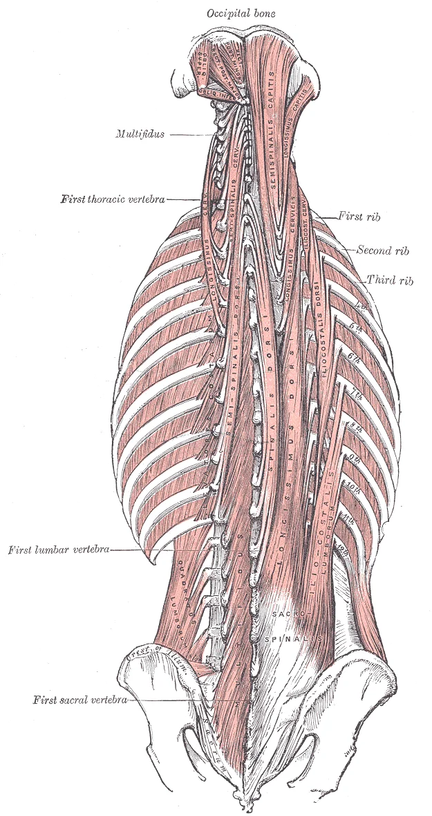
This group maintains the lumbar curve and is divided into three vertical columns. The iliocostalis group is the most lateral, the longissimus is intermediate, and the spinalis is the most medial. [1]
The transversospinalis group lies deep to the erector spinae. These muscles run from the transverse processes to the spinous processes of the vertebrae. This group includes the semispinalis, multifidus, and rotatores muscles. The semispinalis muscles are named for the areas of the body with which they are associated. [1]
This triangle contains the vertebral artery and the suboccipital nerve. The muscles here are crucial for fine movements of the head. [1]
DefinitionErector Spinae
The primary extensor of the vertebral column, comprising the iliocostalis, longissimus, and spinalis groups.
| Group | Components | Primary Action |
|---|---|---|
| Iliocostalis | Cervicis, Thoracis, Lumborum | Lateral flexion and extension |
| Longissimus | Capitis, Cervicis, Thoracis | Extension and rotation |
| Spinalis | Capitis, Cervicis, Thoracis | Extension of the head and neck |
| Muscle | Origin | Insertion | Action |
|---|---|---|---|
| Semispinalis | Transverse processes | Spinous processes | Extension and rotation |
| Multifidus | Sacrum and transverse processes | Spinous processes | Extension and lateral flexion |
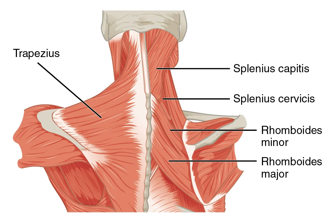
When the head turns, this muscle becomes taut and easily palpable. The trapezius muscle forms the broadest superficial layer of the upper back, extending from the occipital bone and nuchal ligament down to the thoracic spine and laterally to the clavicle and scapula. Its upper fibres are readily felt when shrugging the shoulders. Deep to the trapezius, the latissimus dorsi covers the lower back, while the erector spinae group runs vertically along the vertebral column. The splenius capitis and cervicis lie superficial to the deeper transversospinales muscles in the upper back. The floor of this triangle is formed by the posterior atlanto-occipital membrane and the posterior arch of the atlas. Palpation of the spinous processes of the cervical vertebrae provides a midline reference. The nuchal ligament, a fibrous membrane, extends from the external occipital protuberance to the spinous process of C7, serving as an attachment point for several neck muscles. Understanding these surface landmarks allows for accurate identification of underlying structures during clinical examination.
DefinitionSuboccipital Triangle
A deep anatomical space at the base of the skull bounded by three small muscles, containing the vertebral artery and suboccipital nerve.
The back muscles stabilize and move the vertebral column, and are grouped according to the lengths and direction of the fascicles[1]. The posterior muscles of the neck are primarily concerned with head movements, like extension[1]. The major muscle that laterally flexes and rotates the head is the sternocleidomastoid[1].
| Muscle | Origin | Insertion | Action | Nerve Supply |
|---|---|---|---|---|
| Sternocleidomastoid | Sternum, Clavicle | Mastoid Process | Head rotation, flexion | Accessory Nerve |
| Trapezius | Occipital Bone, Nuchal Ligament | Clavicle, Scapula | Shoulder elevation, head extension | Accessory Nerve |
| Splenius Capitis | Nuchal Ligament | Mastoid Process, Occipital Bone | Head extension, rotation | Dorsal Rami |
| Rectus Capitis Posterior Major | Spinous Process C2 | Occipital Bone | Head extension | Suboccipital Nerve |
The central structures are the vertebral column, consisting of the cervical vertebrae superiorly and the upper thoracic vertebrae inferiorly. Key landmarks include the spinous processes, which project posteriorly and serve as attachment points for the trapezius and latissimus dorsi. The transverse processes extend laterally and provide leverage for the rotatores and multifidus muscles. The suboccipital triangle is formed by the rectus capitis posterior major, obliquus capitis superior, and obliquus capitis inferior, surrounding the vertebral artery and the suboccipital nerve. The atlas (C1) and axis (C2) are distinct due to their unique morphology; the atlas lacks a body and spinous process, while the axis features the dens. These bony landmarks are critical for understanding the origin and insertion of the suboccipital muscles, which control fine movements of the head. The nuchal ligament attaches to the external occipital protuberance and the spinous processes of the cervical vertebrae, providing a midline anchor for the trapezius and rhomboids. The ribs articulate with the thoracic vertebrae, forming the posterior thoracic cage. The sternocleidomastoid muscle attaches to the mastoid process of the temporal bone, which is located just posterior to the ear. The splenius capitis and splenius cervicis attach to the spinous processes and the mastoid process, respectively. The semispinalis capitis attaches to the occipital bone, while the semispinalis cervicis attaches to the transverse processes of the upper thoracic vertebrae. The multifidus muscles attach to the spinous processes and the mammillary processes of the lumbar and thoracic vertebrae. The rotatores muscles are the deepest, attaching between adjacent transverse and spinous processes. The suboccipital muscles are situated deep to the semispinalis capitis and the splenius capitis. The rectus capitis posterior major originates from the spinous process of the axis and inserts into the inferior nuchal line of the occipital bone. The obliquus capitis superior originates from the transverse process of the atlas and inserts into the occipital bone between the nuchal lines. The obliquus capitis inferior originates from the spinous process of the axis and inserts into the transverse process of the atlas. These muscles work together to extend, rotate, and stabilize the head. The vertebral artery passes through the transverse foramina of the cervical vertebrae and enters the skull through the foramen magnum, passing through the suboccipital triangle. The suboccipital nerve, a branch of the dorsal ramus of C1, innervates these muscles. Understanding these bony landmarks is essential for clinical examination and surgical procedures involving the posterior neck and upper back. The alignment of the vertebrae and the integrity of the ligaments are crucial for maintaining posture and preventing injury. The suboccipital triangle is a clinically significant area due to the proximity of the vertebral artery and the suboccipital nerve. Compression or irritation of these structures can lead to headaches and dizziness. The bony landmarks also serve as reference points for acupuncture and massage therapy. The spinous processes are palpable and can be used to identify the level of the vertebrae. The transverse processes are less palpable but can be felt laterally. The mastoid process is easily palpable behind the ear. The external occipital protuberance is a prominent bump at the base of the skull. These landmarks are used in physical therapy and chiropractic care to assess and treat musculoskeletal disorders. The suboccipital muscles are often tight in individuals with poor posture, leading to tension headaches. Stretching and strengthening these muscles can alleviate symptoms. The bony landmarks are also important for imaging studies, such as X-rays and MRI, to identify fractures or degenerative changes. The alignment of the cervical spine is critical for neurological function. Any deviation from normal alignment can compress the spinal cord or nerve roots. The suboccipital muscles play a key role in maintaining the lordotic curve of the cervical spine. Weakness or imbalance in these muscles can lead to forward head posture. Correcting posture through exercise and ergonomic adjustments can reduce strain on the suboccipital muscles. The bony landmarks provide a framework for understanding the complex anatomy of the back and neck. They are essential for medical students to master for clinical practice. [2]
Figure 1Gray409
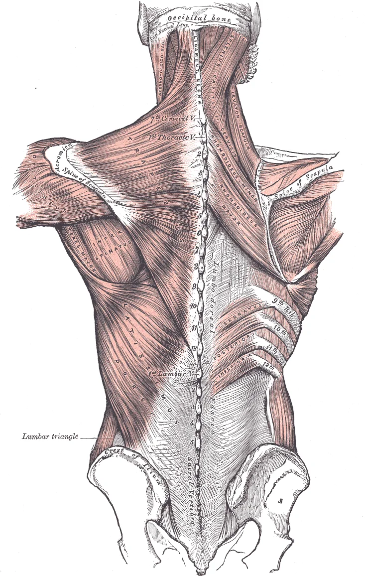
Long description
Muscles connecting the upper extremity to the vertebral column.

Figure 1: Muscles connecting the upper extremity to the vertebral column.
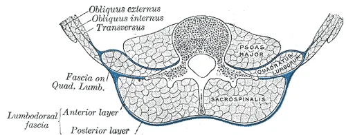
The erector spinae muscles lie deep to the superficial back muscles and are the most prominent group in the posterior trunk. They run vertically along the vertebral column. The thoracolumbar fascia encloses the erector spinae and quadratus lumborum muscles. It has three layers: posterior, intermediate, and anterior. The posterior layer covers the erector spinae. The anterior layer covers the quadratus lumborum. The suboccipital triangle is located at the base of the skull. It is bounded by the rectus capitis posterior major, the obliquus capitis superior, and the obliquus capitis inferior. The floor of the triangle is formed by the posterior atlanto-occipital membrane and the posterior arch of the atlas.
DefinitionThoracolumbar Fascia
A dense fibrous sheet that encloses the deep muscles of the back and provides attachment for the abdominal muscles.
The back muscles are divided into superficial, intermediate, and deep layers. The superficial layer includes the trapezius, latissimus dorsi, levator scapulae, and rhomboids. These muscles move the upper limb. The deep layer includes the erector spinae and transversospinalis groups. These muscles extend and rotate the vertebral column. The erector spinae consists of the iliocostalis, longissimus, and spinalis muscles. The transversospinalis group includes the semispinalis, multifidus, and rotatores muscles. These muscles are innervated by the dorsal rami of the spinal nerves. [1] [2]
| Muscle | Origin | Insertion | Action | Nerve Supply |
|---|---|---|---|---|
| Trapezius | Occipital bone, nuchal ligament, spinous processes of C7-T12 | Clavicle, acromion, spine of scapula | Elevates, retracts, rotates scapula | Accessory nerve (CN XI) |
| Latissimus Dorsi | Spinous processes of T7-L5, iliac crest | Intertubercular groove of humerus | Extends, adducts, medially rotates humerus | Thoracodorsal nerve |
| Erector Spinae | Sacrum, iliac crest, spinous processes | Ribs, vertebrae, mastoid process | Extends vertebral column | Dorsal rami of spinal nerves |
The suboccipital muscles are four small muscles located at the suboccipital region. They include the rectus capitis posterior major, rectus capitis posterior minor, obliquus capitis superior, and obliquus capitis inferior. These muscles extend and rotate the head. They are innervated by the suboccipital nerve, which is the dorsal ramus of C1. The suboccipital triangle contains the vertebral artery and the suboccipital nerve. The artery supplies the brainstem and cerebellum. Lesions in this area can cause vertigo and nystagmus.
Exam trapSuboccipital Triangle
Do not confuse the suboccipital triangle with the posterior triangle of the neck. The suboccipital triangle is deep and located at the skull base.
The thoracolumbar fascia is crucial for transmitting forces from the upper limbs to the lower limbs. It connects the latissimus dorsi to the gluteus maximus. This connection allows for efficient movement during activities such as running and climbing. The fascia also provides protection for the deep back muscles. It helps maintain the posture of the vertebral column.
Figure 2Gray388
Long description
Diagram of a transverse section of the posterior abdominal wall, to show the disposition of the lumbodorsal fascia.

Figure 2: Diagram of a transverse section of the posterior abdominal wall, to show the disposition of the lumbodorsal fascia.
Figure 31117 Muscles of the Neck Upper Back
Long description
Caption: The large, complex muscles of the neck and back move the head, shoulders, and vertebral column. URL: https://cnx.org/contents/FPtK1zmh@8.108:_xq2eUyd@5/Axial-Muscles-of-the-Head-Neck Other versions: Original

Figure 3: Caption: The large, complex muscles of the neck and back move the head, shoulders, and vertebral column (url: https://cnx.org/contents/fptk1zmh@8.108:_xq2euyd@5/axial-muscles-of-the-head-neck other versions: original).
| Muscle Group | Origin | Insertion | Action | Nerve Supply |
|---|---|---|---|---|
| Superficial | Scapula, Iliac Crest, Spinous Processes | Humerus, Ribs, Skull | Move upper limb, extend trunk | Ventral rami (Brachial Plexus) |
| Intermediate | Ribs | Ribs, Vertebrae | Elevate/Depress ribs, Extend trunk | Dorsal rami |
| Deep (Erector Spinae) | Sacrum, Iliac Crest, Vertebrae | Vertebrae, Ribs, Skull | Extend and laterally flex spine | Dorsal rami |
| Deep (Transversospinalis) | Transverse Processes | Spinous Processes | Rotate and extend spine | Dorsal rami |
| Suboccipital | Atlas, Axis | Occipital Bone | Extend and rotate head | Suboccipital nerve (C1) |
DefinitionSuboccipital Triangle
A small anatomical space in the upper neck, bounded by the rectus capitis posterior major, obliquus capitis superior, and obliquus capitis inferior, containing the vertebral artery and suboccipital nerve.
The suboccipital muscles are a specific group of four small muscles located at the junction of the head and neck. They form the floor of the suboccipital triangle. These muscles are unique in that they are innervated by the dorsal ramus of the first cervical nerve, also known as the suboccipital nerve. Their primary action is the extension and rotation of the head. The rectus capitis posterior major extends and rotates the head. The obliquus capitis superior extends and laterally flexes the head. The obliquus capitis inferior rotates the head. The rectus capitis posterior minor extends the head. These muscles work in concert to maintain the posture of the head and allow for precise movements of the skull on the vertebral column. [2]
Clinical pearlClinical Note
Damage to the suboccipital nerve can lead to weakness in head extension and rotation, potentially causing compensatory strain on other neck muscles.
The superficial muscles, such as the trapezius and latissimus dorsi, are innervated by the spinal accessory nerve and the dorsal scapular nerve, respectively. These muscles are crucial for movements of the shoulder girdle and upper limb. The trapezius extends, retracts, and rotates the scapula. The latissimus dorsi extends, adducts, and medially rotates the humerus. The deep muscles, collectively known as the erector spinae and transversospinalis groups, are the primary extensors of the vertebral column. They maintain upright posture and prevent flexion of the trunk. The erector spinae runs vertically along the spine, while the transversospinalis muscles run obliquely, connecting transverse processes to spinous processes. [1]
Figure 4Gray389
Long description
Deep muscles of the back. Labelled illustration from Wikimedia Commons, used under its open licence.

Figure 4: Deep muscles of the back.
The vascular network supplying the deep muscles of the back and the suboccipital region is robust, ensuring adequate perfusion for postural maintenance and head movement. The primary source is the vertebral artery, a major branch of the subclavian artery. It ascends through the transverse foramina of the cervical vertebrae, typically from C6 upwards. As it courses through the suboccipital triangle, it gives off the posterior spinal arteries and the anterior spinal artery, which supply the spinal cord and surrounding meninges.
DefinitionSuboccipital Triangle
A deep anatomical space in the upper neck bounded by the rectus capitis posterior major, obliquus capitis superior, and obliquus capitis inferior muscles.
The dorsal scapular artery, often arising from the transverse cervical artery, plays a significant role in supplying the rhomboid muscles and the levator scapulae. It runs deep to the trapezius and rhomboids, anastomosing with the suprascapular artery. This anastomotic network ensures collateral circulation if one vessel is compromised. It descends behind the neck, piercing the ligamentum nuchae to reach the erector spinae group.
The posterior spinal arteries branch medially from the vertebral artery, entering the spinal canal. Labels identify the rectus capitis posterior major, obliquus capitis inferior, and the vertebral artery itself.
| Vessel | Origin | Primary Supply |
|---|---|---|
| Vertebral Artery | Subclavian Artery | Spinal cord, suboccipital muscles |
| Dorsal Scapular Artery | Transverse Cervical Artery | Rhomboids, levator scapulae |
| Deep Cervical Artery | Costocervical Trunk | Deep back muscles |
Clinical relevance arises when these vessels are compressed or injured. Trauma to the suboccipital region can compromise the vertebral artery, leading to vertebrobasilar insufficiency. Symptoms may include dizziness, vertigo, and visual disturbances. Understanding the precise anatomical course is crucial for surgical approaches to the upper cervical spine. The anastomoses between the vertebral, deep cervical, and dorsal scapular arteries provide redundancy, but significant stenosis in one vessel can still lead to ischaemic symptoms if collateral flow is insufficient.
Clinical pearlClinical Pearl
The vertebral artery is vulnerable during posterior cervical spine surgery due to its close proximity to the lateral masses of C1 and C2.
The arterial supply is not uniform; it varies with individual anatomy. Some individuals may have a dominant deep cervical artery, while others rely more heavily on the vertebral artery branches. This variability must be considered in diagnostic imaging and surgical planning. The rich vascular network supports the high metabolic demand of the postural muscles, which are active continuously to maintain head and neck position.
The venous drainage of the deep back muscles and the suboccipital region follows a hierarchical pattern, moving from small tributaries into larger named vessels that ultimately connect with the systemic circulation. Understanding this pathway requires visualising the spatial relationship between the muscular layers and the vertebral column.
At the deepest level, within the suboccipital triangle, small emissary veins drain the rectus capitis posterior major and minor, as well as the obliquus capitis superior and inferior muscles. These tiny vessels pierce the posterior atlanto-occipital membrane and the dura mater to enter the internal vertebral venous plexus. This plexus is a valveless network of veins located within the epidural space, running longitudinally along the vertebral canal. Because it is valveless, blood can flow in either direction, allowing for collateral circulation between the cranial and pelvic venous systems.
Superficial to the deep plexus, the muscles of the upper back drain into the vertebral veins. It ascends through the transverse foramina of the upper six cervical vertebrae, accompanied by the vertebral artery. Along its course, it receives tributaries from the deep cervical muscles and the spinal cord. [1]
The middle and lower back muscles drain into the intercostal veins. The posterior intercostal veins accompany the posterior intercostal arteries. The azygos vein ascends along the right side of the vertebral column and arches anteriorly over the right main bronchus to drain into the superior vena cava. [1]
Clinical pearlClinical Relevance
The valveless nature of the internal vertebral venous plexus allows metastatic cancer cells from pelvic organs to spread to the vertebral column and brain without passing through the heart or lungs.
| Structure | Origin | Termination | Key Relations |
|---|---|---|---|
| Vertebral Vein | Posterior aspect of internal jugular vein | Brachiocephalic vein | Ascends through transverse foramina C6-C1 |
| Azygos Vein | Right ascending lumbar vein | Superior vena cava | Ascends on right side of vertebral column |
| Hemiazygos Vein | Left ascending lumbar vein | Azygos vein | Crosses midline at T9 level |
The suboccipital region also has direct connections to the dural sinuses. Emissary veins connect the superficial veins of the scalp with the dural sinuses, providing another route for potential spread of infection.
The innervation of the back muscles is derived from the posterior rami of the spinal nerves. These nerves provide both motor supply to the intrinsic back muscles and cutaneous supply to the skin of the back. The suboccipital triangle is a specific anatomical region at the base of the skull, formed by the rectus capitis posterior major, obliquus capitis superior, and obliquus capitis inferior muscles. This triangle houses the suboccipital nerve and the vertebral artery.
The floor of the triangle is formed by the posterior arch of the atlas and the dura mater.
DefinitionSuboccipital Triangle
A deep anatomical space at the base of the skull containing the suboccipital nerve and vertebral artery.
The motor supply to the muscles of the back is primarily from the dorsal rami of all spinal nerves. The cutaneous supply is provided by the medial and lateral cutaneous branches of the dorsal rami. The medial branches supply the skin overlying the erector spinae muscles. The lateral branches supply the skin over the scapular region.
| Structure | Nerve Supply |
|---|---|
| Suboccipital muscles | Suboccipital nerve (Dorsal ramus of C1) |
| Deep back muscles | Dorsal rami of spinal nerves |
| Superficial back muscles | Ventral rami of spinal nerves |
| Skin of back | Medial and lateral cutaneous branches of dorsal rami |
It provides motor innervation to the four suboccipital muscles: rectus capitis posterior major, rectus capitis posterior minor, obliquus capitis superior, and obliquus capitis inferior. These muscles are responsible for fine movements of the head, including extension, rotation, and lateral flexion. The vertebral artery is closely related to these muscles. It passes through the transverse foramen of C1 and loops around the posterior arch of the atlas. This relationship is clinically significant as compression of the vertebral artery can occur during excessive rotation of the head.
Exam trapNerve Supply
Remember that superficial back muscles are innervated by ventral rami, not dorsal rami.
The dorsal rami of the spinal nerves also supply the erector spinae and transversospinalis groups. These muscles maintain posture and facilitate extension of the vertebral column. The cutaneous branches of the dorsal rami provide sensory innervation to the skin of the back. The medial cutaneous branches supply the skin adjacent to the vertebral column. The lateral cutaneous branches supply the skin over the scapula and lateral back.
| Region | Nerve Branch |
|---|---|
| Skin near spine | Medial cutaneous branch of dorsal ramus |
| Lateral back | Lateral cutaneous branch of dorsal ramus |
| Suboccipital region | No cutaneous branch (Suboccipital nerve is motor only) |
Lesions of the suboccipital nerve are rare but can result in weakness of head extension and rotation. The vertebral artery is susceptible to injury in this region due to its close proximity to the suboccipital muscles. Understanding the anatomical relationships in the suboccipital triangle is essential for clinical procedures and understanding cervical spine pathology.
The lymphatic drainage of the muscles of the back and the suboccipital triangle follows a predictable pattern that mirrors the superficial and deep venous systems. Understanding this pathway is essential for predicting the spread of infection or malignancy from the posterior trunk and head. The lymph vessels generally accompany the major blood vessels and nerves, draining into regional nodal groups.
The suboccipital triangle is bounded by the rectus capitis posterior major, obliquus capitis superior, and obliquus capitis inferior. Lymph from the muscles within this triangle, such as the rectus capitis posterior minor, drains into the deep cervical nodes. The jugular trunk then drains into the venous angle, where the internal jugular vein meets the subclavian vein. This anatomical arrangement ensures that lymph from the back and head returns to the systemic circulation efficiently.
The primary drainage site for the majority of the back muscles is the deep cervical lymph nodes. These nodes are located along the internal jugular vein. They receive lymph from the head, neck, and upper back. The suboccipital muscles, being small and deep, contribute to this pool. Infection in the suboccipital region, such as from a scalp abscess, can lead to swelling in the deep cervical chain. This is clinically significant because enlarged nodes may be palpable along the sternocleidomastoid muscle.
For the more superficial muscles of the back, such as the trapezius and latissimus dorsi, drainage is more complex. The upper part of the trapezius drains into the posterior auricular and occipital nodes, which then feed into the deep cervical nodes. The lower part of the back drains into the lumbar and para-aortic nodes. However, the suboccipital triangle remains firmly within the domain of the deep cervical chain. This distinction is crucial for clinical examination. When assessing a patient with suspected pathology in the suboccipital region, one must palpate the deep cervical nodes along the anterior border of the sternocleidomastoid.
Clinical pearlClinical Pearl
Always examine the deep cervical nodes when investigating pathology of the suboccipital triangle, as they are the primary drainage site.
The lymphatic vessels are thin-walled and valved, ensuring unidirectional flow. They are closely associated with the vertebral artery and the suboccipital nerve. This close relationship means that inflammation in the suboccipital triangle can sometimes irritate the nerve, causing referred pain. The lymphatic system acts as a filter, trapping pathogens and abnormal cells before they enter the bloodstream. Therefore, the integrity of the deep cervical nodes is vital for immune surveillance of the posterior head and neck region.
Figure 5Suboccipital triangle
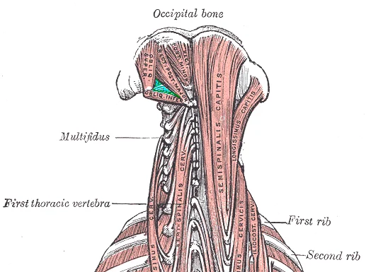
Long description
Suboccipital triangle.PNG. Labelled illustration from Wikimedia Commons, used under its open licence.

Figure 5: Suboccipital triangle.PNG.
PNG.
PNG.
PNG.
PNG.
PNG. Within this triangular space lies the vertebral artery and the suboccipital nerve. Understanding these relations is critical for interpreting clinical signs and surgical approaches to the craniocervical junction.
The suboccipital triangle is a deep anatomical region located beneath the semispinalis capitis muscle. Its roof is formed by the semispinalis capitis and the deep cervical fascia. The floor is formed by the posterior atlanto-occipital membrane and the posterior arch of the atlas. The medial boundary is the rectus capitis posterior major muscle. The lateral boundary is the obliquus capitis superior muscle. The inferior boundary is the obliquus capitis inferior muscle. This arrangement creates a protected corridor for neurovascular structures.
At the level of the atlas, it exits the transverse foramen and curves posteriorly. It then passes behind the lateral mass of the atlas. It lies on the posterior atlanto-occipital membrane. This position makes it susceptible to compression or injury during neck extension or rotation. The artery then pierces the dura mater to enter the cranial cavity.
The suboccipital nerve, which is the dorsal ramus of the first cervical nerve (C1), also traverses this space. It emerges from under the inferior border of the obliquus capitis inferior muscle. The nerve supplies motor innervation to the four suboccipital muscles: rectus capitis posterior major, rectus capitis posterior minor, obliquus capitis superior, and obliquus capitis inferior. The nerve lies superficial to the vertebral artery within the triangle. This close proximity means that pathology affecting one structure may impact the other.
| Muscle | Origin | Insertion | Action | Nerve Supply |
|---|---|---|---|---|
| Rectus capitis posterior major | Spinous process of C2 | Inferior nuchal line | Extends and rotates head | Suboccipital nerve (C1) |
| Rectus capitis posterior minor | Posterior tubercle of C1 | Inferior nuchal line | Extends head | Suboccipital nerve (C1) |
| Obliquus capitis inferior | Spinous process of C2 | Transverse process of C1 | Rotates head | Suboccipital nerve (C1) |
| Obliquus capitis superior | Transverse process of C1 | Inferior nuchal line | Extends head | Suboccipital nerve (C1) |
Clinical pearlClinical Pearl
The vertebral artery is vulnerable to injury in the suboccipital triangle due to its fixed position against the posterior arch of the atlas.
Understanding the radiological anatomy of the back and suboccipital triangle requires correlating cross-sectional imaging with surface landmarks. The muscles of the back are grouped into superficial, intermediate, and deep layers, each with distinct signal characteristics on different modalities.
Plain radiographs provide limited soft-tissue detail but are essential for assessing bony landmarks that anchor these muscles. The spinous processes of the cervical vertebrae, particularly the prominent spinous process of C7 (vertebra prominens), serve as key reference points. On a lateral cervical spine X-ray, the alignment of the spinous processes helps identify the level of the suboccipital triangle. The triangle itself is bounded superiorly by the rectus capitis posterior major and inferiorly by the obliquus capitis inferior. While the muscles themselves are not directly visualized, their attachments to the posterior arch of the atlas (C1) and the axis (C2) are critical. Fractures of the dens or posterior elements can disrupt the integrity of this triangle. The angle of the neck on a lateral view should be neutral to avoid superimposition of structures.
Computed tomography offers superior bony detail and can infer muscle bulk from surrounding fat planes. On axial CT slices through the upper cervical spine, the suboccipital muscles appear as soft-tissue densities posterior to the vertebral arches. The rectus capitis posterior minor is the most medial, lying adjacent to the posterior arch of the atlas. Lateral to it lies the rectus capitis posterior major. The obliquus capitis superior is located superiorly, between the atlas and the occiput. Fat stranding in these regions may indicate inflammation or trauma. The spinal cord is visible within the spinal canal, surrounded by cerebrospinal fluid, which appears dark on CT.
Magnetic resonance imaging is the gold standard for evaluating soft tissue pathology in the back. On T1-weighted images, muscle tissue appears intermediate in signal intensity, while subcutaneous fat is hyperintense. On T2-weighted or STIR sequences, fluid and edema appear bright, allowing detection of muscle strain or tear. The suboccipital muscles are clearly delineated from the overlying trapezius and semispinalis capitis. The rectus capitis posterior major and minor are small but distinct. The obliquus capitis inferior is a broad, flat muscle. Pathology such as muscle atrophy or fatty infiltration appears as increased signal intensity within the muscle belly. The atlanto-occipital and atlantoaxial joints are also visualized, with the transverse ligament of the atlas stabilizing the dens.
Ultrasound provides dynamic assessment of the suboccipital muscles. The high-frequency probe allows visualization of muscle architecture in real-time. The rectus capitis posterior major and minor are superficial and easily identified. The obliquus capitis inferior is deeper and may require careful angling. Ultrasound can detect muscle tears, hematomas, or trigger points. The echotexture of healthy muscle is homogeneous. Disruption of this pattern suggests injury. The proximity to the vertebral artery is a critical consideration, as the artery courses through the transverse foramina of C1-C6. Doppler imaging can assess blood flow in the vertebral artery, which is vital before any invasive procedures in this region.
The rectus capitis posterior minor is medial, adjacent to the posterior arch of C1. The rectus capitis posterior major is lateral to it. The obliquus capitis inferior is the most lateral muscle, connecting C2 to C1. The obliquus capitis superior is superior to the inferior oblique. The spinal cord is central. The caption states: Axial MRI at C1-C2 level showing suboccipital muscle anatomy and vertebral artery course.
The muscles of the back and the suboccipital region are critical for posture, head movement, and respiratory mechanics. Understanding their anatomy is essential for diagnosing pain syndromes and performing safe clinical procedures.
Clinically, the spinous processes of the vertebrae serve as key surface landmarks. This landmark is vital for identifying the correct intervertebral space for lumbar puncture, a procedure aimed at collecting cerebrospinal fluid or administering anaesthesia. The suboccipital triangle, located at the base of the skull, is palpable as a small depression between the head and the upper cervical spine. It contains the suboccipital muscles and the vertebral artery.
Back pain often arises from strain of the erector spinae muscles or ligamentous injury. The erector spinae group runs vertically along the vertebral column. Injury to these muscles can result from sudden twisting or heavy lifting. The suboccipital muscles, including the rectus capitis posterior major and minor, obliquus capitis superior and inferior, are involved in fine head movements. Tension in these muscles can contribute to cervicogenic headache. This figure shows the lesion sites and injury patterns associated with back pain. It highlights the relationship between the deep back muscles and the vertebral column. The view is posterior, showing the orientation of the muscle fibres relative to the spinous processes.
Nerve injuries to the back muscles can lead to weakness and sensory loss. The dorsal rami of the spinal nerves supply the intrinsic back muscles. Injury to these nerves can result from penetrating trauma or compression. For example, compression of the dorsal ramus can cause pain in the corresponding dermatome. The suboccipital nerve, a branch of the first cervical nerve (C1), supplies the suboccipital muscles. Palsy of this nerve can lead to difficulty in stabilizing the head.
Lumbar puncture is a common procedure that relies on knowledge of back anatomy. The needle is inserted between the L3 and L4 or L4 and L5 vertebrae to avoid injury to the spinal cord, which typically ends at the L1 or L2 level in adults. The needle passes through the skin, supraspinous ligament, interspinous ligament, and ligamentum flavum before entering the subarachnoid space.
DefinitionSuboccipital Triangle
A anatomical region at the base of the skull bounded by the rectus capitis posterior major, obliquus capitis superior, and obliquus capitis inferior muscles.
| Muscle | Action | Nerve Supply |
|---|---|---|
| Erector Spinae | Extends vertebral column | Dorsal rami of spinal nerves |
| Suboccipital Muscles | Extends and rotates head | Suboccipital nerve (C1) |
The trapezius is a broad, flat, superficial muscle that extends from the occipital bone to the lower thoracic vertebrae. It is innervated by the spinal accessory nerve. Injury to the spinal accessory nerve results in a drooping shoulder and difficulty elevating the scapula. The trapezius muscle has three distinct parts: upper, middle, and lower fibres. The upper fibres elevate the scapula, while the lower fibres depress it. The middle fibres retract the scapula against the thoracic wall. The sternocleidomastoid muscle rotates the head to the opposite side. It also flexes the neck when both sid [1]
The erector spinae group consists of the iliocostalis, longissimus, and spinalis muscles. These muscles extend the vertebral column and maintain upright posture. The transversospinalis group includes the semispinalis, multifidus, and rotatores muscles. These muscles are responsible for rotating and stabilising the spine. [1]
The rhomboid major and minor muscles retract and rotate the scapula. They are innervated by the dorsal scapular nerve. The levator scapulae muscle elevates the scapula and tilts its glenoid cavity inferiorly. It is innervated by the dorsal scapular nerve and cervical nerves. [1]
The multifidus muscle spans multiple vertebral segments. It extends and rotates the vertebral column. The semispinalis capitis extends the head and neck. It is innervated by the dorsal rami of spinal nerves. The rectus capitis posterior major extends the head at the atlanto-occipital joint. The obliquus capitis superior rotates the head. The obliquus capitis inferior rotates the atlas. [2]
| Muscle | Primary Action | Nerve Supply |
|---|---|---|
| Trapezius | Scapular retraction/elevation | Spinal accessory nerve |
| Latissimus Dorsi | Arm extension/adduction | Thoracodorsal nerve |
| Rhomboids | Scapular retraction | Dorsal scapular nerve |
| Levator Scapulae | Scapular elevation | Dorsal scapular/Cervical nerves |
| Erector Spinae | Spinal extension | Dorsal rami |
| Suboccipitals | Head extension/rotation | Suboccipital nerve (C1) |
Exam trapNerve Injury
Remember that the spinal accessory nerve (CN XI) is vulnerable during neck dissections. Damage causes shoulder drop, not arm weakness.
Students frequently confuse the anatomical boundaries of the suboccipital triangle with those of the posterior cervical triangle. The suboccipital triangle is a deep, small space located at the very base of the skull, whereas the posterior cervical triangle is a much larger superficial region of the neck. A common error is misidentifying the medial boundary. The medial boundary is formed by the rectus capitis posterior major muscle, not the semispinalis capitis. The lateral boundary is the obliquus capitis inferior muscle. The roof is formed by the semispinalis capitis muscle, which covers the triangle superficially. The floor is formed by the posterior atlanto-occipital membrane and the posterior arch of the atlas.
Another frequent mistake involves the contents of the triangle. Students often forget that the vertebral artery and the suboccipital nerve (the first cervical nerve, C1) pass through this space. The vertebral artery arises from the subclavian artery and ascends through the transverse foramina of the cervical vertebrae. It then winds posteriorly around the lateral mass of the atlas before entering the foramen magnum. The suboccipital nerve supplies the four suboccipital muscles: rectus capitis posterior major, rectus capitis posterior minor, obliquus capitis superior, and obliquus capitis inferior. These muscles are responsible for extension and rotation of the head.
A third pitfall is confusing the action of the suboccipital muscles. While they all contribute to head extension, the obliquus capitis superior and inferior are primarily involved in rotation. The rectus capitis posterior major and minor are primarily involved in extension. Students often assume all muscles in the triangle have identical actions, which is incorrect. Understanding the specific action of each muscle is crucial for interpreting clinical signs of nerve injury.
Finally, students may overlook the clinical significance of the suboccipital triangle. Tightness or spasm in these muscles can lead to cervicogenic headaches. The vertebral artery is susceptible to compression or dissection in this region, particularly during extreme neck rotation or extension. This can lead to vertebrobasilar insufficiency, causing dizziness, visual disturbances, or even stroke. Recognising the anatomical relationships is vital for safe manual therapy and surgical approaches to the craniovertebral junction.
Knowledge of the suboccipital triangle is essential in clinical practice, particularly in neurology, orthopaedics, and radiology. At the bedside, clinicians assess suboccipital muscle tone during the examination of patients with chronic neck pain or cervicogenic headaches. Palpation of the triangle can reveal trigger points or tenderness, which may indicate muscle spasm or myofascial pain syndrome. Understanding the innervation by the C1 nerve helps in localising the source of pain, as C1 radiculopathy can present with occipital neuralgia.
In the operating theatre, surgeons must navigate the suboccipital triangle carefully during posterior fossa craniotomies or cervical spine surgeries. The vertebral artery is at risk of injury during procedures involving the C1-C2 complex. Surgeons must identify the artery’s course as it winds around the lateral mass of the atlas. Preoperative imaging is often used to map the arterial path and plan the surgical approach to avoid vascular complications.
On imaging, particularly MRI and CT angiography, the suboccipital triangle is a key landmark for evaluating the vertebral artery. Radiologists look for signs of vertebral artery dissection, stenosis, or aneurysm in this region. The triangle also helps in localising lesions of the C1 nerve root. Abnormal signal intensity in the suboccipital muscles on MRI may indicate inflammation, infection, or neoplasm.
In examinations, medical students are often tested on the boundaries, contents, and actions of the suboccipital triangle. Questions may ask students to identify the muscles forming the triangle or to describe the course of the vertebral artery. Understanding the clinical correlations, such as the link between suboccipital muscle spasm and headaches, is also frequently tested. Students should be able to explain the anatomical basis for these clinical conditions and relate them to relevant imaging findings.
DefinitionSuboccipital Triangle
A deep anatomical space at the base of the skull bounded by the rectus capitis posterior major, obliquus capitis inferior, and obliquus capitis superior muscles, containing the vertebral artery and C1 nerve.
| Muscle | Origin | Insertion | Action | Nerve Supply |
|---|---|---|---|---|
| Rectus capitis posterior major | Spinous process of C2 | Inferior nuchal line | Head extension | Suboccipital nerve (C1) |
| Rectus capitis posterior minor | Posterior tubercle of C1 | Inferior nuchal line | Head extension | Suboccipital nerve (C1) |
| Obliquus capitis inferior | Spinous process of C2 | Transverse process of C1 | Head rotation | Suboccipital nerve (C1) |
| Obliquus capitis superior | Transverse process of C1 | Inferior nuchal line | Head extension | Suboccipital nerve (C1) |
Suboccipital Triangle Assessment
- Patient presents with occipital headacheAssess neck range of motion
- Question: Pain on rotation?
Yes
- Suspect obliquus capitis involvement
No
- Suspect rectus capitis involvement
The suboccipital triangle is bounded by the rectus capitis posterior major, obliquus capitis inferior, and obliquus capitis superior. The roof is formed by the semispinalis capitis muscle. The floor is formed by the posterior atlanto-occipital membrane and the posterior arch of the atlas. The vertebral artery and the suboccipital nerve (C1) are the key contents. The suboccipital muscles are responsible for head extension and rotation. Spasm in these muscles can cause cervicogenic headaches. The vertebral artery is at risk during C1-C2 surgery. Imaging is crucial for evaluating vertebral artery pathology in this region.
Answer each one in your head first, then open the answer.
What forms the medial boundary of the suboccipital triangle?
Show answerHide answer
Rectus capitis posterior majorWhich nerve supplies the suboccipital muscles?
Show answerHide answer
Suboccipital nerve (C1)What is the primary action of the obliquus capitis inferior?
Show answerHide answer
Head rotationWhich artery passes through the suboccipital triangle?
Show answerHide answer
Vertebral arteryWhat muscle forms the roof of the suboccipital triangle?
Show answerHide answer
Semispinalis capitis
- Betts JG, Young KA, Wise JA, et al. Axial Muscles of the Head, Neck, and Back. OpenStax, Anatomy and Physiology 2e (CC BY-NC-SA 4.0). Accessed .
- Betts JG, Young KA, Wise JA, et al. Axial Muscles of the Abdominal Wall, and Thorax. OpenStax, Anatomy and Physiology 2e (CC BY-NC-SA 4.0). Accessed .
Figure credits
- Figure 1: Henry Vandyke Carter, via Wikimedia Commons, Public domain, Public domain.
- Figure 2: Henry Vandyke Carter, via Wikimedia Commons, Public domain, Public domain.
- Figure 3: OpenStax College, via Wikimedia Commons, CC BY 3.0, CC BY 3.0.
- Figure 4: Henry Vandyke Carter, via Wikimedia Commons, Public domain, Public domain.
- Figure 5: Mikael Häggström. When using this image in external works, it may be cited as: Häggström, Mikael (2014). "Medical gallery of Mikael Häggström 2014". WikiJournal of Medicine 1 (2). DOI:10.15347/wjm/2014.008. ISSN 2002-4436. Public Domain. or By Mikael Häggström, used with permission., via Wikimedia Commons, Public domain, Public domain.
Educational content for medical students and doctors. It is not a substitute for clinical judgement or local guidelines.