Intervertebral discs and joints of the column
Anatomy of intervertebral discs and associated spinal joints.
Figure 1Gray304
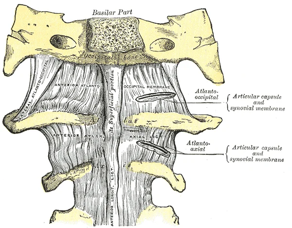
Long description
Anterior atlantoöccipital membrane and atlantoaxial ligament.

Figure 1: Anterior atlantoöccipital membrane and atlantoaxial ligament.
The vertebral column, also known as the spinal column or spine, is a flexible structure that supports the head, neck, and body while protecting the spinal cord. It consists of a sequence of vertebrae separated and united by intervertebral discs. These discs are fibrocartilaginous pads that fill the gap between adjacent vertebral bodies, strongly uniting them. This arrangement forms an intervertebral symphysis, a wide symphysis where bones are joined by fibrocartilage. Unlike synchondroses, which use hyaline cartilage, symphyses allow for small movements and provide cushioning during weight bearing. The discs account for approximately 25 percent of body height between the top of the pelvis and the base of the skull. [1] [2]
Each disc comprises two distinct parts: the anulus fibrosus and the nucleus pulposus. The anulus fibrosus is the tough, fibrous outer layer that forms a circle and is firmly anchored to the outer margins of adjacent vertebral bodies. Inside lies the nucleus pulposus, a softer, gel-like material with high water content that resists compression. This water content declines with age, causing discs to become thinner and reducing flexibility.
DefinitionSymphysis
A cartilaginous joint where bones are joined by fibrocartilage, allowing limited movement and providing shock absorption.
The vertebral column also features zygapophyseal joints between articular processes and uncovertebral joints in the cervical region. These joints, along with the discs, permit the column's flexibility. The intervertebral foramina are openings in the vertebrae through which spinal nerves exit. Compression of a spinal nerve at the foramen can cause pain or muscle weakness in supplied regions.
Figure 2Gray308
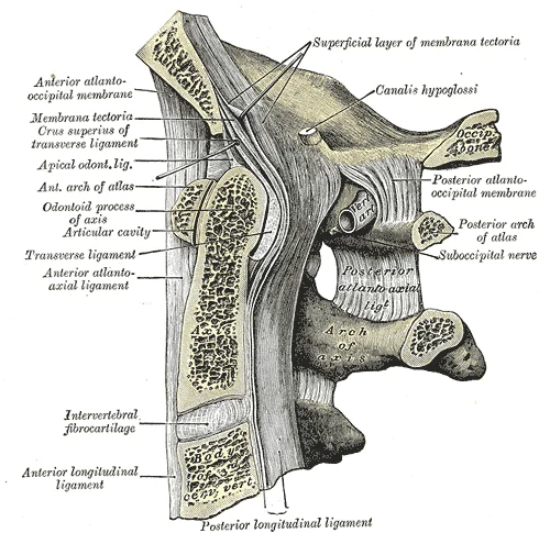
Long description
Median sagittal section through the occipital bone and first three cervical vertebræ. (Spalteholz.)

Figure 2: Median sagittal section through the occipital bone and first three cervical vertebræ (Spalteholz.).
The gel-like nucleus pulposus allows the disc to change shape during movement. Bending forward compresses the anterior disc and expands the posterior disc. If the posterior anulus fibrosus is weakened, the nucleus may protrude posteriorly, resulting in a herniated disc. This posterior bulging can compress a spinal nerve at the intervertebral foramen. The most common sites for herniation are the L4/L5 or L5/S1 discs, causing sciatica. Similar injuries at C5/C6 or C6/C7 can produce pain in the neck, shoulder, and upper limb. Spondylolisthesis and stenosis are associated pathologies involving vertebral alignment and canal narrowing, respectively, often exacerbated by disc degeneration. [1]
The superior articular process of one vertebra joins with the corresponding inferior articular process from the next higher vertebra.
The vertebral column is composed of vertebrae that articulate with each other through two main types of joints: the intervertebral symphyses and the zygapophyseal (facet) joints. The intervertebral symphysis is formed by the bodies of adjacent vertebrae and the intervertebral disc. The body is the anterior portion of each vertebra and is the part that supports the body weight. Because of this, the vertebral bodies progressively increase in size and thickness going down the vertebral column. The bodies of adjacent vertebrae are strongly anchored to each other by an intervertebral disc. This disc allows for movement between the vertebrae. Although the total amount of movement available between any two adjacent vertebrae is small, when these movements are summed together along the entire length of the vertebral column, large body movements can be produced. [1]
The zygapophyseal joints are formed by the articular processes. Seven processes arise from the vertebral arch. A superior articular process extends or faces upward, and an inferior articular process faces or projects downward on each side of a vertebrae. The paired superior articular processes of one vertebra join with the corresponding paired inferior articular processes from the next higher vertebra. The shape and orientation of the articular processes vary in different regions of the vertebral column and play a major role in determining the type and range of motion available in each region. [1]
In the cervical region, the superior and inferior articular processes of the cervical vertebrae are flattened and largely face upward or downward, respectively. The first cervical (C1) vertebra is also called the atlas. The superior articular processes face upward and are deeply curved for articulation with the occipital condyles on the base of the skull. The inferior articular processes are flat and face downward to join with the superior articular processes of the C2 vertebra. [1]
Figure 3Gray306
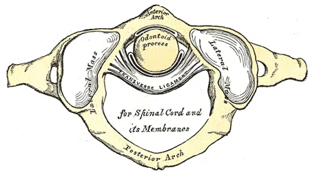
Long description
Articulation between odontoid process and atlas.

Figure 3: Articulation between odontoid process and atlas.
In the thoracic region, the characteristic feature for a typical midthoracic vertebra is the spinous process, which is long and has a pronounced downward angle that causes it to overlap the next inferior vertebra. These orientations are important determinants for the type and range of movements available to the thoracic region of the vertebral column. [1]
At the lumbosacral junction, the superior articular process of the sacrum, one of which is found on either side of the superior opening of the sacral canal, articulates with the inferior articular processes from the L5 vertebra. [1]
DefinitionIntervertebral Disc
A fibrocartilaginous structure that separates and unites the bodies of adjacent vertebrae, providing padding and allowing movement.
DefinitionZygapophyseal Joint
A synovial joint formed by the articulation of the superior articular process of one vertebra with the inferior articular process of the vertebra above it.
| Region | Superior Articular Facet Orientation | Inferior Articular Facet Orientation |
|---|---|---|
| Cervical | Upward | Downward |
| Thoracic | Posteriorly | Anteriorly |
| Lumbosacral | Articulates with L5 inferior processes | N/A (Sacrum) |
Ligaments that extend along the length of the vertebral column also contribute to its overall support and stability. Adjacent vertebrae are united by ligaments that run the length of the vertebral column along both its posterior and anterior aspects. These serve to resist excess forward or backward bending movements of the vertebral column, respectively. The supraspinous ligament is located on the posterior side of the vertebral column, where it interconnects the spinous processes of the thoraci [1]
The vertebral arch forms the posterior portion of each vertebra, consisting of four parts: the right and left pedicles and the right and left laminae[1].
The intervertebral joints are reinforced by several key ligaments. Protection against this movement is particularly important in the neck, where extreme posterior bending of the head and neck can stretch or tear this ligament, resulting in a painful whiplash injury[1].
Figure 4Gray307
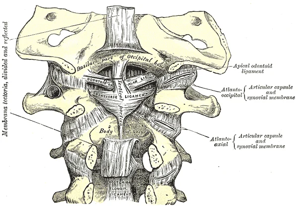
Long description
Membrana tectoria, transverse, and alar ligaments.

Figure 4: Membrana tectoria, transverse, and alar ligaments.
Figure 5Gray305
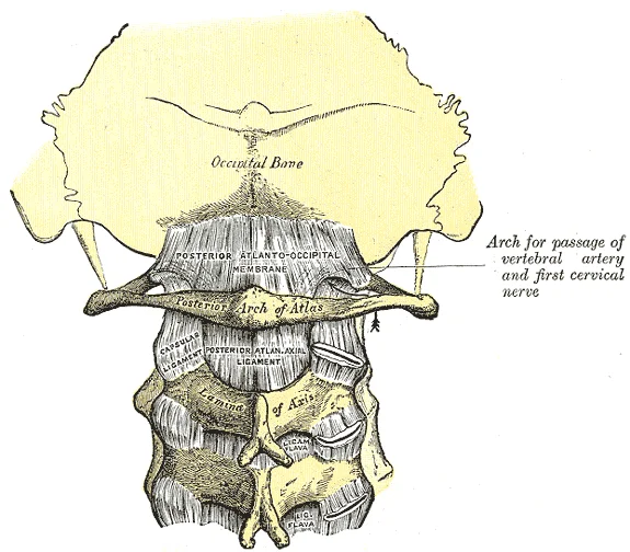
Long description
Posterior atlantoöccipital membrane and atlantoaxial ligament.

Figure 5: Posterior atlantoöccipital membrane and atlantoaxial ligament.
DefinitionLigamentum Flavum
A series of short, paired ligaments interconnecting the lamina regions of adjacent vertebrae, rich in elastic fibers[1].
The supraspinous ligament is located posteriorly and interconnects the spinous processes of the thoracic and lumbar vertebrae[1]. In the neck, this ligament expands to become the nuchal ligament[1]. The nuchal ligament is attached to the cervical spinous processes and superiorly to the base of the skull, out to the external occipital protuberance[1].
NoteClinical Note
Whiplash injuries were common for passengers involved in a rear-end automobile collision prior to the mandatory installation of seat headrests[1].
The vertebral column is subdivided into five regions, with the vertebrae in each area named for that region and numbered in descending order[1]. In the neck, there are seven cervical vertebrae, each designated with the letter “C” followed by its number[1]. Superiorly, the C1 vertebra articulates (forms a joint) with the occipital condyles of the skull[1]. Inferiorly, C1 articulates with the C2 vertebra, and so on[1]. Below these are the 12 thoracic vertebr
DefinitionVertebral Foramen
The large opening between the vertebral arch and body, which contains the spinal cord[1].
Disorders associated with the curvature of the spine include kyphosis (an excessive posterior curvature of the thoracic region), lordosis (an excessive anterior curvature of the lumbar region), and scoliosis (an abnormal, lateral curvature, accompanied by twisting of the vertebral column)[1]. The adult vertebral column does not form a straight line, but instead has four curvatures along its length[1]. These curves increase the vertebral column's strength, flexibility, and abi
Exam trapCurvature Types
Primary curves are retained from the original fetal curvature, while secondary curvatures develop after birth[1].
Within the different regions of the vertebral column, vertebrae vary in size and shape, but they all follow a similar structural pattern[1]. A typical vertebra will consist of a body, a vertebral arch, and processes[1]. In addition to the general characteristics of a typical vertebra described above, vertebrae also display characteristic size and structural features that vary between the different vertebral column regions[1]. Thus, cervical vertebrae are small
| Ligament | Location | Function |
|---|---|---|
| Anterior Longitudinal | Anterior side of entire vertebral column | Resists excess backward bending[1] |
| Posterior Longitudinal | Anterior to spinal cord | Provides support when bending forward[1] |
| Ligamentum Flavum | Posterior to spinal cord, interconnects laminae | Provides support when bending forward[1] |
| Supraspinous | Posteriorly, interconnects spinous processes | N/A |
| Nuchal | Neck, attached to cervical spinous processes | N/A |
The ligaments of the vertebral column provide crucial stability while allowing for controlled movement. These structures can be categorized according to their location and function.
Figure 6Gray301
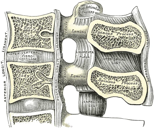
Long description
Median sagittal section of two lumbar vertebræ and their ligaments.

Figure 6: Median sagittal section of two lumbar vertebræ and their ligaments.
| Ligament | Attachments | Function |
|---|---|---|
| Anterior Longitudinal Ligament | Anterior surfaces of vertebral bodies and intervertebral discs from the sacrum to the axis | Resists hyperextension and limits forward flexion |
| Posterior Longitudinal Ligament | Posterior surfaces of vertebral bodies and intervertebral discs within the vertebral canal | Resists hyperflexion and limits backward extension |
| Ligamentum Flavum | Connects laminae of adjacent vertebrae from C7 to sacrum | Assists in maintaining upright posture and limits flexion |
| Interspinous Ligaments | Connects adjacent spinous processes | Limits forward flexion |
| Supraspinous Ligament | Connects tips of spinous processes from C7 to sacrum | Limits forward flexion and assists with upright posture |
| Intertransverse Ligaments | Connects adjacent transverse processes | Limits lateral flexion |
The intervertebral disc is a complex structure composed of two main components. The annulus fibrosus forms the outer ring and consists of concentric layers of fibrocartilage arranged in a crisscross pattern. This arrangement provides tensile strength and resistance to multidirectional forces. The nucleus pulposus forms the central core and has a gel-like consistency composed of water, proteoglycans, and collagen fibers. The nucleus pulposus functions as a shock absorber, distributing compressive forces evenly across the disc.
The relationship between these structures is particularly important in understanding the mechanisms of disc herniation, which often occurs when the annulus fibrosus is weakened, allowing the nucleus pulposus to protrude posteriorly.
This variation reflects the different mechanical demands placed on the spine in different regions. The ligamentum flavum contains elastic fibers that allow it to return to its original length after stretching, which contributes to the upright posture of the spine. [1]
The intervertebral column is a complex mechanical assembly designed to support weight, allow movement, and protect the neural elements. While the intervertebral discs and zygapophyseal joints are the primary articulations, the surrounding soft tissues include bursae and other intra-articular structures that facilitate smooth motion and reduce friction.
For instance, the subcutaneous bursa over the external occipital protuberance can become inflamed, leading to pain and swelling. These structures are generally small and may not be visible on standard imaging unless pathological.
The zygapophyseal (facet) joints are synovial joints formed by the articulation of the superior and inferior articular processes of adjacent vertebrae. The joint capsule is thin and loose, allowing for gliding movements. Inside the joint, the synovial membrane produces fluid for lubrication. They can sometimes become entrapped in the joint space, causing pain and restricted movement. The joint capsule is reinforced by ligaments, including the ligamentum flavum, which connects the laminae of adjacent vertebrae and helps maintain upright posture.
The thoracic curve is convex posteriorly. This secondary curve develops gradually after birth as the child learns to sit upright, stand, and walk.
The intervertebral discs and joints allow for specific movements. Flexion and extension occur primarily in the cervical and lumbar regions. Lateral flexion and rotation are limited but present. The muscles producing these movements originate from the vertebrae and insert onto adjacent structures.
| Muscle | Origin | Insertion | Action | Nerve Supply |
|---|---|---|---|---|
| Sternocleidomastoid | Manubrium and clavicle | Mastoid process | Flexes neck, rotates head | Accessory nerve |
| Trapezius | Occipital bone and spinous processes | Clavicle and scapula | Extends neck, retracts scapula | Accessory nerve |
| Erector Spinae | Iliac crest and sacrum | Ribs and vertebrae | Extends vertebral column | Dorsal rami |
| Rectus Abdominis | Pubic symphysis | Sternum and costal cartilages | Flexes vertebral column | Intercostal nerves |
| Structure | Feature | Clinical Note |
|---|---|---|
| Cervical Vertebrae | Small body, bifid spinous process | Carry least body weight |
| Thoracic Vertebrae | Heart-shaped body, costal facets | Articulate with ribs |
| Lumbar Vertebrae | Large body, no transverse foramen | Carry most body weight |
| Lesion | Deficit | Cause |
|---|---|---|
| Cervical Radiculopathy | Numbness, weakness | Disc herniation |
| Lumbar Radiculopathy | Sciatica | Nerve root compression |
Typical cervical vertebrae, such as C4 or C5, have several characteristic features that differentiate them from thoracic or lumbar vertebrae. Cervical vertebrae have a small body, reflecting the fact that they carry the least amount of body weight. Cervical vertebrae usually have a bifid (Y-shaped) spinous process. The spinous processes of the C3–C6 vertebrae are short, but the spine of C7 is much longer. You can find these vertebrae by running your finger down the midline of the posterior neck until you encounter the prominent C7 spine located at the base of the neck. Each transverse process also has an opening called the transverse foramen. [1]
Figure 7Gray303
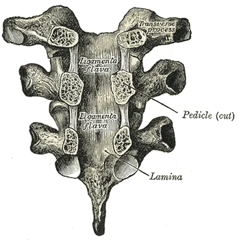
Long description
Vertebral arches of three thoracic vertebræ viewed from the front.

Figure 7: Vertebral arches of three thoracic vertebræ viewed from the front.
The intervertebral discs consist of an outer annulus fibrosus and an inner nucleus pulposus. The annulus fibrosus is composed of concentric layers of fibrocartilage. The nucleus pulposus is a gelatinous core. These structures absorb shock and allow for movement. The joints between vertebral bodies are symphyses, a type of cartilaginous joint. A synchondrosis (“joined by cartilage”) is a cartilaginous joint where bones are joined together by hyaline cartilage, or where bone is united to hyaline cartilage. A synchondrosis may be temporary or permanent. A temporary synchondrosis is the epiphyseal plate (growth plate) of a growing long bone. The epiphyseal plate is the region of growing hyaline cartilage that unites the diaphysis (shaft) of the bone to the epiphysis (end of the bone). Bone lengthening involves growth of the epiphyseal plate cartilage and its replacement by bone, which adds to the diaphysis. For many years during childhood growth, the rates of cartilage growth and bone formation are equal and thus the epiphyseal plate does not change in overall thickness as the bone lengthens. During the late teens and early 20s, growth of the cartilage slows and eventually stops. [2]
The muscles of the back are divided into superficial, intermediate, and deep layers. The superficial muscles move the upper limb. The intermediate muscles are involved in respiration. The deep muscles maintain posture and produce movements of the vertebral column. The transversospinalis group rotates and extends the vertebral column. The segmental muscles stabilize individual vertebrae.
Clinical pearlClinical Pearl
The cervical curve develops as the infant begins to hold their head upright when sitting. [1]
DefinitionSynchondrosis
A cartilaginous joint where bones are joined together by hyaline cartilage.
The stability of the vertebral column is maintained through a complex interplay of osseous, ligamentous, and muscular structures. The intervertebral discs, located between adjacent vertebrae, contribute to stability through their unique composition and structure. Each disc consists of a central gel-like nucleus pulposus surrounded by the annulus fibrosus, which is composed of concentric layers of fibrocartilage. The nucleus pulposus contains approximately 80% water in young adults, providing tur [3]
The facet joints, formed by the articulation of superior and inferior articular processes of adjacent vertebrae, provide stability through their orientation and surrounding ligaments. In the cervical region, these joints are angled to allow for greater mobility, while in the thoracic and lumbar regions, they are oriented more coronally, providing greater stability. [3]
The ligamentous system plays a crucial role in spinal stability. The anterior and posterior longitudinal ligaments run along the anterior and posterior aspects of the vertebral bodies, respectively, providing resistance to excessive flexion and extension. The ligamentum flavum, connecting adjacent laminae, becomes thickened with age, contributing to the maintenance of upright posture. [3]
The multifidus and erector spinae muscles form the primary muscular support system of the spine. The erector spinae group, consisting of the iliocostalis, longissimus, and spinalis muscles, originates from the sacrum, iliac crest, and lumbar vertebrae, inserting into the ribs and vertebrae, providing global stability. [3]
These curves help distribute mechanical forces and maintain balance during movement. [3]
The intervertebral disc and the associated joints of the vertebral column rely on a specific vascular and neural architecture that supports their structural integrity and sensory feedback. Understanding this supply requires examining the principles of tissue perfusion and innervation in the axial skeleton.
The disc itself is avascular in its central nucleus pulposus but receives limited diffusion from the capillary loops in the peripheral annulus fibrosus.
The blood supply to the vertebral column is segmental. The segmental arteries, such as the intercostal and lumbar arteries, give off spinal branches. These branches enter the vertebral canal through the intervertebral foramina. They supply the vertebral bodies, the posterior elements, and the dura mater. This diffusion is critical for the health of the chondrocytes in the annulus fibrosus and the proteoglycans in the nucleus pulposus. As we age, the vascular supply to the endplates may diminish, contributing to disc degeneration.
The nerve supply to the intervertebral disc and the joints of the column is complex. It re-enters the vertebral canal through the intervertebral foramen. It supplies the outer third of the annulus fibrosus, the posterior longitudinal ligament, and the dura mater. This nerve is a branch of the spinal nerve. It carries both sensory and sympathetic fibres. The sensory fibres transmit pain from the disc. The sympathetic fibres regulate blood flow. The joints of the column, including the facet joints, are supplied by the medial branches of the dorsal rami of the spinal nerves. These nerves provide proprioceptive information and pain sensation from the joint capsules.
Hilton's law states that the nerve supplying a joint also supplies the muscles that move the joint and the skin overlying it. This principle helps explain the referred pain patterns associated with spinal pathology. For example, irritation of the sinuvertebral nerve can cause pain that radiates to the buttock or leg. This is because the spinal nerve roots that give rise to the sinuvertebral nerve also supply the lower limb. Understanding this relationship is crucial for diagnosing the source of back pain.
DefinitionSinuvertebral Nerve
The sinuvertebral nerve is a branch of the spinal nerve that re-enters the vertebral canal to supply the outer annulus fibrosus, posterior longitudinal ligament, and dura mater.
Clinical pearlHilton's Law
Hilton's law states that the nerve supplying a joint also supplies the muscles that move the joint and the skin overlying it.
The intervertebral disc serves as the primary articulation between adjacent vertebral bodies. It consists of two distinct components: the nucleus pulposus and the annulus fibrosus. The nucleus pulposus is a gelatinous core located centrally within the disc. It is composed primarily of water and proteoglycans, which provide hydration and resilience. The annulus fibrosus surrounds the nucleus. It consists of concentric layers of fibrocartilage. These layers are oriented at alternating angles to resist tensile forces.
Figure 8718 Vertebra
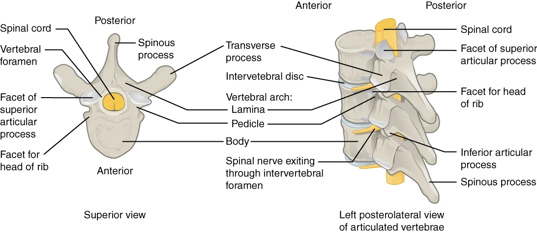
Long description
A typical vertebra consists of a body and a vertebral arch. The arch is formed by the paired pedicles and paired laminae. Arising from the vertebral arch are the transverse, spinous, superior articular, and inferior articular processes. The vertebral foramen provides for passage of the spinal cord.

Figure 8: A typical vertebra consists of a body and a vertebral arch.
Radiologically, the disc appears as a hypointense structure on T1-weighted MRI sequences and hyperintense on T2-weighted sequences due to its high water content. The height of the disc is crucial for maintaining the intervertebral foramen size. A reduction in disc height can lead to narrowing of the foramen, potentially compressing nerve roots. The disc acts as a shock absorber, distributing loads across the vertebral column.
They guide and limit the movement of the spine. In the lumbar region, the facet joints are oriented in the sagittal plane. This orientation allows for flexion and extension while limiting rotation. The articular surfaces are covered with hyaline cartilage. The joint capsule surrounds the articulation and contains synovial fluid. [1]
Radiologically, the facet joints are visible as paired structures posterior to the vertebral body. Degenerative changes in these joints, such as osteophyte formation or joint space narrowing, are common in older adults. These changes can contribute to spinal stenosis. The alignment of the facet joints is critical for spinal stability. Misalignment can lead to spondylolisthesis, where one vertebra slips forward over another.
Several ligaments support the intervertebral discs and joints. The anterior longitudinal ligament runs along the anterior surface of the vertebral bodies. It prevents hyperextension of the spine. The posterior longitudinal ligament runs along the posterior surface of the vertebral bodies within the spinal canal. It prevents hyperflexion and protects the spinal cord from posterior disc herniation. The ligamentum flavum connects the laminae of adjacent vertebrae. It helps maintain upright posture and returns the spine to extension after flexion. [1]
These ligaments are less distinct on standard radiographs but are clearly visible on MRI. Thickening of the ligamentum flavum can contribute to spinal stenosis by reducing the volume of the spinal canal. The integrity of these ligaments is essential for maintaining the structural integrity of the vertebral column.
The thoracic spine is distinct due to its numerous articulation sites for the ribs, which are critical for thoracic stability and respiratory mechanics. Each thoracic vertebra possesses specific facets where rib attachment occurs. Most thoracic vertebrae feature two costal facets located on the lateral aspects of the vertebral body. These costal facets, derived from the Latin for 'rib', articulate specifically with the head or end of a rib. This articulation forms the costovertebral joint, a key component in the structural integrity of the thoracic cage.
An additional facet is present on the transverse process of each thoracic vertebra. This transverse costal facet articulates with the tubercle of the corresponding rib. This connection forms the costotransverse joint. The presence of these dual articulation points per vertebra distinguishes the thoracic region from the cervical and lumbar spines. Clinically, understanding these articulation sites is vital for interpreting imaging and assessing trauma patterns involving the posterior thoracic wall. Dislocation or fracture of these facets can compromise spinal stability and potentially injure adjacent neural structures or pleural spaces.
The sacrum is a large, triangular bone formed by the fusion of five sacral vertebrae. Its posterior surface features the median sacral crest, which is formed by the fused spinous processes of these vertebrae. Lateral to this is the lateral sacral crest, derived from the fused transverse processes. These bony landmarks serve as attachment points for ligaments and muscles of the lower back and pelvis. [1]
Anteriorly and posteriorly, the sacrum contains foramina that allow branches of the sacral spinal nerves to exit the vertebral canal. The auricular surfaces are located on the lateral aspects of the sacrum. These rough, ear-shaped surfaces serve as articulation sites that anchor the sacrum to the hipbones, thereby forming the pelvic girdle. This sacroiliac joint is crucial for weight transmission from the spine to the lower limbs.
Distal to the sacrum lies the coccyx, commonly known as the tailbone. It is a small, rudimentary structure derived from the fusion of four small vertebrae. Injury to the coccyx, often resulting from a fall onto the buttocks, can cause significant pain but typically heals with conservative management.
Injury patterns in the spine often involve the intervertebral discs and associated joints. The discs act as shock absorbers and allow for movement between vertebral bodies. Degeneration or herniation of these discs can lead to nerve root compression, causing radicular pain. Examination tests such as the straight leg raise test are used to assess for lumbar disc herniation affecting the sciatic nerve.
Aspiration sites for diagnostic or therapeutic purposes are chosen based on anatomical landmarks. For example, lumbar puncture is typically performed at the L3-L4 or L4-L5 interspace to avoid injury to the spinal cord, which usually terminates at the L1-L2 level in adults. Understanding the precise anatomy of the vertebral column, including the facets, foramina, and disc spaces, is essential for safe clinical procedures and accurate diagnosis of spinal pathology.
The intervertebral disc consists of a central nucleus pulposus and an outer annulus fibrosus. The nucleus is gelatinous and rich in proteoglycans, providing hydrostatic pressure resistance. The annulus comprises concentric lamellae of type I collagen, oriented at alternating angles to resist torsion.
The vertebral body articulates with the disc via the cartilaginous endplate. This hyaline cartilage layer facilitates nutrient diffusion from the subchondral bone marrow. It acts as a semi-permeable barrier, restricting large molecule movement while allowing water and ions to pass.
The annulus fibrosus attaches firmly to the vertebral body margins. This attachment is reinforced by the anterior and posterior longitudinal ligaments. These ligaments limit excessive flexion and extension, respectively, while stabilising the column.
The nucleus pulposus contains chondrocyte-like cells embedded in a matrix. This matrix has high water content in youth, decreasing with age. The loss of proteoglycans reduces osmotic pressure, leading to disc dehydration and height loss.
Intervertebral joints are classified as symphyses, a type of secondary cartilaginous joint. They allow limited movement, primarily compression and slight rotation. The joint space is maintained by the tensile strength of the annulus.
Degeneration begins with fissuring in the outer annulus. These tears allow nucleus material to protrude. If the tear extends through the entire annulus, a herniation may occur. This can compress adjacent nerve roots, causing radiculopathy.
The blood supply to the disc is limited. The outer third of the annulus receives vascular ingrowth from the periosteum. The inner two-thirds and the nucleus are avascular in adults. Nutrients diffuse from the vertebral bodies through the endplates.
Lymphatic drainage is minimal. Waste products diffuse back into the vertebral body vasculature. This slow turnover explains why disc injuries heal poorly compared to other tissues. The low metabolic rate requires careful load management.
They are synovial joints with hyaline cartilage covering the articular surfaces. The joint capsule is thin and loose, allowing gliding movements.
It is bounded anteriorly by the vertebral body and disc, and posteriorly by the facet joints. The superior and inferior vertebral notches form the boundaries.
Osteophytes often form at the disc margins. These bony outgrowths are a response to instability. They can narrow the intervertebral foramen, contributing to nerve root compression. This is a common feature of spondylosis.
Clinical pearlDisc Herniation
A herniation occurs when nucleus pulposus material protrudes through a tear in the annulus fibrosus, potentially compressing neural structures.
Exam trapBlood Supply
Do not assume the disc has a direct blood supply; the nucleus and inner annulus are avascular in adults, relying on diffusion.
Students frequently confuse the structural composition of intervertebral discs with that of typical synovial joints. A common error is assuming the disc allows for free movement like a knee or shoulder. In reality, the intervertebral disc is a symphysis, a type of cartilaginous joint where bones are joined by fibrocartilage. Fibrocartilage is distinct because it contains numerous bundles of thick collagen fibers, giving it a much greater ability to resist pulling and bending forces when compared with hyaline cartilage. This structural difference means the joint is functionally classified as an amphiarthrosis, allowing only limited movement while strongly uniting adjacent bones. [2]
Another frequent mistake involves the anatomy of the upper cervical spine, specifically the relationship between the atlas (C1) and the axis (C2). Students often overlook the specific mechanism of rotation. The second cervical (C2) vertebra is called the axis because it serves as the axis for rotation when turning the head toward the right or left. This rotation occurs because the dens (odontoid process), a bony projection extending upward from the vertebral body of C2, joins with the inner aspe [1]
Learners also frequently misinterpret the weight-bearing roles of the lower vertebral column. It is a misconception that the coccyx bears significant weight during standing. The coccyx, or tailbone, is derived from the fusion of four very small coccygeal vertebrae and articulates with the inferior tip of the sacrum. It is not weight bearing in the standing position, but may receive some body weight when sitting. In contrast, the sacrum is thick and wide across its superior base where it is weight bearing and then tapers down to an inferior, non-weight bearing apex. [1]
A further pitfall relates to the developmental timeline of the sacrum. Students may assume the sacral segments are fused at birth. The sacrum is formed by the fusion of five sacral vertebrae, a process that does not begin until after the age of 20. On the anterior surface of the older adult sacrum, the lines of vertebral fusion can be seen as four transverse ridges. Similarly, the fused transverse processes form the lateral sacral crest, while the median sacral crest is the remnant of the fused spinous processes. [1]
Finally, students often confuse the general principle of vertebral counts with specific anatomical variations. While almost all mammals have seven cervical vertebrae, regardless of body size, this does not imply uniformity in structure. In a full-grown giraffe, each cervical vertebra is 11 inches tall, demonstrating large variations in size ranging from the very small cervical vertebrae of a shrew to the greatly elongated vertebrae in the neck of a giraffe. However, in human anatomy, the count remains constant, and errors arise when applying mammalian variation logic to human clinical presentations. [1]
Knowledge of intervertebral discs and spinal joints is applied at the bedside to assess spinal stability and pain sources. When examining a patient with neck pain, understanding that C2 serves as the axis for rotation helps localise symptoms to the atlantoaxial joint if rotation is painful. The integrity of the transverse ligament holding the dens is critical; injury here can lead to atlantoaxial instability, a life-threatening condition requiring immediate recognition.
In the operating theatre, surgeons rely on the structural properties of the symphysis. Because fibrocartilage resists pulling and bending forces, intervertebral discs are key shock absorbers. During spinal fusion procedures, surgeons must account for the amphiarthrotic nature of these joints, ensuring that instrumentation does not compromise the limited but essential movement allowed by the fibrocartilaginous union.
On imaging, particularly CT and MRI, radiologists look for signs of degeneration or trauma. The transverse ridges on the anterior sacrum indicate fusion lines, which should be symmetrical in adults. The median sacral crest, a bumpy ridge on the posterior surface, serves as a landmark for midline procedures. Abnormalities in the dens or its relationship with the atlas are visible on lateral cervical spine views, aiding in the diagnosis of fractures or ligamentous tears.
In examinations, students are tested on the functional classification of joints. The intervertebral disc is a classic example of a symphysis, functionally classified as an amphiarthrosis. Questions often ask for the tissue type (fibrocartilage) and its mechanical advantage (resistance to pulling and bending) over hyaline cartilage. Understanding that the coccyx is non-weight bearing in standing but may bear weight when sitting is a frequent multiple-choice question topic.
Assessment of Cervical Instability
- Patient presents with neck painHistory and physical exam
- Question: Pain on rotation?
Yes
- Suspect atlantoaxial involvementCheck C2 axis function
No
- Evaluate other cervical levelsCheck flexion/extension
- Imaging requiredCT/MRI for dens and ligaments
- Question: Dens displaced?
Yes
- Transverse ligament injuryHigh risk of instability
No
- Stable jointConservative management
Clinical pearlSacral Landmarks
The median sacral crest is the remnant of fused spinous processes, while the lateral sacral crest is formed by fused transverse processes. These landmarks are crucial for identifying the midline during lumbar punctures or surgical approaches to the sacrum.
DefinitionSymphysis
A cartilaginous joint where bones are joined by fibrocartilage, functionally classified as an amphiarthrosis, allowing limited movement while strongly uniting adjacent bones.
Intervertebral discs are symphyses joined by fibrocartilage, which resists pulling and bending forces better than hyaline cartilage. The C2 vertebra, or axis, serves as the pivot for head rotation, with the dens held by the transverse ligament. The sacrum is formed by the fusion of five vertebrae, a process starting after age 20. The coccyx is non-weight bearing in standing but may bear weight when sitting. The sacral crest landmarks help identify the midline for clinical procedures. Symphyses are functionally amphiarthroses, allowing limited movement. The dens articulates with the anterior arch of the atlas (C1). Fibrous connections in symphyses provide strong union with slight mobility.
Answer each one in your head first, then open the answer.
What type of cartilage joins the bones in a symphysis?
Show answerHide answer
FibrocartilageWhich vertebra is known as the axis?
Show answerHide answer
C2What ligament holds the dens in place against the atlas?
Show answerHide answer
Transverse ligamentIs the coccyx weight bearing in the standing position?
Show answerHide answer
NoHow many sacral vertebrae fuse to form the sacrum?
Show answerHide answer
Five
- Betts JG, Young KA, Wise JA, et al. The Vertebral Column. OpenStax, Anatomy and Physiology 2e (CC BY-NC-SA 4.0). Accessed .
- Betts JG, Young KA, Wise JA, et al. Cartilaginous Joints. OpenStax, Anatomy and Physiology 2e (CC BY-NC-SA 4.0). Accessed .
- Menshawi K, Mohr JP, Gutierrez J. A Functional Perspective on the Embryology and Anatomy of the Cerebral Blood Supply. Journal of stroke 2015;17(2):144-158. doi:10.5853/jos.2015.17.2.144. Accessed .
Figure credits
- Figure 1: Henry Vandyke Carter, via Wikimedia Commons, Public domain, Public domain.
- Figure 2: Henry Vandyke Carter, via Wikimedia Commons, Public domain, Public domain.
- Figure 3: Henry Vandyke Carter, via Wikimedia Commons, Public domain, Public domain.
- Figure 4: Henry Vandyke Carter, via Wikimedia Commons, Public domain, Public domain.
- Figure 5: Henry Vandyke Carter, via Wikimedia Commons, Public domain, Public domain.
- Figure 6: Henry Vandyke Carter, via Wikimedia Commons, Public domain, Public domain.
- Figure 7: Henry Vandyke Carter, via Wikimedia Commons, Public domain, Public domain.
- Figure 8: OpenStax College, via Wikimedia Commons, CC BY 3.0, CC BY 3.0.
Educational content for medical students and doctors. It is not a substitute for clinical judgement or local guidelines.