The vertebral column and vertebrae
The bony axial skeleton supporting the head and trunk, protecting the spinal cord.
The vertebral column, also known as the spinal column or spine, is a flexible structure that supports the head, neck, and body while allowing for movement[1]. It protects the spinal cord, which passes down the back through openings in the vertebrae[1]. The column originally develops as a series of 33 vertebrae, but this number is eventually reduced to 24 vertebrae, plus the sacrum and coccyx[1]. The vertebral column is subdivided into five regions, with the vertebra
Figure 1Gray112
Long description
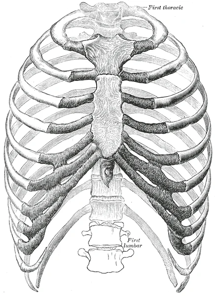
The thorax from in front. (Spalteholz.). Labelled illustration from Wikimedia Commons, used under its open licence.

Figure 1: The thorax from in front (Spalteholz.).
The adult vertebral column does not form a straight line, but instead has four curvatures along its length[1]. These curves increase the vertebral column’s strength, flexibility, and ability to absorb shock[1]. When the load on the spine is increased, by carrying a heavy backpack for example, the curvatures increase in depth to accommodate the extra weight[1]. They then spring back when the weight is removed[1]. The four adult curvatures are classified as either primary or secondary curvatures[1]. Primary curves are retained from the original fetal curvature, while secondary curvatures develop after birth[1].
During fetal development, the body is flexed anteriorly into the fetal position, giving the entire vertebral column a single curvature that is concave anteriorly[1]. In the adult, this fetal curvature is retained in two regions of the vertebral column as the thoracic curve, which involves the thoracic vertebrae, and the sacrococcygeal curve, formed by the sacrum and coccyx[1]. Each of these is thus called a primary curve because they are retained from the original fetal curvature of the vertebral column[1]. The cervical and lumbar curves develop after birth and thus are secondary curves[1]. The cervical curve develops as the infant begins to hold up the head, and the lumbar curve appears with standing and walking[1].
The vertebral column forms the neck and back[1]. The vertebrae are divided into the cervical region (C1–C7 vertebrae), the thoracic region (T1–T12 vertebrae), and the lumbar region (L1–L5 vertebrae)[1]. The sacrum arises from the fusion of five sacral vertebrae and the coccyx from the fusion of four small coccygeal vertebrae[1]. The vertebral column has four curvatures, the cervical, thoracic, lumbar, and sacrococcygeal curves[1].
DefinitionVertebral Foramen
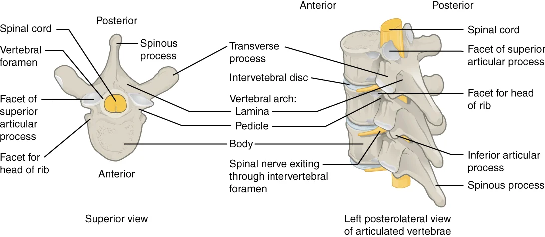
The large opening between the vertebral arch and body is the vertebral foramen, which contains the spinal cord[1].
The vertebral arch forms the posterior portion of each vertebra[1]. It consists of four parts, the right and left pedicles and the right and left laminae[1]. Each pedicle forms one of the lateral sides of the vertebral arch[1]. The pedicles are anchored to the posterior side of the vertebral body[1]. Each lamina forms part of the posterior roof of the vertebral arch[1]. In the intact vertebral column, the vertebral foramina of all of the vertebrae align to form the vertebral (spinal) canal, which serves as the bony protection and passageway for the spinal cord down the back[1].
NoteSacral Fusion
The sacrum is formed by the fusion of five sacral vertebrae, a process that does not begin until after the age of 20[1].
The sacrum is a triangular-shaped bone that is thick and wide across its superior base where it is weight bearing and then tapers down to an inferior, non-weight bearing apex[1]. On the anterior surface of the older adult sacrum, the lines of vertebral fusion can be seen as four transverse ridges[1]. On the posterior surface, running down the midline, is the median sacral crest, a bumpy ridge that is the remnant of the fused spinous processes[1]. Similarly, the fused transverse processes of the sacral vertebrae form the lateral sacral crest[1].
Lymph from the thoracic vertebrae follows a more complex route. [1]
Figure 2716 Intervertebral Disk
Long description
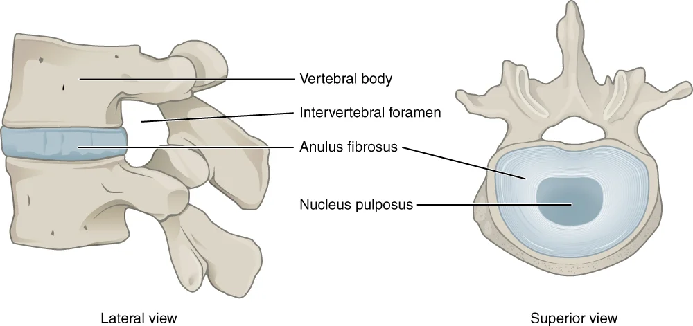
The bodies of adjacent vertebrae are separated and united by an intervertebral disc, which provides padding and allows for movements between adjacent vertebrae. The disc consists of a fibrous outer layer called the anulus fibrosus and a gel-like center called the nucleus pulposus. The intervertebral

Figure 2: The bodies of adjacent vertebrae are separated and united by an intervertebral disc, which provides padding and allows for movements between adjacent vertebrae.
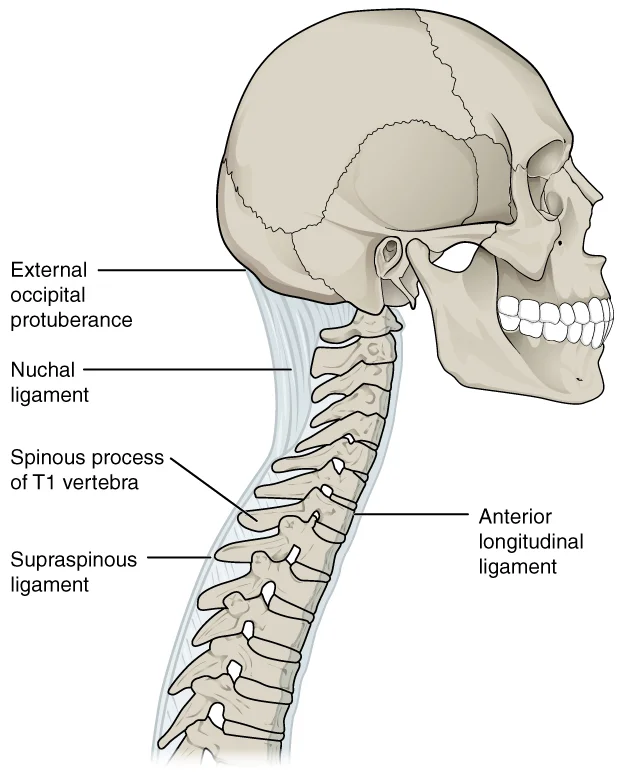
You can trace the midline from the base of the skull downwards. The most prominent landmark is the spinous process of C7. This is often called the vertebra prominens. You find it by running your finger down the midline of the posterior neck. It is located at the base of the neck. The spinous processes of C3 to C6 are short. The spine of C7 is much longer. This makes it easy to identify. Below C7, the spinous processes of the thoracic vertebrae angle downwards. They overlap the next inferior vertebra. This creates a stepped appearance. The supraspinous ligament interconnects these processes. In the posterior neck, this ligament expands into the nuchal ligament. It attaches to the cervical spinous processes and extends to the external occipital protuberance. This supports the skull. [1]
Palpable landmarks include the iliac crests. These correspond to the level of L4. The posterior superior iliac spines mark the level of S2. These are useful for identifying lumbar and sacral levels. The vertebral bodies progressively increase in size and thickness going down the column. This reflects the increasing weight bearing. The intervertebral discs are thin in the cervical region. They are thickest in the lumbar region. In total, the intervertebral discs account for approximately 25 percent of your body height between the top of the pelvis and the base of the skull.
Ossification begins in the fetal period. Primary ossification centres appear in the vertebral arches. Secondary centres appear in the body. The vertebral column grows from these centres. The surface anatomy reflects the underlying bony structures. The spinous processes are palpable. The transverse processes are less distinct. The bodies are deep. The intervertebral foramina are not palpable. They are formed by notches in the margins of the pedicles. These foramina allow spinal nerves to exit. The shape and orientation of the articular processes vary in different regions. This determines the type and range of motion. The cervical vertebrae have small bodies. They carry the least amount of body weight. The thoracic vertebrae have larger bodies. The lumbar vertebrae have the largest bodies. They carry the most body weight. The surface anatomy helps in clinical examination. It allows identification of vertebral levels. It helps in locating spinal nerves. It helps in assessing spinal alignment.
Clinical pearlVertebra Prominens
The spinous process of C7 is much longer than those of C3–C6, making it a key landmark for identifying the cervical-thoracic junction.
| Landmark | Vertebral Level |
|---|---|
| External Occipital Protuberance | Base of Skull |
| Vertebra Prominens (C7) | C7 |
| Iliac Crests | L4 |
| Posterior Superior Iliac Spines | S2 |
The vertebral spines project posteriorly at the midline and can be felt as a series of bumps under the skin. Each vertebra possesses seven processes arising from the vertebral arch. The paired transverse processes project laterally from the junction of the pedicle and lamina. The paired superior articular processes face upward, while the inferior articular processes face downward. These articular processes join to form slightly moveable joints between adjacent vertebrae. [1]
The vertebral column is divided into regions. The cervical region includes the atlas (C1) and axis (C2). The atlas lacks a body and consists of a bony ring formed by anterior and posterior arches. The axis is distinguished by the dens, a bony projection extending upward from the vertebral body. The dens joins the anterior arch of the atlas, held by the transverse ligament. Typical cervical vertebrae have small bodies, bifid spinous processes, and U-shaped transverse processes with transverse foramina. [1]
Figure 3Gray82
Long description
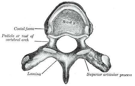
A typical thoracic vertebra, viewed from above.

Figure 3: A typical thoracic vertebra, viewed from above.
The thoracic vertebrae are distinguished by long, downward-projecting spinous processes and facets for rib attachment. The lumbar vertebrae support the greatest body weight and have large, thick bodies with short, blunt spinous processes. The sacrum is triangular and articulates with the ilium via the auricular surface to form immobile sacroiliac joints. The sacral promontory is the anterior lip of the superior base. The sacral canal terminates at the sacral hiatus, with foramina allowing nerve exit. The coccyx consists of fused small vertebrae and articulates with the sacrum. [1]
Intervertebral discs fill gaps between vertebral bodies, providing padding and strong attachments. The disc comprises an outer anulus fibrosus and a gel-like nucleus pulposus. The anterior longitudinal ligament runs down the anterior side, uniting vertebral bodies and resisting backward bending. End plates interface between rigid bodies and pliant discs, balancing strength and porosity. The bony end plate thickness is generally between 0.2 and 0.8 mm. [1] [2]
The vertebral column is a complex structure where stability and mobility are balanced through specific articulations. This view shows the alignment of the vertebrae, highlighting the intervertebral discs and the facet joints. The labelled structures include the vertebral bodies, the intervertebral foramina, and the articular processes. The intervertebral discs lie between the vertebral bodies, while the facet joints connect the superior and inferior articular processes of adjacent vertebrae. Understanding these relationships is crucial for recognising pathology on imaging.
DefinitionIntervertebral Disc
A fibrocartilaginous joint connecting adjacent vertebral bodies, composed of a central nucleus pulposus and an outer anulus fibrosus.
The intervertebral discs act as shock absorbers and allow movement. The gel-like nature of the nucleus pulposus allows the disc to change shape as one vertebra rocks side to side or forward and back in relation to its neighbors during movements of the vertebral column. Bending forward causes compression of the anterior portion of the disc but expansion of the posterior disc. If the posterior anulus fibrosus is weakened due to injury or increasing age, the pressure exerted on the disc when bending forward and lifting a heavy object can cause the nucleus pulposus to protrude posteriorly through the anulus fibrosus, resulting in a herniated disc. This posterior bulging can cause compression of a spinal nerve at the point where it exits through the intervertebral foramen, with resulting pain and/or muscle weakness in those body regions supplied by that nerve. [1]
The facet joints, or zygapophyseal joints, are synovial joints between the articular processes. The superior and inferior articular processes of the cervical vertebrae are flattened and largely face upward or downward, respectively. In the cervical region, the superior articular processes face upward and are deeply curved for articulation with the occipital condyles on the base of the skull. The inferior articular processes are flat and face downward to join with the superior articular processes of the C2 vertebra. The first cervical (C1) vertebra is also called the atlas, because this is the vertebra that supports the skull on top of the vertebral column. The C1 vertebra does not have a body or spinous process. Instead, it is ring-shaped, consisting of an anterior arch and a posterior arch. The transverse processes of the atlas are longer and extend more laterally than do the transverse processes of any other cervical vertebrae. [1]
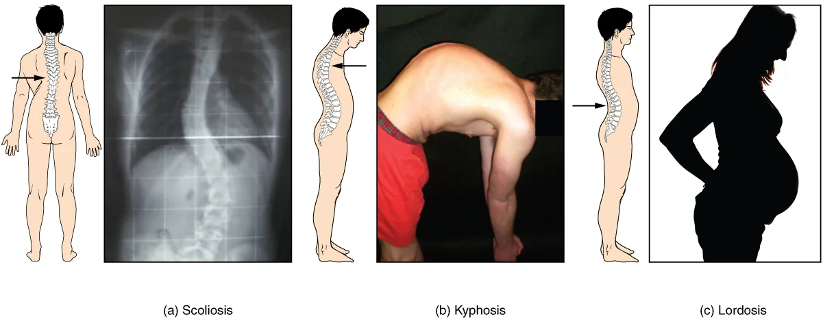
Disorders associated with the curvature of the spine include kyphosis (an excessive posterior curvature of the thoracic region), lordosis (an excessive anterior curvature of the lumbar region), and scoliosis (an abnormal, lateral curvature, accompanied by twisting of the vertebral column). These conditions reflect abnormalities in the normal alignment and function of the vertebral joints. [1]
Lymph from the thoracic vertebrae follows a more complex route.
DefinitionThoracic Vertebrae
Thoracic vertebrae have several additional articulation sites, each of which is called a facet, where a rib is attached. [1]
Most thoracic vertebrae have two facets located on the lateral sides of the body, each of which is called a costal facet (costal = “rib”). These are for articulation with the head (end) of a rib. An additional facet is located on the transverse process for articulation with the tubercle of a rib. [1]
DefinitionLumbar Vertebrae
Lumbar vertebrae carry the greatest amount of body weight and are thus characterized by the large size and thickness of the vertebral body. [1]
They have short transverse processes and a short, blunt spinous process that projects posteriorly. The articular processes are large, with the superior process facing medially and the inferior facing laterally. [1]
DefinitionEnd Plate Structure
The end plate is a bilayer of cartilage and bone that separates the intervertebral disks from the adjacent vertebrae. [2]
During prenatal development, the future vertebra starts as a cartilage anlagen that arises from chondrification centers of the sclerotomes during the sixth embryonic week. [2]
By age 18, the epiphyseal cartilage has thinned and a subchondral bone plate has formed, thus creating the adult end plate bilayer. Simultaneously, the ring apophysis fuses to the vertebral body. [2]
DefinitionNutritional Pathway
Nutritionally, the end plate is the primary pathway for transport between vertebral capillaries and cells within the disk nucleus. [2]
The typical marrow contact area (or effective exchange area) is between 10 and 40%, with the central end plate (adjacent the nucleus) being more permeable than at the periphery. [2]
DefinitionEnd Plate Vulnerability
End plates play a central role in maintaining disk and vertebral health. [2]
Their structure and composition reflect a balance between competing requirements for porosity and strength. As a result, end plates are particularly vulnerable to damage. End plate regions weakened by damage facilitate communication between the disk nucleus and vertebral marrow, which can cause an adverse combination of end plate nerve proliferation plus chemical sensitization and mechanical stimulation. [2]
Figure 41117 Muscles of the Back
Long description
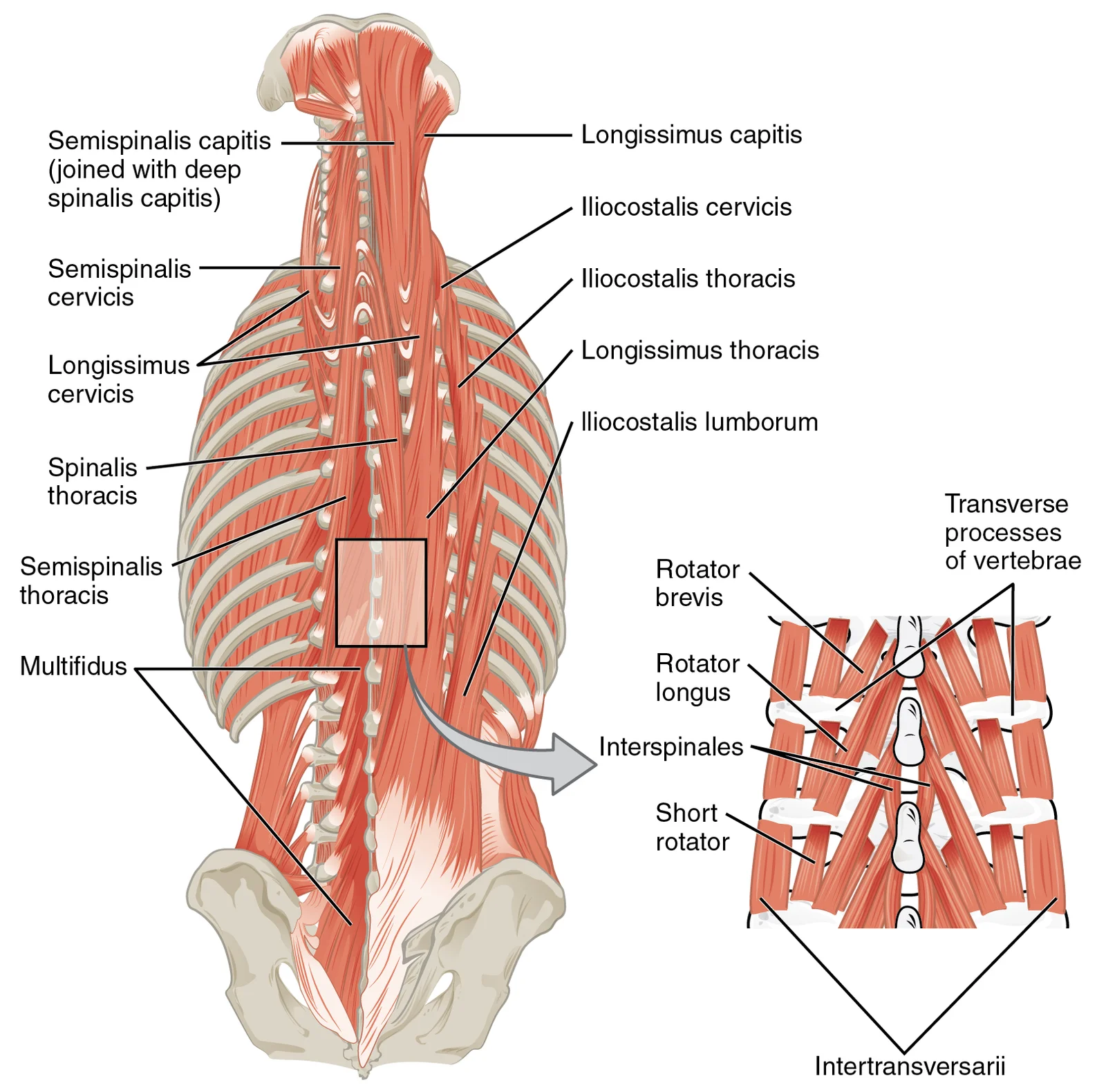
Caption: The large, complex muscles of the neck and back move the head, shoulders, and vertebral column. URL: https://cnx.org/contents/FPtK1zmh@8.108:_xq2eUyd@5/Axial-Muscles-of-the-Head-Neck Other versions: Original

Figure 4: Caption: The large, complex muscles of the neck and back move the head, shoulders, and vertebral column (url: https://cnx.org/contents/fptk1zmh@8.108:_xq2euyd@5/axial-muscles-of-the-head-neck other versions: original).
The muscles of the vertebral column are grouped by compartment. The posterior compartment contains the intrinsic back muscles, which are innervated by the dorsal rami of the spinal nerves. These muscles maintain posture and extend the vertebral column.
| Muscle Group | Origin | Insertion | Action | Nerve Supply |
|---|---|---|---|---|
| Iliocostalis | Sacrum, iliac crest, spinous processes | Ribs, transverse processes | Extends and laterally flexes vertebral column | Dorsal rami |
| Longissimus | Sacrum, iliac crest, spinous processes | Transverse processes, ribs, mastoid process | Extends and laterally flexes vertebral column | Dorsal rami |
| Spinalis | Spinous processes | Spinous processes | Extends vertebral column | Dorsal rami |
| Semispinalis | Transverse processes | Spinous processes | Extends and rotates vertebral column | Dorsal rami |
| Multifidus | Sacrum, articular processes, transverse processes | Spinous processes | Extends and rotates vertebral column | Dorsal rami |
| Rotatores | Transverse processes | Spinous processes | Rotates vertebral column | Dorsal rami |
DefinitionPosterior Compartment
The intrinsic back muscles lie deep to the superficial extrinsic muscles and are responsible for fine control of spinal movement and posture.
The anterior and lateral compartments contain extrinsic muscles that connect the trunk to the upper limbs and pelvis. These are innervated by the ventral rami. They are not primarily responsible for vertebral movement but for limb movement and trunk stability.
Exam trapInnervation Distinction
Remember that intrinsic back muscles are innervated by dorsal rami, whereas extrinsic muscles are innervated by ventral rami.
The vertebral column relies on these muscular groups for dynamic stability. The erector spinae group acts as a primary extensor, counteracting the forward flexion moment. The transversospinalis group provides segmental stability. Weakness in these muscles can lead to poor posture and increased load on the intervertebral discs.
The vertebral column receives its blood supply from a complex network of segmental arteries, which ensure adequate perfusion to the vertebral bodies, arches, and intervertebral discs. Understanding this vascular architecture is fundamental to appreciating the metabolic demands of the spinal column, particularly given the avascular nature of the intervertebral disc nucleus.
The vertebral bodies are supplied by anterior segmental medullary arteries, which enter the vertebral body and anastomose with branches from the posterior circulation.
The end plate cartilage consists of chondrocytes interspersed throughout an extracellular matrix of proteoglycans, collagen (types I and II), and water. [2]
Movement of small solutes such as glucose, lactate, and oxygen occurs primarily by diffusion through the disk matrix. This diffusion is driven by the concentration gradient between blood plasma and tissue matrix. The end plate must balance conflicting demands: it must be strong to resist mechanical failure yet porous to facilitate chemical transport. When the spine is compressed, the bony end plate is subjected to high tensile strains. Damage often occurs to the central end plate, which is the thinnest and weakest region. Accumulation of damage can cause focal weak points that progress to circ [2]
The venous drainage of the vertebral column is a complex network that facilitates the return of blood from the spinal structures to the systemic circulation. This system is characterized by its lack of valves, allowing for bidirectional flow, which has significant clinical implications for the spread of infection or metastatic disease.
The internal vertebral venous plexuses are located within the vertebral canal, lying between the dura mater and the periosteum of the vertebral bones. These plexuses are divided into anterior and posterior components. The anterior internal vertebral venous plexus runs along the posterior aspect of the vertebral bodies and intervertebral discs. The posterior internal vertebral venous plexus is situated on the inner surface of the laminae and ligamenta flava. These plexuses are valveless and commu [1]
The external vertebral venous plexuses are located outside the vertebral column, surrounding the posterior aspects of the vertebrae. These plexuses include the anterior external vertebral venous plexus, which lies on the anterior longitudinal ligament, and the posterior external vertebral venous plexus, which is found on the posterior elements of the spine. These external plexuses communicate with the internal plexuses through intervertebral foramina. They also connect with the segmental veins, such as the intercostal and lumbar veins. The valveless nature of these connections allows for retrograde flow, particularly when intra-abdominal or intrathoracic pressure increases. This mechanism can facilitate the spread of pathological processes from the pelvis or abdomen to the spine.
Figure 5Gray83
Long description
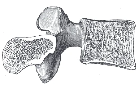
Sagittal section of a lumbar vertebra. Labelled illustration from Wikimedia Commons, used under its open licence.

Figure 5: Sagittal section of a lumbar vertebra.
The vertebral veins are major tributaries that drain the cervical region of the vertebral column. They originate from the suboccipital venous plexus and the internal vertebral venous plexus. The vertebral veins ascend through the transverse foramina of the cervical vertebrae, accompanying the vertebral arteries. They receive contributions from the deep cervical veins and the occipital veins. The vertebral veins terminate by draining into the brachiocephalic veins. This drainage pathway is distinct from the azygos system, which drains the thoracic and lumbar regions. The vertebral veins play a key role in draining the brainstem and cerebellum via connections with the basilar and inferior petrosal sinuses. [1]
The termination of these veins into the brachiocephalic veins is also demonstrated. Understanding this anatomy is essential for interpreting imaging studies and managing conditions involving venous congestion or metastasis.
The innervation of the vertebral column is complex, involving both motor supply to the supporting musculature and cutaneous supply to the overlying skin. However, a critical and often overlooked component is the sensory innervation of the vertebrae themselves, particularly the intervertebral discs and vertebral bodies. This internal sensory network plays a significant role in generating pain signals during pathological conditions.
Figure 6718 Vertebra
Long description
A typical vertebra consists of a body and a vertebral arch. The arch is formed by the paired pedicles and paired laminae. Arising from the vertebral arch are the transverse, spinous, superior articular, and inferior articular processes. The vertebral foramen provides for passage of the spinal cord.

Figure 6: A typical vertebra consists of a body and a vertebral arch.
The basivertebral nerve provides the primary sensory input from the end plates and the marrow space. Understanding this pathway is essential for interpreting back pain that originates from within the bone rather than the muscles or joints.
The basivertebral nerve innervates the end plate subchondral bone. The fibers of this nerve reach the bone marrow along with nutrient arteries. These arteries enter the vertebra through the posterior basivertebral foramen. This anatomical arrangement ensures that the inner structures of the vertebra are monitored for damage. The innervation of the end plate is comparable to that of the peripheral annulus. This similarity suggests a shared mechanism for detecting structural integrity. Furthermore, this innervation is increased in areas of bone damage. This upregulation likely serves as a protective mechanism to limit movement in unstable segments.
Nearly all marrow pain fibers express calcitonin gene-related peptide (CGRP). These fibers also coexpress TrkA and p74 receptors. These receptors are sensitized by nerve growth factor (NGF). This molecular profile makes the marrow fibers highly responsive to inflammatory mediators. These marrow fibers are the first to encounter pathologic processes occurring in the bone space. Consequently, patients can experience bone pain from elevated interosseous pressures even when the pathology is confined to the marrow. This pain can be ameliorated when bone innervation is ablated, such as after vertebroplasty. This clinical observation underscores the importance of the basivertebral nerve in chronic back pain syndromes.
DefinitionBasivertebral Nerve
A sensory nerve that innervates the vertebral body and intervertebral disc end plates, entering via the posterior basivertebral foramen.
| Structure | Innervation Source | Key Receptors | Clinical Relevance |
|---|---|---|---|
| End Plate Subchondral Bone | Basivertebral Nerve | CGRP, TrkA, p74 | Pain in bone damage |
| Bone Marrow | Basivertebral Nerve | CGRP, TrkA, p74 | Pain from interosseous pressure |
| Peripheral Annulus | Similar to End Plate | Information insufficient | Structural integrity detection |
The lymphatic drainage of the vertebral column and vertebrae follows a pattern that reflects the embryological origin and anatomical relations of the spinal structures. Understanding this drainage is critical for tracing the spread of infection or malignancy from the axial skeleton to regional nodal groups.
Lymph from the vertebral bodies and intervertebral discs drains primarily into the deep cervical and thoracic duct systems. The initial lymphatic vessels are located in the periosteum and the surrounding soft tissues. These vessels converge to form larger collecting trunks that accompany the segmental arteries and veins. [1]
Lymph from the thoracic vertebrae follows a more complex route.
Lymph from the thoracic vertebrae follows a more complex route.
Lymph from the thoracic vertebrae follows a more complex route.
The vertebral column forms the posterior axis of the trunk, providing structural support and protection for the spinal cord. It consists of thirty-three vertebrae arranged in five regions: cervical, thoracic, lumbar, sacral, and coccygeal. The spaces between these bones are critical for neural and vascular passage.
Between adjacent vertebral bodies lie the intervertebral discs, which act as shock absorbers and allow for limited movement. Each disc comprises a central nucleus pulposus surrounded by an annulus fibrosus. Lateral to the vertebral bodies are the intervertebral foramina, formed by the notches of adjacent vertebrae. These openings serve as the exit routes for spinal nerves and accompanying blood vessels.
The vertebral foramina of all vertebrae align to form the vertebral (spinal) canal. This bony tunnel houses the spinal cord, meninges, and cerebrospinal fluid. The spinal cord itself is protected by three meningeal layers: the dura mater, arachnoid mater, and pia mater. The subarachnoid space, located between the arachnoid and pia mater, contains cerebrospinal fluid, which cushions the cord against mechanical shock.
Figure 7729 Ligaments of Vertebral Column
Long description
The anterior longitudinal ligament runs the length of the vertebral column, uniting the anterior sides of the vertebral bodies. The supraspinous ligament connects the spinous processes of the thoracic and lumbar vertebrae. In the posterior neck, the supraspinous ligament enlarges to form the nuchal

Figure 7: The anterior longitudinal ligament runs the length of the vertebral column, uniting the anterior sides of the vertebral bodies.
The stability of the column relies on several key ligaments. The anterior longitudinal ligament runs down the anterior surface of the vertebral bodies, preventing hyperextension. The posterior longitudinal ligament lies within the vertebral canal, anterior to the spinal cord, and prevents hyperflexion. Laterally, the ligamenta flava connect the laminae of adjacent vertebrae, helping to maintain upright posture and assist in returning the spine to neutral after flexion. [1]
The cervical vertebrae are smaller and possess transverse foramina for the vertebral arteries. The thoracic vertebrae articulate with ribs and have heart-shaped bodies. The lumbar vertebrae are large and robust, designed to bear weight, and lack rib articulations. The sacrum is a fused triangular bone at the base of the column, articulating with the pelvis. The coccyx, or tailbone, is a small, rudimentary structure at the very bottom.
The vertebral column is assessed using plain radiography, computed tomography (CT), magnetic resonance imaging (MRI), and ultrasound. Plain X-rays provide a quick overview of alignment, curvature, and bony integrity but offer limited soft tissue detail. CT excels in visualising complex bony anatomy, fractures, and degenerative changes such as osteophytes or facet joint arthritis. It provides high-resolution cross-sectional images of the vertebral bodies, pedicles, and posterior elements.
MRI is the modality of choice for evaluating soft tissues, including intervertebral discs, spinal cord, nerve roots, and bone marrow. It is particularly sensitive to inflammatory and degenerative changes. As noted in evidence, Modic type I and II changes adjacent to the end plate are among the most specific of all MRI observations for predicting concordant PD-induced pain. In one prospective study, moderate to severe Modic type I or II end plate abnormalities correlated 100% of the time with positive concordant pain at the adjacent disk. However, the presence of Modic changes are not very sensitive (15 to 65%) to PD-confirmed disk pain. Poor sensitivity may reflect the categorical and subjective techniques used to classify BML, rather than ones that are quantitative and objective. It may also be that innervated end plate damage is not well visualized using standard MRI techniques, because the end plate has a short T2 that shows little signal with pulse sequences that have long echo times. Newer imaging sequences with ultrashort echo times may therefore help discriminate between patients with and without end plate pathologies. [2]
Ultrasound is rarely used for the deep spinal column but can assess superficial structures or guide interventions.
The vertebral bodies are aligned vertically. The intervertebral discs appear hyperintense (bright) due to high water content in the nucleus pulposus. The Modic changes, if present, would appear as signal alterations in the vertebral end plates adjacent to the disc. Schmorl nodes, if present, appear as herniations of disc material into the vertebral body. For example, Schmorl nodes are relatively common in asymptomatic individuals.
Clinical pearlMRI Specificity
Modic changes are highly specific for pain but have poor sensitivity.
| Modality | Best For | Limitations |
|---|---|---|
| X-ray | Alignment, fractures | Poor soft tissue detail |
| CT | Bony detail, complex fractures | Radiation exposure |
| MRI | Soft tissue, discs, marrow | Cost, time, contraindications |
| Ultrasound | Superficial structures, guidance | Limited deep penetration |
The vertebral column provides structural support and protects the spinal cord, but its biomechanical complexity makes it susceptible to specific injury patterns. Understanding the clinical anatomy requires linking structural landmarks to neurological deficits and common pathologies.
Figure 8717 Abnormal Curves of Vertebral Column
Long description
(a) Scoliosis is an abnormal lateral bending of the vertebral column. (b) An excessive curvature of the upper thoracic vertebral column is called kyphosis. (c) Lordosis is an excessive curvature in the lumbar region of the vertebral column.

Figure 8: (a) Scoliosis is an abnormal lateral bending of the vertebral column.
In the cervical section, the C5/C6 and C6/C7 levels are marked. These areas are prone to injury following forcible hyperflexion of the neck, such as in collision accidents or football injuries. The resulting damage produces pain in the neck, shoulder, and upper limb.
Injuries to the vertebral column often involve nerve palsies corresponding to the level of damage. For instance, cervical disc injuries at C5/C6 or C6/C7 affect the upper limb innervation. Lumbar disc herniations at L4/L5 or L5/S1 affect the lower limb, causing sciatica. Surface landmarks such as the spinous processes help clinicians localize these injuries during physical examination.
Deformities of the spine, such as scoliosis and kyphosis, alter the normal curvature and biomechanics of the column. Scoliosis involves a lateral curvature, while kyphosis involves an excessive forward rounding. Osteoporotic fractures are another critical clinical concern, particularly in the elderly. These fractures often occur in the vertebral bodies due to reduced bone density, leading to compression fractures. The clinical anatomy of these conditions requires careful assessment of spinal alignment and neurological status.
Chronic low back pain is a difficult clinical problem to diagnose and treat. While axial back pain is often attributed to sensitized nociceptors within the annulus fibrosus of degenerating disks (annulogenic pain), there is growing evidence that end plate damage may represent a common painful pathology (vertebrogenic pain). The end plates are richly innervated, and damage to these areas can cause significant pain. However, current diagnostic tools do not depict subtle end plate damage that associates with neoinnervation. This underappreciation complicates the identification of the pain generator, which is requisite for optimal treatment. Properly distinguishing between annulogenic and vertebrogenic pain is important for improving patient outcomes. [2]
Clinical pearlClinical Pearl
Sciatica typically results from herniation at L4/L5 or L5/S1, radiating pain down the leg.
| Level | Mechanism | Symptoms |
|---|---|---|
| L4/L5, L5/S1 | Disc herniation | Sciatica, pain radiating down thigh and leg |
| C5/C6, C6/C7 | Forcible hyperflexion | Pain in neck, shoulder, and upper limb |
The vertebral column provides the central axial support for the skeleton and protects the spinal cord. It consists of thirty-three vertebrae grouped into five regions: cervical, thoracic, lumbar, sacral, and coccygeal. Understanding the structural differences between these regions is critical for clinical anatomy and radiological interpretation.
The cervical region comprises seven vertebrae (C1–C7). C1, the atlas, lacks a vertebral body and a spinous process, consisting instead of anterior and posterior arches that support the skull. C2, the axis, is distinguished by the dens (odontoid process), which serves as a pivot for rotation of the atlas. Typical cervical vertebrae (C3–C6) are characterised by small, bifid spinous processes and transverse foramina that transmit the vertebral arteries. The transverse processes are also perforated by these foramina, a key distinguishing feature from thoracic and lumbar vertebrae. [1]
There are twelve thoracic vertebrae (T1–T12). They are distinguished by their articulation with the ribs. Each typical thoracic vertebra has demi-facets on the lateral aspects of the body for rib head articulation and full facets on the transverse processes for rib tubercle articulation (except T11 and T12). The spinous processes are long, slender, and point inferiorly, often overlapping the vertebra below. The vertebral foramen is smaller and more circular than in the cervical region. [1]
The lumbar region contains five large vertebrae (L1–L5). They are designed to bear weight, featuring massive, kidney-shaped bodies and short, thick, blunt spinous processes that project horizontally. The transverse processes are long and arise from both the body and the neural arch. Unlike cervical vertebrae, they lack transverse foramina. The superior articular processes face medially, while the inferior articular processes face laterally, restricting rotation but allowing flexion and extension.
The sacrum is a large, triangular bone formed by the fusion of five sacral vertebrae. It articulates with the ilium at the sacroiliac joints and with L5 above. The sacral canal is the continuation of the vertebral canal. The superior articular process of the sacrum, one of which is found on either side of the superior opening of the sacral canal, articulates with the inferior articular processes from the L5 vertebra. The coccyx consists of four fused rudimentary vertebrae and serves as an attachment point for pelvic floor muscles. [1]
Students frequently confuse the structural composition of the vertebral column with that of the intervertebral disc, leading to errors in understanding degenerative changes. A critical distinction is that the vertebral body contains bone marrow and cortical bone, whereas the disc is fibrocartilaginous. A common error is assuming that the disc remains vascular throughout life. In reality, the nucleus pulposus is avascular in adults, relying on diffusion through the cartilage end plates for nutrition. This reliance on diffusion makes the disc highly susceptible to metabolic stress when end plate permeability declines. [2]
Proteoglycan content decreases from 300 µg/mg at age 2 to 150 µg/mg by age 80. [2]
A third pitfall is misinterpreting the role of collagen in disc degeneration. Students may think collagen increases as the disc ages. However, type I collagen decreases from 0.9 ng/mg to 0.25 ng/mg. This loss of structural integrity is accompanied by an increase in type X collagen, a marker of chondrocyte hypertrophy. This suggests that the cells within the disc are undergoing a transformation similar to that seen in endochondral ossification, which is pathological in this context. [2]
A fourth error is assuming that the cartilage end plates remain permeable during degeneration. In fact, the end plates undergo calcification and thinning with age. This calcification further restricts the diffusion of nutrients to the disc cells. It is unclear whether end plate permeability and vascularity in degenerated disks is sufficient to support increases in cell density and metabolism. This limitation is crucial because it means that the disc cannot easily repair itself once the nutrient supply is compromised. [2]
It is unclear whether end plate permeability and vascularity in degenerated disks is sufficient to support increases in cell density and metabolism (because poor nutrition may have led to the degeneration in the first place). These uncertainties may ultimately limit or prevent the successful extrapolation of disk repair technologies from small animals to man. Students must remember that animal discs often have different vascular patterns than human discs. [2]
If it proves true that disk cell density and ultimately disk degeneration are tightly coupled to end plate permeability, then by definition, disk cellularity cannot be enhanced without commensurate increases in end plate permeability and vascularity. [2]
A seventh error is assuming that gene therapy can easily reverse degeneration. Efforts to increase synthesis rates of existing cells (by gene or growth factor therapy) may create excessive demands on a tenuous nutrition supply and thereby promote cell death. This highlights the importance of the metabolic environment in disc health. [2]
An eighth mistake is overlooking the clinical significance of the cartilage end plate. The end plate is not just a passive barrier; it is an active participant in disc nutrition. Its deterioration is a key driver of disc degeneration. Understanding this helps in interpreting MRI findings, where changes in the end plate signal often precede visible disc height loss.
Knowledge of vertebral column anatomy and disc physiology is essential at the bedside for diagnosing back pain. Clinicians use this understanding to differentiate between mechanical back pain and radicular pain. Radicular pain often results from disc herniation compressing a nerve root. The location of the herniation determines which nerve root is affected. For example, a posterolateral herniation at L4-L5 typically compresses the L5 nerve root. This is because the L5 root exits above the L5 vertebra but below the L4 vertebra, and the herniation pushes into the lateral recess where the L5 root travels.
In the operating theatre, surgeons must navigate the complex anatomy of the spine. They must identify the correct vertebral level before making an incision. This is often done using fluoroscopy or pre-operative imaging. Surgeons must also be aware of the blood supply to the vertebral bodies and discs. The anterior longitudinal ligament provides stability and contains blood vessels that supply the outer annulus fibrosus. During discectomy, surgeons must avoid damaging the nerve roots and thecal sac. Understanding the relations of the disc to the spinal cord and nerve roots is critical to prevent iatrogenic injury.
On imaging, radiologists look for signs of disc degeneration. MRI is the gold standard for evaluating disc pathology. Degenerative changes are visible as decreased signal intensity in the T2-weighted images. This corresponds to the loss of water content and proteoglycans described in the passages. Radiologists also assess the height of the disc space. Loss of height indicates advanced degeneration. They also look for Modic changes, which are signal changes in the vertebral end plates. These changes reflect bone marrow oedema or fatty replacement, which are associated with chronic disc degeneration.
In examinations, medical students are tested on the segmental anatomy of the spine. They must know the number of vertebrae in each region: seven cervical, twelve thoracic, five lumbar, five sacral (fused), and four coccygeal (fused). They must also know the distinguishing features of each type. Cervical vertebrae have transverse foramina for the vertebral arteries. Thoracic vertebrae have facets for rib articulation. Lumbar vertebrae have large bodies to bear weight. Sacral vertebrae are fused to form the sacrum.
Students must also understand the biomechanics of the spine. The intervertebral discs act as shock absorbers. They allow for movement in all planes. The annulus fibrosus resists tensile forces, while the nucleus pulposus resists compressive forces. This distribution of forces is maintained by the hydrostatic pressure within the nucleus. When this pressure is lost due to degeneration, the disc becomes less effective at absorbing shock. This can lead to increased stress on the facet joints, contributing to osteoarthritis.
Furthermore, knowledge of disc physiology is relevant in the context of spinal fusion. Fusion eliminates motion at the fused segment, which increases stress on the adjacent segments. This can accelerate degeneration at the levels above and below the fusion. Understanding the natural history of disc degeneration helps in counselling patients about the risks and benefits of surgery.
Finally, this knowledge is used in physiotherapy and rehabilitation. Exercises are designed to strengthen the core muscles that support the spine. This helps to reduce the load on the intervertebral discs. Patients are advised to maintain good posture to minimize stress on the spine. Understanding the role of proteoglycans and water content in disc health helps in explaining why hydration and nutrition are important for spinal health.
The vertebral column consists of 24 movable vertebrae, the sacrum, and the coccyx. Intervertebral discs are composed of the annulus fibrosus and the nucleus pulposus. The cartilage end plates facilitate nutrient diffusion to the avascular disc. Proteoglycan content decreases from 300 µg/mg at age 2 to 150 µm/mg by age 80. Water content decreases from 78 to 67% with age, reducing disc height and shock absorption. [2] Type I collagen decreases from 0.9 ng/mg to 0.25 ng/mg, weakening structural integrity. Degeneration is associated with calcification and thinning of the cartilage end plates. Poor nutrition and reduced end plate permeability limit disc repair and cell metabolism.
Answer each one in your head first, then open the answer.
What is the primary function of the nucleus pulposus?
Show answerHide answer
Resist compressive forces through hydrostatic pressure.Which collagen type increases during disc degeneration?
Show answerHide answer
Type X collagen.Why is the adult disc avascular?
Show answerHide answer
It relies on diffusion through the cartilage end plates.What happens to proteoglycan content with age?
Show answerHide answer
It decreases from 300 µg/mg at age 2 to 150 µm/mg by age 80.What is a common consequence of disc height loss?
Show answerHide answer
Increased stress on the facet joints.
- Betts JG, Young KA, Wise JA, et al. The Vertebral Column. OpenStax, Anatomy and Physiology 2e (CC BY-NC-SA 4.0). Accessed .
- Lotz JC, Fields AJ, Liebenberg EC. The role of the vertebral end plate in low back pain. Global spine journal 2013;3(3):153-164. doi:10.1055/s-0033-1347298. Accessed .
Figure credits
- Figure 1: Henry Vandyke Carter, via Wikimedia Commons, Public domain, Public domain.
- Figure 2: OpenStax College, via Wikimedia Commons, CC BY 3.0, CC BY 3.0.
- Figure 3: Henry Vandyke Carter, via Wikimedia Commons, Public domain, Public domain.
- Figure 4: OpenStax College, via Wikimedia Commons, CC BY 3.0, CC BY 3.0.
- Figure 5: Henry Vandyke Carter, via Wikimedia Commons, Public domain, Public domain.
- Figure 6: OpenStax College, via Wikimedia Commons, CC BY 3.0, CC BY 3.0.
- Figure 7: OpenStax College, via Wikimedia Commons, CC BY 3.0, CC BY 3.0.
- Figure 8: OpenStax College, via Wikimedia Commons, CC BY 3.0, CC BY 3.0.
Educational content for medical students and doctors. It is not a substitute for clinical judgement or local guidelines.