Gait and applied anatomy of the lower limb
Anatomical structures and biomechanics of the lower limb gait cycle.
The lower limb is divided into three regions: the thigh between the hip and knee joints, the leg between the knee and ankle joints, and the foot distal to the ankle. Each lower limb contains 30 bones. These include the femur, patella, tibia, fibula, seven tarsal bones, five metatarsal bones, and 14 phalanges. The femur is the single bone of the thigh and is the longest and strongest bone of the body. It accounts for approximately one-quarter of a person’s total height. The patella is the kneecap and articulates with the distal femur. The tibia is the larger, weight-bearing bone located on the medial side of the leg. The fibula is the thin bone of the lateral leg. [2]
Figure 1Gray1239
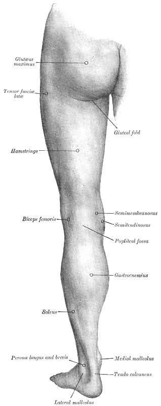
Long description
Back of left lower extremity. Labelled illustration from Wikimedia Commons, used under its open licence.

Figure 1: Back of left lower extremity.
The hip joint is a ball-and-socket joint whose motions are more restricted than at the shoulder to provide greater stability during weight bearing. It is the articulation between the head of the femur and the acetabulum of the hip bone. The acetabulum is the socket portion of the hip joint. This space is deep and has a large articulation area for the femoral head, thus giving stability and weight bearing ability to the joint. The acetabulum is further deepened by the acetabular labrum, a fibrocartilage lip attached to the outer margin of the acetabulum. The surrounding articular capsule is strong, with several thickened areas forming intrinsic ligaments. These ligaments arise from the hip bone, at the margins of the acetabulum, and attach to the femur at the base of the neck. The ligaments are the iliofemoral ligament, pubofemoral ligament, and ischiofemoral ligament, all of which spiral around the head and neck of the femur. The ligaments are tightened by extension at the hip, thus pulling the head of the femur tightly into the acetabulum when in the upright, standing position. [1]
The knee includes three articulations. The femoropatellar joint is between the patella and distal femur. The patella, a sesamoid bone incorporated into the tendon of the quadriceps femoris muscle of the anterior thigh, serves to protect this tendon from rubbing against the distal femur during knee movements. The medial and lateral tibiofemoral joints, between the condyles of the femur and condyles of the tibia, are modified hinge joints that allow for knee extension and flexion. During these movements, the condyles of the femur both roll and glide over the surface of the tibia. As the knee comes into full extension, a slight medial rotation of the femur serves to “lock” the knee into its most stable, weight-bearing position. The reverse motion, a small lateral rotation of the femur, is required to initiate knee flexion. When the knee is flexed, some rotation of the leg is available. [1] [2]
The talocrural joint forms the ankle. It consists of the articulation between the talus bone and the medial malleolus of the tibia, the distal end of the tibia, and the lateral malleolus of the fibula. This is a uniaxial hinge joint that allows only dorsiflexion and plantar flexion of the foot. Gliding motions at the subtalar and intertarsal joints of the foot allow for inversion/eversion of the foot. The ankle joint is supported on the medial side by the deltoid ligament, which prevents side-to-side motions of the talus at the talocrural joint and resists excessive eversion of the foot. The lateral ankle is supported by the anterior and posterior talofibular ligaments and the calcaneofibular ligament. These support the ankle joint and also resist excess inversion of the foot. An inversion ankle sprain, a common injury, will result in injury to one or more of these lateral ankle ligaments. [1]
DefinitionTrendelenburg Gait
A gait abnormality caused by weakness of the hip abductors, leading to pelvic drop on the contralateral side during stance phase.
Fractures of the femoral neck are a common injury in elderly individuals, particularly those with weakened bones due to osteoporosis. This may result from a fall, or it may cause the fall. This can happen as one lower limb is taking a step and all of the body weight is placed on the other limb, causing the femoral neck to break and producing a fall. Any accompanying disruption of the blood supply to the femoral neck or head can lead to necrosis of these areas, resulting in bone and cartilage death. Femoral fractures usually require surgical treatment, after which the patient will need mobility assistance for a prolonged period, either from family members or in a long-term care facility. [1]
| Structure | Location/Function |
|---|---|
| Femur | Thigh bone, longest and strongest |
| Tibia | Medial leg, main weight-bearing bone |
| Fibula | Lateral leg, thin bone |
| Acetabulum | Hip socket, deepened by labrum |
| Talocrural Joint | Ankle, allows dorsiflexion/plantar flexion |
The lower limb joints are specialized for stability and strength rather than the large range of motion seen in the upper limb[1]. This robust design supports body weight during running, jumping, or kicking activities[1]. During these activities, the foot absorbs tremendous impact. When running, the force applied to each foot upon ground contact can be up to 2.5 times your body weight[1]. The bones, joints, ligaments, and muscles of the foot absorb this force, greatly reducing the shock passed superiorly into the lower limb and body[1].
Palpable landmarks begin with the pelvic girdle. The appendicular muscles of the lower body position and stabilize the pelvic girdle, which serves as a foundation for the lower limbs[1]. There is very little movement of the pelvic girdle because of its connection with the sacrum at the base of the axial skeleton[1]. The pelvic girdle has less range of motion because it was designed to stabilize and support the body[1].
Moving distally, the femur presents the patellar surface. Anteriorly, the smooth surfaces of the condyles join together to form a wide groove called the patellar surface, which provides for articulation with the patella bone[1]. The combination of the medial and lateral condyles with the patellar surface gives the distal end of the femur a horseshoe (U) shape[1]. The patella is a sesamoid bone located within a muscle tendon[1]. It articulates with the patellar surface on the anterior side of the distal femur, thereby protecting the muscle tendon from rubbing against the femur[1]. [2]
In the leg, the anterior compartment contains key muscles. The tibialis anterior is a long and thick muscle on the lateral surface of the tibia[1]. The extensor hallucis longus lies deep under it, and the extensor digitorum longus is lateral to it[1]. These muscles contribute to raising the front of the foot when they contract[1]. The fibularis tertius is a small muscle that originates on the anterior surface of the fibula[1]. It is associated with the extensor digitorum longus and sometimes fused to it, but is not present in all people[1]. Thick bands of connective tissue called the superior extensor retinaculum (transverse ligament of the ankle) and the inferior extensor retinaculum hold the tendons of these muscles in place during dorsiflexion[1].
DefinitionSesamoid Bone
A sesamoid bone is a bone embedded within a tendon, such as the patella, which protects the tendon from excessive wear and tear.
The foot arches play a critical role in shock absorption. When weight is applied to the foot, these arches will flatten somewhat, thus absorbing energy[1]. When the weight is removed, the arch rebounds, giving “spring” to the step[1]. The arches also serve to distribute body weight side to side and to either end of the foot[1].
| Muscle | Origin | Insertion | Action | Nerve Supply |
|---|---|---|---|---|
| Tibialis Anterior | Lateral surface of tibia | Medial cuneiform and base of first metatarsal | Dorsiflexion and inversion | Deep fibular nerve |
| Extensor Hallucis Longus | Anterior fibula | Distal phalanx of great toe | Extension of great toe | Deep fibular nerve |
| Extensor Digitorum Longus | Anterior tibia | Dorsal digital expansions of toes 2-5 | Extension of toes | Deep fibular nerve |
| Fibularis Tertius | Anterior fibula | Base of fifth metatarsal | Dorsiflexion and eversion | Deep fibular nerve |
The skeleton is divided into the posterior foot, anterior foot, and the connecting segments that facilitate weight transmission.
The posterior foot comprises seven tarsal bones. The talus sits superiorly, articulating with the distal tibia, the medial malleolus of the tibia, and the lateral malleolus of the fibula to form the ankle joint. Inferiorly, the talus articulates with the calcaneus bone. The sustentaculum tali of the calcaneus helps to support the talus. Anterior to the talus lies the navicular bone. Anterior to the navicular are the medial, intermediate, and lateral cuneiform bones. The cuboid bone is anterior to the calcaneus. [2]
The anterior foot is formed by five metatarsal bones. These elongated bones are numbered 1–5, starting with the medial side of the foot. The first metatarsal bone is shorter and thicker than the others. The second metatarsal is the longest. The base of the metatarsal bone is the proximal end of each metatarsal bone. These articulate with the cuboid or cuneiform bones. The base of the fifth metatarsal has a large, lateral expansion that provides for muscle attachments. This expanded base of the fifth metatarsal can be felt as a bony bump at the midpoint along the lateral border of the foot. The expanded distal end of each metatarsal is the head of the metatarsal bone. The heads of the metatarsal bones also rest on the ground and form the ball (anterior end) of the foot. [2]
The toes contain a total of 14 phalanx bones (phalanges). The toes are numbered 1–5, starting with the big toe (hallux). The big toe has two phalanx bones, the proximal and distal phalanges. The remaining toes all have proximal, middle, and distal phalanges. A joint between adjacent phalanx bones is called an interphalangeal joint. [2]
The foot has a transverse arch, a medial longitudinal arch, and a lateral longitudinal arch. The transverse arch forms the medial-lateral curvature of the mid-foot. It is formed by the wedge shapes of the cuneiform bones and bases (proximal ends) of the first to fourth metatarsal bones. This arch helps to distribute body weight from side to side within the foot, thus allowing the foot to accommodate uneven terrain. [2]
The longitudinal arches run down the length of the foot. The lateral longitudinal arch is relatively flat, whereas the medial longitudinal arch is larger (taller). The longitudinal arches are formed by the tarsal bones posteriorly and the metatarsal bones anteriorly. These arches are supported at either end, where they contact the ground. Posteriorly, this support is provided by the calcaneus bone and anteriorly by the heads (distal ends) of the metatarsal bones. The talus bone, which receives the weight of the body, is located at the top of the longitudinal arches. Body weight is then conveyed from the talus to the ground by the anterior and posterior ends of these arches. Strong ligaments unite the adjacent foot bones to prevent disruption of the arches during weight bearing. On the bottom of the foot, additional ligaments tie together the anterior and posterior ends of the arches. [2]
The talocrural joint of the ankle is supported by several strong ligaments located on the sides of the joint. These ligaments extend from the medial malleolus of the tibia or lateral malleolus of the fibula and anchor to the talus and calcaneus bones. Since they are located on the sides of the ankle joint, they allow for dorsiflexion and plantar flexion of the foot. They also prevent abnormal side-to-side and twisting movements of the talus and calcaneus bones during eversion and inversion of the foot. On the medial side is the broad deltoid ligament. The deltoid ligament supports the ankle joint and also resists excessive eversion of the foot. The lateral side of the ankle has several smaller ligaments. [1]
Additional joints between the tarsal bones of the posterior foot allow for the movements of foot inversion and eversion. Most important for these movements is the subtalar joint, located between the talus and calcaneus bones. The joints between the talus and navicular bones and the calcaneus and cuboid bones are also important contributors to these movements. All of the joints between tarsal bones are plane joints. Together, the small motions that take place at these joints all contribute to the production of inversion and eversion foot motions. [1]
The structural classification of a joint determines its permitted movements. A multiaxial ball-and-socket joint allows significantly greater mobility than a uniaxial hinge joint. However, the total range of motion is restricted by supporting ligaments and muscles. The shoulder joint possesses minimal ligamentous support, granting it a very large range of motion. In contrast, the hip joint movements are restricted by strong ligaments, which reduce its range of motion but confer stability during standing and weight bearing. This stability is crucial because the body’s center of gravity lies within the pelvic area. If the pelvic girdle allowed the same range of motion as the pectoral girdle, walking would expend more energy. The leg muscles compensate for limited versatility with size and power, facilitating stabilization and posture. [1] [3]
The knee joint relies on intrinsic ligaments to resist hyperextension. In the fully extended position, both collateral ligaments are taut, stabilizing the knee and preventing side-to-side or rotational motions between the femur and tibia. The posterior articular capsule is thickened by intrinsic ligaments that help to resist knee hyperextension. Inside the knee, the anterior and posterior cruciate ligaments anchor inferiorly to the tibia at the intercondylar eminence. These ligaments run diagonally upward to attach to the inner aspect of a femoral condyle, forming an X-shape. The posterior cruciate ligament is the stronger ligament, supporting the knee when it is flexed and weight bearing, as when walking downhill. [1]
The vertebral column joints include symphysis joints formed by intervertebral discs and plane synovial joints between articular processes. These provide limited motions that sum to produce flexion, extension, lateral flexion, and rotation. The cervical region allows all these motions, while the lumbar region has considerable extension, flexion, and lateral flexion, but rotation is prevented. The atlanto-occipital joint allows for flexion and extension of the head, while the atlantoaxial joint provides for rotation of the head. [1]
Stretching of the ligaments supporting the longitudinal arches can lead to pain, particularly in individuals who stand for long periods or walk long distances[1]. Prolonged or excessive stretching can result in gradual lengthening of the supporting ligaments, with subsequent depression or collapse of the longitudinal arches, particularly on the medial side of the foot[1]. This condition is called pes planus[1]. [2]
Deep to the gluteus maximus, the piriformis, obturator internus, obturator externus, superior gemellus, inferior gemellus, and quadratus femoris laterally rotate the femur at the hip. [3]
Deep fascia in the thigh separates it into medial, anterior, and posterior compartments. The muscles in the medial compartment of the thigh are responsible for adducting the femur at the hip. Along with the adductor longus, adductor brevis, adductor magnus, and pectineus, the strap-like gracilis adducts the thigh in addition to flexing the leg at the knee. [3]
Similar to the thigh muscles, the muscles of the leg are divided by deep fascia into compartments, although the leg has three: anterior, lateral, and posterior. The foot also has intrinsic muscles, which originate and insert within it. These muscles primarily provide support for the foot and its arch, and contribute to movements of the toes. The principal support for the longitudinal arch of the foot is a deep fascia called plantar aponeurosis, which runs from the calcaneus bone to the toes. Inflammation of this tissue is the cause of “plantar fasciitis,” which can affect runners. The intrinsic muscles of the foot consist of two groups. The dorsal group includes only one muscle, the extensor digitorum brevis. The second group is the plantar group, which consists of four layers, starting with the most superficial. [3] [1]
DefinitionCompartment Syndrome
A condition where increased pressure within a closed fascial compartment compromises circulation and function of the tissues within that space.
The thigh muscles that move the femur, tibia, and fibula are divided into medial, anterior, and posterior compartments. The medial compartment includes the adductors, pectineus, and the gracilis. The anterior compartment comprises the quadriceps femoris, quadriceps tendon, patellar ligament, and the sartorius. The quadriceps femoris is made of four muscles: the rectus femoris, the vastus lateralis, the vastus medius, and the vastus intermedius, which together extend the knee. The posterior compartment of the thigh includes the hamstrings: the biceps femoris, semitendinosus, and the semimembranosus, which all flex the knee. [3]
The muscles of the leg that move the foot and toes are divided into anterior, lateral, superficial- and deep-posterior compartments. The anterior compartment includes the tibialis anterior, the extensor hallucis longus, the extensor digitorum longus, and the fibularis (peroneus) tertius. The lateral compartment houses the fibularis (peroneus) longus and the fibularis (peroneus) brevis. The superficial posterior compartment has the gastrocnemius, soleus, and plantaris. The deep posterior compartment has the popliteus, tibialis posterior, flexor digitorum longus, and flexor hallucis longus. [3]
| Compartment | Key Muscles |
|---|---|
| Thigh Medial | Adductors, Pectineus, Gracilis |
| Thigh Anterior | Quadriceps Femoris, Sartorius |
| Thigh Posterior | Hamstrings (Biceps Femoris, Semitendinosus, Semimembranosus) |
| Leg Anterior | Tibialis Anterior, Extensor Digitorum Longus |
| Leg Lateral | Fibularis Longus, Fibularis Brevis |
| Leg Posterior (Superficial) | Gastrocnemius, Soleus, Plantaris |
| Leg Posterior (Deep) | Popliteus, Tibialis Posterior, Flexor Digitorum Longus |
Understanding these anatomical relationships is vital for recognising how muscle imbalances or injuries affect walking mechanics. [3]
The muscles of the lower limb are grouped by compartment, each with specific origins, insertions, actions, and nerve supplies. The anterior compartment of the thigh contains the quadriceps femoris group, which extends the leg at the knee. The rectus femoris originates from the anterior inferior iliac spine and inserts into the patella via the quadriceps tendon. The vastus lateralis, vastus medialis, and vastus intermedius originate from the femur and also insert into the patella. All four muscles are innervated by the femoral nerve. The sartorius, a band-like muscle, extends from the anterior superior iliac spine to the medial side of the proximal tibia, flexing and laterally rotating the thigh. [3]
The posterior compartment of the thigh includes the hamstring group: biceps femoris, semitendinosus, and semimembranosus. These muscles flex the leg and extend the thigh. They originate from the ischial tuberosity and insert into the tibia and fibula. The biceps femoris inserts into the head of the fibula, while the semitendinosus and semimembranosus insert into the medial tibia. These muscles are innervated by the tibial division of the sciatic nerve. [3]
The lateral compartment of the leg includes the fibularis longus and fibularis brevis, which evert the foot and plantar flex the ankle. They originate from the fibula and insert into the medial cuneiform and first metatarsal (longus) and the fifth metatarsal (brevis). These muscles are innervated by the superficial fibular nerve. [1]
The posterior compartment of the leg contains superficial and deep muscles. The superficial muscles, gastrocnemius and soleus, insert into the calcaneal tendon (Achilles tendon), which attaches to the calcaneus. These muscles plantar flex the ankle. The gastrocnemius originates from the femur, while the soleus originates from the tibia and fibula. The deep muscles include the popliteus, flexor digitorum longus, flexor hallucis longus, and tibialis posterior. These muscles flex the toes and invert the foot. They are innervated by the tibial nerve. [3]
| Muscle | Origin | Insertion | Action | Nerve Supply |
|---|---|---|---|---|
| Rectus Femoris | Anterior inferior iliac spine | Patella (via quadriceps tendon) | Extends leg, flexes thigh | Femoral nerve |
| Vastus Lateralis | Femur (greater trochanter, linea aspera) | Patella (via quadriceps tendon) | Extends leg | Femoral nerve |
| Vastus Medialis | Femur (intertrochanteric line, linea aspera) | Patella (via quadriceps tendon) | Extends leg | Femoral nerve |
| Vastus Intermedius | Femur (anterior and lateral surfaces) | Patella (via quadriceps tendon) | Extends leg | Femoral nerve |
| Sartorius | Anterior superior iliac spine | Medial proximal tibia | Flexes, laterally rotates thigh | Femoral nerve |
| Biceps Femoris | Ischial tuberosity | Head of fibula | Flexes leg, extends thigh | Tibial nerve |
| Semitendinosus | Ischial tuberosity | Medial proximal tibia | Flexes leg, extends thigh | Tibial nerve |
| Semimembranosus | Ischial tuberosity | Medial condyle of tibia | Flexes leg, extends thigh | Tibial nerve |
| Gastrocnemius | Femur (medial and lateral condyles) | Calcaneus (via Achilles tendon) | Plantar flexes ankle | Tibial nerve |
| Soleus | Tibia and fibula | Calcaneus (via Achilles tendon) | Plantar flexes ankle | Tibial nerve |
| Fibularis Longus | Fibula | Medial cuneiform and first metatarsal | Everts foot, plantar flexes ankle | Superficial fibular nerve |
| Fibularis Brevis | Fibula | Fifth metatarsal | Everts foot, plantar flexes ankle | Superficial fibular nerve |
| Popliteus | Lateral condyle of femur | Posterior tibia | Laterally rotates tibia | Tibial nerve |
| Flexor Digitorum Longus | Tibia | Distal phalanges of toes 2-5 | Flexes toes | Tibial nerve |
| Flexor Hallucis Longus | Fibula | Distal phalanx of big toe | Flexes big toe | Tibial nerve |
| Tibialis Posterior | Tibia, fibula, interosseous membrane | Navicular and other tarsal bones | Inverts foot, plantar flexes ankle | Tibial nerve |
The arterial supply to the lower limb is a continuous conduit that ensures adequate perfusion during the dynamic demands of gait. Understanding this vascular architecture is fundamental, as any compromise can lead to significant functional impairment or tissue loss. The primary vessel is the femoral artery, which serves as the main trunk for the thigh and proximal leg. It originates from the external iliac artery as it passes beneath the inguinal ligament. The course of the femoral artery is initially anterior to the hip joint, running through the femoral triangle. Within this triangle, it lies lateral to the femoral vein and medial to the femoral nerve. This spatial relationship is critical for clinical procedures such as arterial puncture. [1]
As the artery passes under the sartorius muscle, it enters the adductor canal. The main femoral artery continues distally, piercing the adductor hiatus to become the popliteal artery. [1]
The popliteal artery is the direct continuation of the femoral artery. It courses through the popliteal fossa, lying deep to the popliteal fascia. Its relations are vital for surgical safety. The artery lies anterior to the knee joint capsule. It is accompanied by the tibial nerve and popliteal veins. The popliteal artery gives off several branches. These include the superior medial and lateral genicular arteries, which anastomose around the knee. This anastomosis is crucial for collateral circulation if the femoral artery is obstructed. The inferior genicular arteries also arise here. They supply the distal femur and proximal tibia.
Distally, the popliteal artery terminates by dividing into the anterior and posterior tibial arteries. The anterior tibial artery passes anterior to the interosseous membrane. It runs down the anterior compartment of the leg. It supplies the extensor muscles of the leg and foot. The posterior tibial artery descends behind the tibia. It runs deep to the soleus muscle. This vessel supplies the posterior compartment of the leg. It gives rise to the fibular artery. The fibular artery supplies the lateral compartment of the leg.
| Vessel | Origin | Course | Supply Region |
|---|---|---|---|
| Femoral Artery | External Iliac | Through femoral triangle | Thigh anterior compartment |
| Profunda Femoris | Femoral | Lateral branch | Deep thigh muscles |
| Popliteal Artery | Femoral (via hiatus) | Popliteal fossa | Knee joint and anastomosis |
| Anterior Tibial | Popliteal | Anterior interosseous membrane | Anterior leg and dorsum foot |
| Posterior Tibial | Popliteal | Deep posterior leg | Posterior leg and sole foot |
The arterial supply must withstand the mechanical stresses of walking. The vessels are protected by surrounding muscles and fascia. The anastomoses around the knee provide redundancy. This network ensures blood flow even if one major vessel is compromised. The distal pulses, such as the dorsalis pedis and posterior tibial pulses, are key clinical indicators of this supply. Palpation of these pulses assesses the integrity of the entire arterial tree.
The venous system of the lower limb is divided into superficial and deep compartments, which communicate via perforating veins. This arrangement is critical for understanding gait mechanics and the applied anatomy of the lower limb, as pressure changes during muscle contraction drive blood flow.
Perforating veins connect the two systems. The relationship between the veins and the bones is clear. The relationship between the veins and the muscles is clear. The relationship between the veins and the nerves is clear.
The superficial veins lie in the subcutaneous tissue and do not accompany arteries. They drain the skin and subcutaneous tissues. The great saphenous vein is the longest vein in the body. It arises from the medial marginal vein of the foot. It ascends anterior to the medial malleolus. It runs up the medial aspect of the leg. It passes posterior to the medial condyle of the femur. It ascends along the medial aspect of the thigh. It terminates in the femoral vein. The small saphenous vein arises from the lateral marginal vein of the foot. It ascends posterior to the lateral malleolus. It runs up the posterior aspect of the leg. It terminates in the popliteal vein. Perforating veins connect the superficial and deep systems. They contain valves that prevent backflow. [1]
The deep veins accompany the arteries of the same name. They lie deep to the fascia. They drain the muscles and bones. The tibial veins accompany the posterior tibial artery. The peroneal veins accompany the peroneal artery. The anterior tibial veins accompany the anterior tibial artery. These veins unite to form the popliteal vein. The popliteal vein passes through the adductor hiatus. It becomes the femoral vein. The femoral vein ascends in the femoral triangle. It passes under the inguinal ligament. It becomes the external iliac vein. The deep veins have numerous valves. They are subject to the muscle pump. [3]
Perforating veins pass through the deep fascia. They connect the superficial and deep systems. They contain one-way valves. The valves direct blood from superficial to deep veins. This prevents venous hypertension in the superficial system. incompetence of these valves can lead to varicose veins. The Cockett perforators are located in the lower leg. The Boyd perforator is located in the thigh.
| Feature | Superficial Veins | Deep Veins |
|---|---|---|
| Location | Subcutaneous tissue | Accompany arteries |
| Valves | Fewer | Numerous |
| Drainage | Skin and subcutaneous tissue | Muscles and bones |
| Termination | Femoral or popliteal vein | External iliac vein |
The obturator nerve travels medially to supply the adductor compartment. The sciatic nerve emerges from the greater sciatic foramen, passing deep to the gluteus maximus. It typically divides into the tibial and common fibular nerves. The tibial nerve runs down the posterior compartment, innervating the hamstrings and all muscles of the leg and foot responsible for plantar flexion and inversion. The common fibular nerve wraps around the neck of the fibula, a site vulnerable to injury, before dividing into superficial and deep branches. These supply the anterior and lateral compartments of the leg, controlling dorsiflexion and eversion. Understanding this distribution is critical for localising lesions based on motor weakness or sensory loss patterns during gait analysis. [1]
DefinitionFemoral Nerve
The femoral nerve supplies the anterior compartment of the thigh, enabling knee extension and hip flexion, with cutaneous branches to the anterior thigh and medial leg.
DefinitionSciatic Nerve
The sciatic nerve is the largest nerve in the body, supplying the posterior thigh and all structures below the knee, including plantar flexors and invertors.
| Nerve | Primary Muscles | Primary Action |
|---|---|---|
| Femoral | Quadriceps femoris | Knee extension |
| Obturator | Adductor longus, brevis, magnus | Hip adduction |
| Tibial | Gastrocnemius, soleus | Plantar flexion |
| Common Fibular | Tibialis anterior | Dorsiflexion |
The adductor longus, adductor brevis, and adductor magnus can both medially and laterally rotate the thigh depending on the placement of the foot. The adductor longus flexes the thigh, whereas the adductor magnus extends it. The pectineus adducts and flexes the femur at the hip as well. The pectineus is located in the femoral triangle, which is formed at the junction between the hip and the leg and also includes the femoral nerve, the femoral artery, the femoral vein, and the deep inguinal lymph nodes. [3]
Exam trapAdductor Function
The adductor magnus extends the thigh, while the adductor longus flexes it; this distinction is vital for understanding hip mechanics during the stance phase of gait.
The lymphatic drainage of the lower limb is a critical component of lower limb anatomy, particularly in the context of gait and applied anatomy. Understanding this drainage is essential for diagnosing and managing conditions such as lymphoedema, which can significantly impact mobility and quality of life.
The superficial lymphatics of the lower limb are divided into two main groups: the medial and lateral groups. The medial group drains the skin and subcutaneous tissues of the medial aspect of the leg and foot. These vessels accompany the great saphenous vein and terminate in the superficial inguinal lymph nodes. The lateral group drains the skin and subcutaneous tissues of the lateral aspect of the leg and foot. These vessels accompany the small saphenous vein and terminate in the popliteal lymph nodes.
The deep lymphatics of the lower limb accompany the deep veins of the leg and foot. These vessels drain the muscles, joints, and bones of the lower limb. The deep lymphatics of the leg terminate in the popliteal lymph nodes, while those of the thigh terminate in the deep inguinal lymph nodes.
The inguinal lymph nodes are divided into superficial and deep groups. The superficial inguinal lymph nodes are located in the subcutaneous tissue of the groin and are divided into horizontal and vertical groups. The horizontal group drains the skin and subcutaneous tissues of the lower abdominal wall, buttocks, and external genitalia. The vertical group drains the skin and subcutaneous tissues of the lower limb. The deep inguinal lymph nodes are located deep to the fascia lata and drain the deep structures of the lower limb.
The popliteal lymph nodes are located in the popliteal fossa and drain the deep structures of the leg and foot. These nodes receive lymph from the superficial lymphatics of the lateral aspect of the leg and foot, as well as from the deep lymphatics of the leg.
Disruption of the lymphatic drainage of the lower limb can lead to lymphoedema, a condition characterized by swelling of the affected limb. Lymphoedema can be primary, due to congenital abnormalities of the lymphatic system, or secondary, due to damage to the lymphatic system from surgery, radiation, or infection. Early diagnosis and management of lymphoedema are essential to prevent complications such as skin infections and impaired mobility.
The lower limb is a complex mechanical unit where bony architecture, muscular leverage, and neurovascular supply intersect to facilitate weight-bearing and propulsion. Understanding the spatial relationships within the thigh, leg, and foot is essential for interpreting gait abnormalities and localising lesions. The structures are organised into compartments defined by deep fascia, each containing specific muscles, vessels, and nerves.
The apex of the triangle points distally towards the adductor hiatus. The base is formed by the inguinal ligament. The medial border is defined by the adductor longus muscle. The lateral border is formed by the sartorius muscle. Within this triangle, from lateral to medial, lie the femoral nerve, femoral artery, and femoral vein. The femoral nerve lies lateral to the artery and provides motor innervation to the anterior compartment muscles. The femoral artery is the direct continuation of the external iliac artery. It passes deep to the inguinal ligament and gives off branches such as the deep femoral artery. The femoral vein lies medial to the artery and receives the great saphenous vein. Posterior to these vessels lies the iliopsoas muscle, which acts as a hip flexor. This arrangement ensures that the neurovascular bundle is protected by the surrounding muscular and fascial structures. [1]
The leg is divided into anterior, lateral, and posterior compartments by the interosseous membrane and deep fascia. The anterior compartment contains the tibialis anterior, extensor digitorum longus, and extensor hallucis longus muscles. These muscles are supplied by the deep fibular nerve. They are responsible for dorsiflexion of the foot and extension of the toes. The lateral compartment contains the fibularis longus and brevis muscles, supplied by the superficial fibular nerve. These muscles evert the foot. The posterior compartment is divided into superficial and deep layers. The superficial layer contains the gastrocnemius, soleus, and plantaris muscles, which form the triceps surae. These muscles are supplied by the tibial nerve and are responsible for plantar flexion. The deep posterior compartment contains muscles such as the tibialis posterior, flexor digitorum longus, and flexor hallucis longus. These muscles assist in plantar flexion and inversion of the foot. The tibial nerve and posterior tibial vessels run through this compartment. [1]
The foot contains numerous small bones and joints that contribute to the arches. The medial longitudinal arch is supported by the tibialis posterior tendon. The lateral longitudinal arch is supported by the fibularis longus tendon. The transverse arch is maintained by the interosseous muscles. These arches act as shock absorbers during gait. The plantar fascia provides additional support. The neurovascular supply to the foot is derived from the tibial and fibular nerves. The tibial nerve divides into the medial and lateral plantar nerves. These nerves supply the intrinsic muscles of the foot. The blood supply is provided by the dorsalis pedis and plantar arteries. Understanding these relationships allows for precise localisation of pathology. [1]
DefinitionFemoral Triangle
A triangular space in the upper thigh bounded by the inguinal ligament, adductor longus, and sartorius, containing the femoral nerve, artery, and vein from lateral to medial.
| Muscle | Origin | Insertion | Action | Nerve Supply |
|---|---|---|---|---|
| Tibialis Anterior | Lateral condyle of tibia | Medial cuneiform and base of 1st metatarsal | Dorsiflexion and inversion | Deep fibular nerve |
| Extensor Digitorum Longus | Lateral condyle of tibia | Dorsal digital expansions of toes 2-5 | Extension of toes | Deep fibular nerve |
| Extensor Hallucis Longus | Middle part of fibula | Base of distal phalanx of great toe | Extension of great toe | Deep fibular nerve |
Understanding the lower limb requires correlating surface landmarks with deep structures visible on imaging. This section outlines the appearance of key anatomical features across different modalities, focusing on the structures relevant to gait and applied anatomy.
The femoral head is spherical and articulates with the lunate surface of the acetabulum. Adjacent to the femoral head lies the acetabular labrum, a fibrocartilaginous rim that deepens the socket. The femoral neck extends laterally and superiorly from the shaft, connecting the head to the trochanters. The greater trochanter is a large, palpable prominence on the lateral aspect, serving as an attachment for the gluteus medius and minimus. The femoral shaft descends vertically, with the linea aspera running along its posterior surface.
DefinitionAcetabulum
The cup-shaped cavity of the pelvis that articulates with the head of the femur to form the hip joint.
On plain X-ray, the joint space represents the cartilage thickness. Normal hip joint space is approximately 3 to 4 mm. Narrowing indicates cartilage loss. The femoral head must remain congruent with the acetabulum. Subchondral sclerosis appears as increased bone density beneath the cartilage. Cysts may form in the subchondral bone. The femoral neck angle is typically 125 degrees in adults. Deviation suggests dysplasia or deformity.
| Structure | Appearance on X-ray | Clinical Significance |
|---|---|---|
| Joint Space | Radiolucent gap | Cartilage thickness; narrowing indicates arthritis |
| Femoral Head | Spherical radiopacity | Congruence with acetabulum is critical |
| Greater Trochanter | Lateral bony prominence | Attachment for abductor muscles |
| Lesser Trochanter | Medial bony prominence | Insertion of iliopsoas tendon |
MRI provides superior soft tissue contrast. It visualises the labrum, ligaments, and marrow. The ligamentum teres connects the femoral head to the acetabular notch. On T2-weighted images, fluid appears bright. Synovial fluid in the joint capsule is normal. Pathological effusion appears as a large bright collection. The labrum appears as a dark triangular structure. Tears appear as high signal intensity extending into the labrum.
Ultrasound is dynamic and useful for guiding injections. It visualises the iliopsoas tendon as it passes over the anterior hip capsule. The tendon appears hypoechoic with a fibrillar pattern. The femoral nerve lies lateral to the artery. The femoral vein is medial. These structures are visible in the femoral triangle. The femoral triangle is bounded by the inguinal ligament superiorly, the sartorius laterally, and the adductor longus medially. The contents from lateral to medial are Nerve, Artery, Vein, Empty space, Lymphatics.
CT arthrography involves injecting contrast into the joint. It highlights labral tears and loose bodies. The contrast outlines the labrum and joint capsule. It is more sensitive than standard MRI for intra-articular pathology. The radiation dose is higher than plain X-ray but lower than multiple CT scans.
NoteImaging Choice
Use X-ray for bone alignment and joint space. Use MRI for soft tissue and marrow pathology. Use ultrasound for dynamic assessment and guidance.
The lower limb functions as a kinetic chain, where precise anatomical relationships dictate gait mechanics and clinical outcomes. Understanding surface landmarks and nerve pathways is essential for diagnosing injuries and planning interventions.
The femur is the single bone of the thigh. Its rounded head articulates with the acetabulum of the hip bone to form the hip joint. The narrow neck joins inferiorly with the greater and lesser trochanters. Passing between these bony expansions are the intertrochanteric line on the anterior femur and the larger intertrochanteric crest on the posterior femur. On the posterior shaft of the femur is the gluteal tuberosity proximally and the linea aspera in the mid-shaft region. The expanded distal end consists of three articulating surfaces: the medial and lateral condyles, and the patellar surface. The outside margins of the condyles are the medial and lateral epicondyles. The adductor tubercle is on the superior aspect of the medial epicondyle. [2]
During initial flexion from a fully extended position, the knee “unlocks” by the tibia internally rotating. The knee is well constructed for weight bearing in its extended position, but is vulnerable to injuries associated with hyperextension, twisting, or blows to the medial or lateral side of the joint, particularly while weight bearing. The knee joint has multiple ligaments that provide support, particularly in the extended position. Outside of the articular capsule, located at the sides of the knee, are two extrinsic ligaments. The fibular collateral ligament (lateral collateral ligament) is on the lateral side and spans from the lateral epicondyle of the femur to the head of the fibula. The tibial collateral ligament (medial collateral ligament) of the medial knee runs from the medial epicondyle of the femur to the medial tibia. As it crosses the knee, the tibial collateral ligament is firmly attached on its deep side to the articular capsule and to the medial meniscus, an important factor when considering knee injuries. [1]
| Ligament | Origin | Insertion | Clinical Significance |
|---|---|---|---|
| Fibular collateral | Lateral epicondyle of femur | Head of fibula | Resists varus stress |
| Tibial collateral | Medial epicondyle of femur | Medial tibia | Attached to medial meniscus |
The posterior half of the foot is formed by seven tarsal bones. The most superior bone is the talus. This has a relatively square-shaped, upper surface that articulates with the tibia and fibula to form the ankle joint. Three areas of articulation form the ankle joint: The superomedial surface of the talus bone articulates with the medial malleolus of the tibia, the top of the talus articulates with the distal end of the tibia, and the lateral side of the talus articulates with the lateral malleolus of the fibula. Inferiorly, the talus articulates with the calcaneus (heel bone), the largest bone of the foot, which forms the heel. Body weight is transferred from the tibia to the talus to the calcaneus, which rests on the ground. The medial calcaneus has a prominent bony extension called the sustentaculum tali (“support for the talus”) that supports the medial side of the talus bone. [2]
Clinical pearlWeight Transfer
Body weight is transferred from the tibia to the talus to the calcaneus, which rests on the ground.
Injuries to these structures often result from specific mechanisms. For instance, blows to the medial or lateral side of the knee joint can damage the respective collateral ligaments.
Surface projection of vessels and nerves is critical for procedures. The femoral nerve, artery, and vein lie in the femoral triangle, with the nerve lateral to the artery. The popliteal vessels lie deep to the popliteal fascia. The tibial nerve runs posterior to the medial malleolus. The common fibular nerve winds around the neck of the fibula, making it susceptible to injury. Understanding these relationships allows clinicians to perform safe injections and assess neurological deficits accurately.
The knee joint relies on a complex arrangement of ligaments to maintain stability during weight-bearing activities. Understanding the specific role of each ligament is crucial for diagnosing injuries. The tibial collateral ligament, located on the medial side, and the fibular collateral ligament, on the lateral side, are extrinsic ligaments. These structures primarily serve to resist hyperextension or rotation of the extended knee joint. This means that valgus or varus stresses in extension are checked by these collateral ligaments. [1]
In contrast, the intracapsular ligaments provide stability during flexion. The anterior cruciate ligament and posterior cruciate ligament span between the tibia and the inner aspects of the femoral condyles. This is a key point for clinical examination, as anterior drawer tests assess its integrity. The posterior cruciate ligament prevents anterior sliding of the femur, thus supporting the knee when it is flexed and weight bearing. This function is critical when descending stairs or squatting. [1]
The medial and lateral menisci are articular discs located between the femoral and tibial condyles. They provide padding and improve the fit between the bones. This cushioning effect distributes load and reduces friction. Injury to the menisci often results from twisting forces on a flexed knee. [1]
Exam trapCommon Trap
Do not confuse the function of the cruciate ligaments. The ACL prevents anterior translation of the tibia relative to the femur, while the PCL prevents posterior translation. Remember that the PCL supports the knee when flexed and weight bearing.
DefinitionCruciate Ligaments
The anterior and posterior cruciate ligaments are intracapsular structures that span between the tibia and the inner aspects of the femoral condyles.
Clinical pearlMeniscus Function
The menisci are articular discs that provide padding and improve the fit between the bones. They are essential for load distribution.
NoteCollateral Ligaments
The tibial and fibular collateral ligaments are extrinsic. They resist hyperextension or rotation of the extended knee joint.
DefinitionTibial Collateral Ligament
This ligament is located on the medial side of the knee. It works with the fibular collateral ligament to stabilize the joint against rotational forces.
DefinitionFibular Collateral Ligament
Located on the lateral side, this ligament resists hyperextension or rotation of the extended knee joint. It is distinct from the iliotibial band.
Exam trapACL Function
It does not primarily prevent anterior sliding of the femur; that is the role of the PCL.
Clinical pearlPCL Function
The posterior cruciate ligament prevents anterior sliding of the femur, thus supporting the knee when it is flexed and weight bearing.
NoteIntracapsular Location
The cruciate ligaments are intracapsular but extrasynovial. They are located within the joint capsule but outside the synovial membrane.
DefinitionKnee Stability
Stability is provided by extrinsic collaterals in extension and intrinsic cruciates in flexion. The menisci improve the fit between the bones.
Students frequently confuse the functional mechanics of the knee with those of the ankle, leading to errors in clinical reasoning. The knee is not a simple hinge; it is a complex joint involving three articulations within a single capsule: the femoropatellar joint, and the medial and lateral tibiofemoral joints. A critical error is assuming the knee allows rotation when fully extended. In reality, the knee must “screw home” to fully extend, meaning the tibia externally rotates to achieve proper alignment, and rotation is only available when the knee is flexed. Another common mistake involves the hip joint’s stability. Students often overlook that the hip’s freedom of movement is offset by a loss of stability due to a lack of structural support, unlike the ankle which is a uniaxial hinge joint allowing only dorsiflexion and plantar flexion. The ankle’s stability comes from the square-shaped superior aspect of the talus being firmly held by the medial and lateral malleoli, preventing side-to-side motion. [1]
A frequent anatomical error concerns the ligaments of the elbow. The medial elbow is supported by the ulnar collateral ligament, while the radial collateral ligament supports the lateral side, preventing side-to-side movements and resisting hyperextension. Students often mistake the annular ligament’s role; it surrounds the head of the radius to hold it in place at the proximal radioulnar joint, a pivot joint allowing rotation during pronation and supination. Similarly, at the hip, the ligament of the head of the femur (ligamentum teres) is intracapsular but normally slack, providing no significant joint support, though it carries an important artery to the femoral head. Confusing this with the strong extrinsic ligaments of the TMJ is a trap; the TMJ ligament anchors the mandible to the skull between the base of the skull and the lingula on the medial side of the mandibular ramus. [1]
Exam trapKnee Rotation
Rotation is only possible when the knee is flexed. Full extension locks the joint via external tibial rotation.
This knowledge is applied at the bedside to assess joint integrity and muscle function. In the gluteal region, the psoas major and iliacus form the iliopsoas, while the gluteus maximus, medius, and minimus extend and abduct the femur[1]. The tensor fascia lata forms the iliotibial tract with the gluteus maximus, and lateral rotators include the piriformis, obturator internus, and quadratus femoris[1]. On the medial thigh, the adductor longus, brevis, and magnus adduct and medially rotate the thigh, while the pectineus adducts and flexes the femur[1]. Understanding these actions helps in diagnosing gait abnormalities, such as a Trendelenburg gait resulting from gluteus medius weakness. [3]
In the operating theatre, precise anatomical landmarks are vital. The proximal tibia features the intercondylar eminence between the condyles and the tibial tuberosity anteriorly, which is continuous with the anterior border. The posterior proximal tibia has the curved soleal line. At the distal tibia, the medial malleolus is the bony expansion, and the fibular notch is the groove on the lateral side. These landmarks guide surgical approaches to the knee and ankle. For the elbow, the lateral epicondyle of the humerus is the origin of the radial collateral ligament, which blends into the annular ligament encircling the radial head. This relationship is crucial in radial head fractures. [3]
On imaging, the ankle’s uniaxial nature is evident. The talus articulates with the inferior tibia, carrying body weight, while its sides are held by the malleoli. The TMJ’s complex motion involves the condyle and articular disc gliding forward from the mandibular fossa onto the articular tubercle during mouth opening. This forward and downward motion is distinct from the hinge motion of the shoulder, which is a ball-and-socket joint formed by the humeral head and glenoid cavity. The shoulder’s lack of structural support explains its high dislocation rate compared to the hip. [1]
Knee Joint Mechanics
- Assess Knee PositionIs the knee flexed or extended?
- Question: Extended?
Yes
- Screw Home MechanismTibia externally rotates for alignment. No rotation allowed.
No
- Flexed PositionRotation is available. Rolling and gliding occur.
In examinations, students must distinguish between joint types. The atlanto-occipital joint allows flexion and extension (shaking head “yes”) due to its U-shaped curvature. The proximal radioulnar joint is a pivot joint for forearm rotation. The hip joint attaches the legs to the axial skeleton via the pelvic girdle[1]. The knee is the largest joint, functioning primarily as a hinge but with rotational capacity when flexed. The ankle is a uniaxial hinge joint for dorsiflexion and plantar flexion.
| Structure | Key Feature |
|---|---|
| Hip Joint | Ball-and-socket; high mobility, low stability. |
| Knee Joint | Three articulations; screws home to extend. |
| Ankle Joint | Uniaxial hinge; talus held by malleoli. |
| Elbow | Hinge joint; ulnar/radial collateral ligaments. |
| TMJ | Gliding and hinge; condyle glides forward. |
The knee consists of three articulations within one capsule and requires external tibial rotation to fully extend. The hip joint offers the greatest range of motion but lacks structural stability, increasing dislocation risk. The ankle is a uniaxial hinge joint where the talus is stabilized by the medial and lateral malleoli. The elbow’s collateral ligaments prevent side-to-side movement and resist hyperextension. The TMJ involves both hinge and gliding motions, with the condyle moving onto the articular tubercle. [1] The proximal radioulnar joint is a pivot joint allowing forearm rotation via the annular ligament. The iliopsoas extends from the psoas major and iliacus to flex the femur at the hip. The ligamentum teres of the hip is intracapsular but slack, carrying an artery to the femoral head.
Answer each one in your head first, then open the answer.
What motion does the U-shaped curvature of the atlanto-occipital joint allow?
Show answerHide answer
Flexion and extension of the headWhich ligament anchors the mandible to the skull at the TMJ?
Show answerHide answer
The extrinsic ligament spanning the base of the skull and the lingulaWhat motion is permitted at the knee when it is fully extended?
Show answerHide answer
No rotation; the tibia externally rotates to screw homeWhich bones form the ankle joint?
Show answerHide answer
The talus, tibia, and fibula
- Betts JG, Young KA, Wise JA, et al. Anatomy of Selected Synovial Joints. OpenStax, Anatomy and Physiology 2e (CC BY-NC-SA 4.0). Accessed .
- Betts JG, Young KA, Wise JA, et al. Bones of the Lower Limb. OpenStax, Anatomy and Physiology 2e (CC BY-NC-SA 4.0). Accessed .
- Betts JG, Young KA, Wise JA, et al. Appendicular Muscles of the Pelvic Girdle and Lower Limbs. OpenStax, Anatomy and Physiology 2e (CC BY-NC-SA 4.0). Accessed .
Figure credits
Educational content for medical students and doctors. It is not a substitute for clinical judgement or local guidelines.