Blood supply and venous drainage: general principles
General principles of vascular anatomy, circulation circuits, and hemodynamics.
Arteries are thick-walled vessels that carry blood away from the heart. Veins are thinner-walled vessels that return blood to the heart. Capillaries are microscopic vessels where gas and nutrient exchange occurs. These structures form the basis of the two distinct but linked circuits in the human circulation called the pulmonary and systemic circuits. The pulmonary circuit transports blood to and from the lungs, where it picks up oxygen and delivers carbon dioxide for exhalation. The systemic circuit typically transports oxygenated blood to most tissues of the body and returns relatively deoxygenated blood and carbon dioxide to the heart. [2]
Arteries and veins transport blood in two distinct circuits: the systemic circuit and the pulmonary circuit. Systemic arteries provide blood rich in oxygen to the body’s tissues. The blood returned to the heart through systemic veins has less oxygen, since much of the oxygen carried by the arteries has been delivered to the cells. In contrast, in the pulmonary circuit, arteries carry blood low in oxygen exclusively to the lungs for gas exchange. Pulmonary veins then return freshly oxygenated blood from the lungs to the heart to be pumped back out into systemic circulation. [3]
The right ventricle pumps deoxygenated blood into the pulmonary trunk, which leads toward the lungs and bifurcates into the left and right pulmonary arteries. These vessels in turn branch many times before reaching the pulmonary capillaries, where gas exchange occurs: Carbon dioxide exits the blood and oxygen enters. The pulmonary trunk arteries and their branches are the only arteries in the post-natal body that carry relatively deoxygenated blood. Highly oxygenated blood returning from the pulmonary capillaries in the lungs passes through a series of vessels that join together to form the pulmonary veins—the only post-natal veins in the body that carry highly oxygenated blood. [2]
Once gas exchange is completed, oxygenated blood flows from the pulmonary capillaries into a series of pulmonary venules that eventually lead to a series of larger pulmonary veins. Four pulmonary veins, two on the left and two on the right, return blood to the left atrium. At this point, the pulmonary circuit is complete. The pulmonary veins conduct blood into the left atrium, which pumps the blood into the left ventricle, which in turn pumps oxygenated blood into the aorta and on to the many branches of the systemic circuit. [1] [2]
Blood flow is the movement of blood through a vessel, tissue, or organ. Blood pressure is the force that blood exerts upon the walls of the blood vessels or chambers of the heart. The components of blood pressure include systolic pressure, which results from ventricular contraction, and diastolic pressure, which results from ventricular relaxation. Pulse pressure is the difference between systolic and diastolic measures, and mean arterial pressure is the “average” pressure of blood in the arterial system, driving blood into the tissues. [4]
Blood flow through vessels can be dramatically influenced by vasoconstriction and vasodilation in their walls. This pressure gradient drives blood back toward the heart. Again, the presence of one-way valves and the skeletal muscle and respiratory pumps contribute to this increased flow. Since approximately 64 percent of the total blood volume resides in systemic veins, any action that increases the flow of blood through the veins will increase venous return to the heart. [3] [4]
DefinitionEnd Artery
An artery that supplies a specific area with no significant anastomoses, meaning occlusion leads to tissue infarction.
DefinitionAnastomosis
A connection between two blood vessels, allowing collateral circulation if one vessel is blocked.
The blood exiting the systemic capillaries is lower in oxygen concentration than when it entered. The capillaries will ultimately unite to form venules, joining to form ever-larger veins, eventually flowing into the two major systemic veins, the superior vena cava and the inferior vena cava, which return blood to the right atrium. The blood in the superior and inferior venae cavae flows into the right atrium, which pumps blood into the right ventricle. This process of blood circulation continues as long as the individual remains alive. [2]
NoteVenous Return
Maintaining vascular tone within the veins prevents the veins from merely distending, dampening the flow of blood, and as you will see, vasoconstriction actually enhances the flow. [4]
| Feature | Pulmonary Circuit | Systemic Circuit |
|---|---|---|
| Origin | Right Ventricle | Left Ventricle |
| Artery Content | Deoxygenated | Oxygenated |
| Vein Content | Oxygenated | Deoxygenated |
| Destination | Lungs | Body Tissues |
Blood vessels are classified by their function and direction of flow relative to the heart. An artery is a blood vessel that carries blood away from the heart, where it branches into ever-smaller vessels. Eventually, the smallest arteries, vessels called arterioles, further branch into tiny capillaries, where nutrients and wastes are exchanged. These capillaries often combine with other vessels exiting capillaries to form venules, which are small blood vessels that typically carry blood to a vein, a larger vessel that returns blood to the heart. [3]
Blood flow refers to the movement of blood through a vessel, tissue, or organ, and is usually expressed in terms of volume of blood per unit of time. It is initiated by the contraction of the ventricles of the heart. Ventricular contraction ejects blood into the major arteries, resulting in flow from regions of higher pressure to regions of lower pressure. As blood encounters smaller arteries and arterioles, then capillaries, then the venules and veins of the venous system, it encounters resistance. [4]
Hydrostatic pressure is the force exerted by a fluid due to gravitational pull, usually against the wall of the container in which it is located. One form of hydrostatic pressure is blood pressure, the force exerted by blood upon the walls of the blood vessels or the chambers of the heart. Blood pressure may be measured in capillaries and veins, as well as the vessels of the pulmonary circulation; however, the term blood pressure without any specific descriptors typically refers to systemic arterial blood pressure—that is, the pressure of blood flowing in the arteries of the systemic circulation. In clinical practice, this pressure is measured in mm Hg and is usually obtained using the brachial artery of the arm. [4]
The relationship between blood volume, blood pressure, and blood flow is intuitively obvious. As blood volume decreases, pressure and flow decrease. As blood volume increases, pressure and flow increase. [4]
DefinitionVenous Reserve
Approximately 21 percent of the venous blood is located in venous networks within the liver, bone marrow, and integument. This volume of blood is referred to as venous reserve. [3]
When blood flow needs to be redistributed to other portions of the body, the vasomotor center located in the medulla oblongata sends sympathetic stimulation to the smooth muscles in the walls of the veins, causing constriction—or in this case, venoconstriction. Less dramatic than the vasoconstriction seen in smaller arteries and arterioles, venoconstriction may be likened to a “stiffening” of the vessel wall. This increases pressure on the blood within the veins, speeding its return to the heart. Through venoconstriction, this “reserve” volume of blood can get back to the heart more quickly for redistribution to other parts of the circulation. [3]
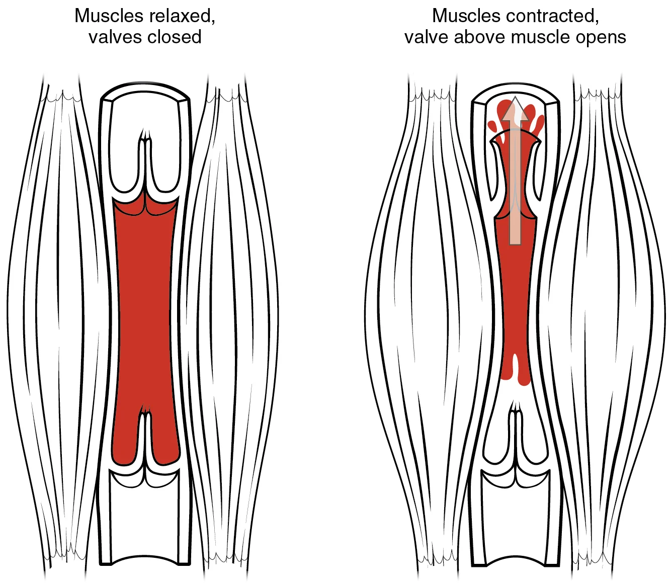
In the venous system, constriction increases blood pressure as it does in arteries; the increasing pressure helps to return blood to the heart. In addition, constriction causes the vessel lumen to become more rounded, decreasing resistance and increasing blood flow. Venoconstriction, while less important than arterial vasoconstriction, works with the skeletal muscle pump, the respiratory pump, and their valves to promote venous return to the heart. [4]
In the arterial system, vasodilation and vasoconstriction of the arterioles is a significant factor in systemic blood pressure. Slight vasodilation greatly decreases resistance and increases flow, whereas slight vasoconstriction greatly increases resistance and decreases flow. In the arterial system, as resistance increases, blood pressure increases and flow decreases. [4]
The internal carotid arteries along with the vertebral arteries are the two primary suppliers of blood to the human brain. Given the central role and vital importance of the brain to life, it is critical that blood supply to this organ remains uninterrupted. Recall that blood flow to the brain is remarkably constant, with approximately 20 percent of blood flow directed to this organ at any given time. When blood flow is interrupted, even for just a few seconds, a transient ischemic attack (TIA), or mini-stroke, may occur, resulting in loss of consciousness or temporary loss of neurological function. In some cases, the damage may be permanent. Loss of blood flow for longer periods, typically between 3 and 4 minutes, will likely produce irreversible brain damage or a stroke, also called a cerebrovascular accident (CVA). [1]
The hepatic portal system carries blood to the liver for processing before it enters circulation. [1]
The right atrium receives all of the systemic venous return. Most of the blood flows into either the superior vena cava or inferior vena cava. If you draw an imaginary line at the level of the diaphragm, systemic venous circulation from above that line will generally flow into the superior vena cava; this includes blood from the head, neck, chest, shoulders, and upper limbs. The exception to this is that most venous blood flow from the coronary veins flows directly into the coronary sinus and from there directly into the right atrium. Beneath the diaphragm, systemic venous flow enters the inferior vena cava, that is, blood from the abdominal and pelvic regions and the lower limbs. [1]
The bronchial arteries follow the same path as the respiratory branches, beginning with the bronchi and ending with the bronchioles. There is considerable, but not total, intermingling of the systemic and pulmonary blood at anastomoses in the smaller branches of the lungs. This may sound incongruous—that is, the mixing of systemic arterial blood high in oxygen with the pulmonary arterial blood lower in oxygen—but the systemic vessels also deliver nutrients to the lung tissue just as they do elsewhere in the body. The mixed blood drains into typical pulmonary veins, whereas the bronchial artery branches remain separate and drain into bronchial veins described later. Each pericardial artery supplies blood to the pericardium, the esophageal artery provides blood to the esophagus, and the mediastinal artery provides blood to the mediastinum. [1]
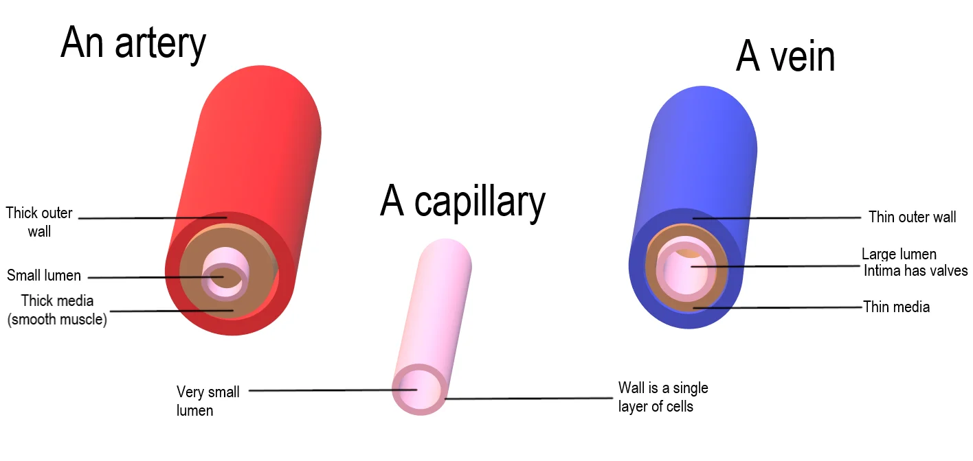
To understand the circulation, one must first distinguish the structural and functional differences between the two main types of vessels. Arteries and arterioles possess thicker walls than veins and venules because they are closer to the heart and receive blood surging at far greater pressure. Each vessel type has a lumen, a hollow passageway through which blood flows. Arteries have smaller lumens than veins, a characteristic that helps maintain the pressure of blood moving through the system. Together, their thicker walls and smaller diameters give arterial lumens a more rounded appearance in cross section than the lumens of veins. [3]
In contrast, a vein is a blood vessel that conducts blood toward the heart. Compared to arteries, veins are thin-walled vessels with large and irregular lumens. Because they are low-pressure vessels, larger veins are commonly equipped with valves that promote the unidirectional flow of blood toward the heart and prevent backflow toward the capillaries caused by the inherent low blood pressure in veins as well as the pull of gravity. In addition to their primary function of returning blood to the heart, veins may be considered blood reservoirs, since systemic veins contain approximately 64 percent of the blood volume at any given time. Their ability to hold this much blood is due to their high capacitance, that is, their capacity to distend (expand) readily to store a high volume of blood, even at a low pressure. The large lumens and relatively thin walls of veins make them far more distensible than arteries; thus, they are said to be capacitance vessels. [3]
Compliance is the ability of any compartment to expand to accommodate increased content. A metal pipe, for example, is not compliant, whereas a balloon is. The greater the compliance of an artery, the more effectively it is able to expand to accommodate surges in blood flow without increased resistance or blood pressure. Veins are more compliant than arteries and can expand to hold more blood. [4]
In the arterial side, the tunica media is thick and prominent, reflecting the high pressure environment. In the venous side, the tunica media is thin, and the lumen is larger and more irregular.
The velocity of blood flow decreases dramatically as the blood moves from arteries to arterioles to capillaries. This slow flow rate allows more time for exchange processes to occur. As blood flows through the veins, the rate of velocity increases, as blood is returned to the heart. [4]
DefinitionCapacitance Vessels
Systemic veins are called capacitance vessels because they contain approximately 64 percent of the blood volume at any given time and can distend readily to store blood at low pressure.
| Feature | Arteries | Veins |
|---|---|---|
| Wall Thickness | Thick | Thin |
| Lumen Size | Smaller, rounded | Larger, irregular |
| Pressure | High | Low |
| Valves | Absent | Present (in larger veins) |
| Blood Volume | Less | ~64% of total volume |
NoteClinical Relevance
Under normal circumstances, blood volume varies little. Low blood volume, called hypovolemia, may be caused by bleeding, dehydration, vomiting, severe burns, or some medications used to treat hypertension. It is important to recognize that other regulatory mechanisms in the body are so effective at maintaining blood pressure that an individual may be asymptomatic until 10–20 percent of the blood volume has been lost. [4]
Understanding the general principles of blood supply and venous drainage is essential for diagnosing and managing ischaemia and thrombosis. Ischaemia occurs when blood flow to a tissue is insufficient to meet metabolic demands, often due to arterial occlusion or reduced perfusion pressure. The heart ejects blood at high pressure, creating a systolic pressure typically around 120 mm Hg, which reflects the arterial pressure resulting from the ejection of blood during ventricular contraction. This high pressure is maintained by the elastic recoil of large arterial walls, which expand during systole and recoil during diastole, ensuring continuous flow even when the heart is relaxed. If these vessels were rigid, resistance would increase, requiring the heart to pump harder to maintain adequate stroke volume and pressure. [4]
Figure 1Artery Vein Capillary Comparison
Long description
Comparison of the structure of the cross section of an artery, capillary and vein.

Figure 1: Comparison of the structure of the cross section of an artery, capillary and vein.
Arteries are vessels that conduct blood away from the heart and possess thick walls to withstand high pressure. Vessels larger than 10 mm in diameter are typically elastic arteries, containing a high percentage of elastic fibers in all three tunics. These elastic fibers allow the vessel to expand as blood passes through and then recoil after the surge has passed. This property is critical for maintaining the pressure gradient that drives blood through the arterial system. The subclavian artery, for example, supplies the arms, chest, and central nervous system, giving rise to the internal thoracic, vertebral, and thyrocervical arteries. [3]
Venous return relies on low-pressure dynamics. By the time blood enters venules, the pressure exerted by heart contractions has diminished significantly. Veins have thinner walls and larger lumens than arteries, allowing more blood to flow with less resistance. Because blood flow becomes sluggish in the extremities due to low pressure and gravity, many veins, particularly in the limbs, contain valves that assist unidirectional flow toward the heart. In the lower limb, the dorsal and plantar venous arches drain into the anterior and posterior tibial veins, which combine to form the popliteal vein. The atria receive this venous blood on a nearly continuous basis, preventing flow from stopping while ventricles contract. [3]
Figure 22114 Skeletal Muscle Vein Pump
Long description
The contraction of skeletal muscles surrounding a vein compresses the blood and increases the pressure in that area. This action forces blood closer to the heart where venous pressure is lower. Note the importance of the one-way valves to assure that blood flows only in the proper direction.

Figure 2: The contraction of skeletal muscles surrounding a vein compresses the blood and increases the pressure in that area.
Ischaemia results from a mismatch between oxygen supply and demand. The total cross-sectional area of capillary beds is far greater than any other vessel type, but blood pressure drops unevenly as it travels through the system. The site of the most precipitous pressure drop and greatest resistance is the arterioles. Thrombosis, the formation of a blood clot within a vessel, can obstruct flow in either arteries or veins. In veins, the sluggish flow and presence of valves make the system susceptible to stasis, a key factor in thrombus formation. The coronary sinus drains the heart muscle into the right atrium, where blood is relatively low in oxygen and high in carbon dioxide before being pumped to the lungs. [4]
| Feature | Arteries | Veins |
|---|---|---|
| Wall Thickness | Thick, with high elastic fiber content | Considerably thinner walls |
| Lumen Diameter | Smaller relative to wall thickness | Correspondingly larger diameter |
| Pressure | High, withstands ejection pressure | Low, pressure diminishes after capillaries |
| Valves | Absent | Present in limbs to assist unidirectional flow |
| Function | Conduct blood away from heart | Return blood to heart against gravity |
Exam trapPressure Drop Location
The greatest resistance and most precipitous drop in blood pressure occur at the arterioles, not the capillaries, despite the capillaries having the largest total cross-sectional area.
The middle cerebral artery territory is clinically significant as it represents the most common site for cerebrovascular accidents.
Venous drainage of the lower limb and pelvis involves the convergence of the external and internal iliac veins. The external iliac vein drains the leg, while the internal iliac vein drains pelvic organs and integument. These combine near the inferior portion of the sacroiliac joint to form the common iliac vein. The common iliac veins unite at the level of L5 to form the inferior vena cava. [1]
The heart’s pumping capacity is substantial; a resting adult heart ejects approximately 70 mL of blood per contraction. Over a 75-year lifespan, the heart contracts nearly 3 billion times. [2]
Structurally, the heart comprises four chambers: two atria acting as receiving chambers and two ventricles serving as primary pumping chambers. The myocardium, the thickest layer, consists of cardiac muscle cells arranged in a complex figure 8 pattern to enhance pumping efficiency. This swirling pattern allows for more effective blood propulsion than a simple linear arrangement. [2]
Atrial contraction contributes approximately 20 percent of ventricular filling, occurring toward the end of ventricular relaxation. The mitral valve guards the opening between the left atrium and ventricle, preventing backflow during ventricular systole. During contraction, the atrioventricular valves close while semilunar valves open to eject blood into the pulmonary trunk and aorta. [2]
Vascular distribution varies by tissue type; adipose tissue contains approximately 200 miles of vessels per pound, whereas skeletal muscle contains more than twice that amount. An individual weighing 150 pounds possesses approximately 60,000 miles of vessels. Gaining 10 pounds adds between 2000 and 4000 miles of vessels, increasing cardiac workload. [4]
Exam trapCommon Trap
Do not confuse the visceral branches of the thoracic aorta with the parietal branches; visceral branches supply organs like the lungs and esophagus, while parietal branches supply the body wall.
DefinitionKey Definition
Cardiac output is the volume of blood pumped by the heart per minute, calculated as stroke volume multiplied by heart rate.
| Branch Type | Examples | Primary Supply |
|---|---|---|
| Visceral | Bronchial arteries | Lungs and visceral pleura |
| Visceral | Esophageal arteries | Esophagus |
| Visceral | Pericardial arteries | Pericardium |
| Parietal | Intercostal arteries | Body wall |
Venous Convergence
- StartLower body venous return
- Question: Which vein drains the leg?
Yes
- External iliac vein
No
- Internal iliac vein
- Combine at sacroiliac jointForms common iliac vein
- L5 LevelCommon iliac veins form IVC
Students frequently struggle with the fundamental direction of flow and the structural differences between arterial and venous systems. A primary error is assuming that increased pressure always increases flow in all vessels. In the arterial system, if you increase pressure (afterload) and cardiac function does not compensate, blood flow will actually decrease. Conversely, in the venous system, increased pressure does not decrease flow; instead, it actually increases flow. This distinction is critical because venous pressure is normally relatively low, requiring atrial pressure during diastole to approach zero to allow blood to flow back into the heart. [4]
Another common mistake is confusing the layers of the vessel wall. The tunica media is generally the thickest layer in arteries, but it is much thicker in arteries than it is in veins. Students often overlook that the tunica media consists of layers of smooth muscle supported by connective tissue primarily made up of elastic fibers, most of which are arranged in circular sheets. A related error is misunderstanding the function of the endothelium. It is not merely a passive boundary; it is physiologically critical for regulating capillary exchange and altering blood flow. Damage to this endothelial lining and exposure of blood to the collagenous fibers beneath is one of the primary causes of clot formation. [3]
Naming conventions also cause confusion. An artery or vein can change names as it passes an anatomical landmark, much like a street changing names at an intersection. For example, the left subclavian artery becomes the axillary artery as it passes through the body wall, and then becomes the brachial artery as it flows into the upper arm. Students often fail to recognize that anastomoses, where two blood vessels that previously branched reconnect, are especially common in veins. These venous anastomoses help maintain blood flow even when one vessel is blocked or narrowed. While there are important arterial anastomoses in the brain, the venous system relies heavily on these connections for collateral flow. [1]
Finally, students misinterpret the timing of coronary circulation. They often assume it is continuous, but it is not; rather, it cycles, reaching a peak when the heart muscle is relaxed and nearly ceasing while it is contracting[1]. This is because the heart is a remarkable pump composed largely of cardiac muscle cells that are incredibly active throughout life, requiring a dedicated, complex, and extensive coronary circulation[1]. The need for this blood supply is even greater than for a typical cell due to the critical and nearly ceaseless activity of the heart[1]. [2]
Exam trapPressure and Flow
In arteries, increased pressure decreases flow if the heart does not compensate; in veins, increased pressure increases flow. [4]
| Layer | Composition | Function |
|---|---|---|
| Tunica intima | Simple squamous epithelium (endothelium) and connective tissue | Lining; damage leads to clot formation |
| Tunica media | Smooth muscle and elastic fibers in circular sheets | Thickest in arteries; controls lumen diameter |
| Tunica adventitia | Connective tissue | Outer support layer |
Understanding these principles is vital at the bedside when assessing circulation. Clinicians must recognize that the aorta is the largest artery in the body, arising from the left ventricle and descending to the abdominal region. It bifurcates at the level of the fourth lumbar vertebra into the two common iliac arteries. The aorta consists of the ascending aorta, the aortic arch, and the descending aorta. The ascending aorta moves in a superior direction for approximately 5 cm and ends at the sternal angle. At the base of the aorta is the aortic semilunar valve that prevents backflow of blood into the left ventricle while the heart is relaxing. [2] [1]
In the operating theatre, knowledge of the atrioventricular septum is essential. It is marked by the presence of four openings that allow blood to move from the atria into the ventricles and from the ventricles into the pulmonary trunk and aorta[1]. Located in each of these openings between the atria and ventricles is a valve, a specialized structure that ensures one-way flow of blood[1]. The valves between the atria and ventricles are known generically as atrioventricular valves[1]. The valves at the openings that lead to the pulmonary trunk and aorta are known generically as semilunar valves[1]. When the right ventricle contracts, it ejects blood into the pulmonary trunk, which branches into the left and right pulmonary arteries that carry it to each lung[1]. At the base of the pulmonary trunk is the pulmonary semilunar valve that prevents backflow from the pulmonary trunk[1]. [2]
On imaging, radiologists trace the course of the abdominal aorta. After crossing through the diaphragm at the aortic hiatus, the thoracic aorta is called the abdominal aorta. This vessel remains to the left of the vertebral column and is embedded in adipose tissue behind the peritoneal cavity. It formally ends at approximately the level of vertebra L4, where it bifurcates to form the common iliac arteries. Before this division, the abdominal aorta gives rise to several important branches. Understanding the relationship between the aorta and the vertebral column helps in identifying landmarks during CT angiography. [1]
In examinations, students are tested on the ability to calculate resistance in the vascular system. While normally this value is extremely difficult to measure, it can be calculated from known relationships involving pressure and flow. The concept of afterload is frequently tested, where increased arterial pressure decreases flow if cardiac function does not compensate. Students must also be able to describe the role of endothelins, local chemicals released by the endothelium that can constrict the smooth muscle within the walls of the vessel to increase blood pressure. [3]
Venous Return Logic
- Blood in veinsPressure is normally relatively low
- Question: Is atrial pressure low?
Yes
- Blood flows to heartAtrial pressure approaches zero during diastole
No
- Flow impededIncreased venous pressure actually increases flow
Clinical pearlCoronary Timing
Coronary circulation peaks when the heart muscle is relaxed and nearly ceases while it is contracting[1].
- The aorta bifurcates at the level of the fourth lumbar vertebra into the common iliac arteries.
- The tunica media is the thickest layer in arteries and is much thicker than in veins.
- In the venous system, increased pressure increases flow, unlike in the arterial system.
- Coronary circulation cycles, peaking during heart relaxation and ceasing during contraction[1].
- The endothelium releases endothelins that constrict smooth muscle to increase blood pressure.
- The abdominal aorta remains to the left of the vertebral column and ends at L4.
- Venous anastomoses are common and help maintain flow when a vessel is blocked.
- The ascending aorta moves superiorly for approximately 5 cm and ends at the sternal angle.
Answer each one in your head first, then open the answer.
What happens to arterial blood flow if afterload increases and the heart does not compensate?
Show answerHide answer
Blood flow will actually decrease.At which vertebral level does the abdominal aorta bifurcate?
Show answerHide answer
It bifurcates at the level of the fourth lumbar vertebra.Which layer of the vessel wall is generally the thickest in arteries?
Show answerHide answer
The tunica media is generally the thickest layer in arteries.What is the function of the pulmonary semilunar valve?
Show answerHide answer
It prevents backflow from the pulmonary trunk[1].When does coronary circulation reach its peak?
Show answerHide answer
It reaches a peak when the heart muscle is relaxed[1].
- Betts JG, Young KA, Wise JA, et al. Circulatory Pathways. OpenStax, Anatomy and Physiology 2e (CC BY-NC-SA 4.0). Accessed .
- Betts JG, Young KA, Wise JA, et al. Heart Anatomy. OpenStax, Anatomy and Physiology 2e (CC BY-NC-SA 4.0). Accessed .
- Betts JG, Young KA, Wise JA, et al. Structure and Function of Blood Vessels. OpenStax, Anatomy and Physiology 2e (CC BY-NC-SA 4.0). Accessed .
- Betts JG, Young KA, Wise JA, et al. Blood Flow, Blood Pressure, and Resistance. OpenStax, Anatomy and Physiology 2e (CC BY-NC-SA 4.0). Accessed .
Figure credits
Educational content for medical students and doctors. It is not a substitute for clinical judgement or local guidelines.