The lymphatic system: general anatomy
The lymphatic system drains interstitial fluid and filters pathogens through vessels nodes and ducts
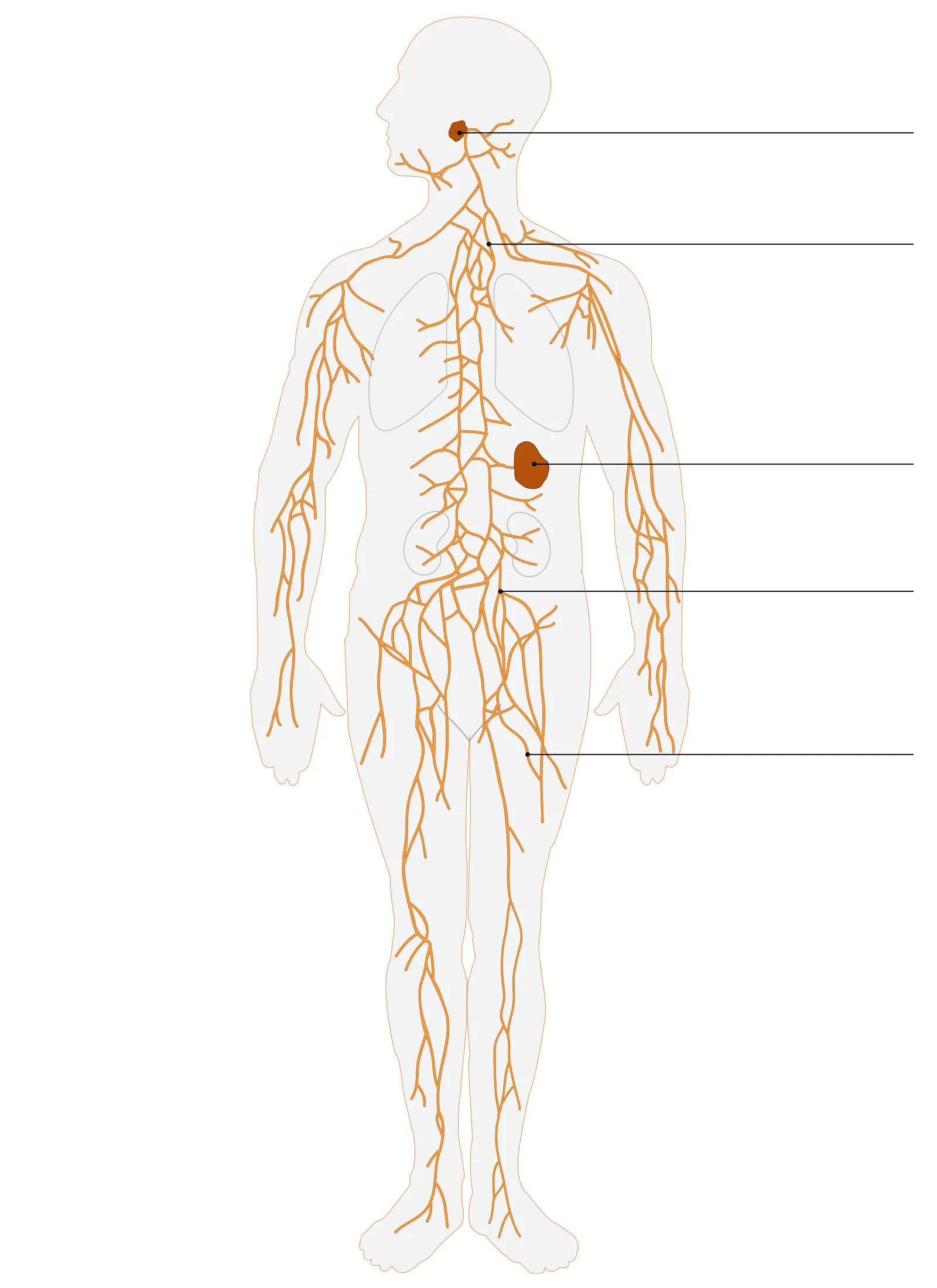
Figure 1Lymphatic system
Long description
Lymphatic system. Labelled anatomical illustration from Wikimedia Commons, used under its open licence.

Figure 1: Lymphatic system.
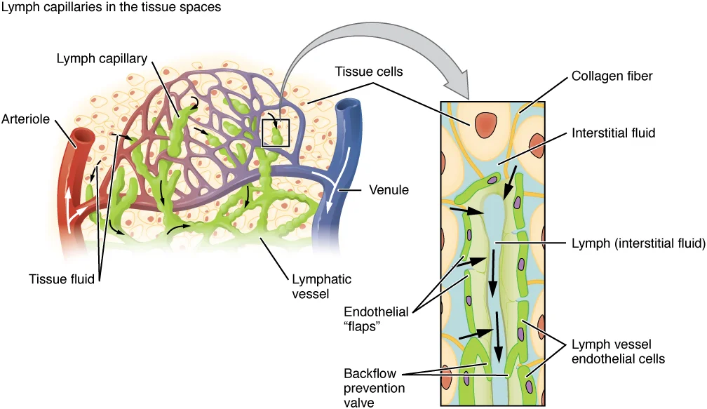
Lymphatic capillaries, also called terminal lymphatics, are blind-ended vessels where interstitial fluid enters the lymphatic system to become lymph[1]. They are located in almost every tissue, interlaced among arterioles and venules in soft connective tissues, but are absent from the central nervous system, bone marrow, bones, teeth, and the cornea[1]. Capillaries merge into larger lymphatic vessels that have a three-tunic structure similar to veins and contain one-way semi-lunar valves spaced close together, giving the vessels a beaded appearance[1]. Lymph is not actively pumped by the heart; it moves by skeletal muscle contraction, body movements, and breathing, with valves ensuring flow toward the heart[1].
Figure 22202 Lymphatic Capillaries
Long description
Lymphatic capillaries are interlaced with the arterioles and venules of the cardiovascular system. Collagen fibers anchor a lymphatic capillary in the tissue (inset). Interstitial fluid slips through spaces between the overlapping endothelial cells that compose the lymphatic capillary.

Figure 2: Lymphatic capillaries are interlaced with the arterioles and venules of the cardiovascular system.
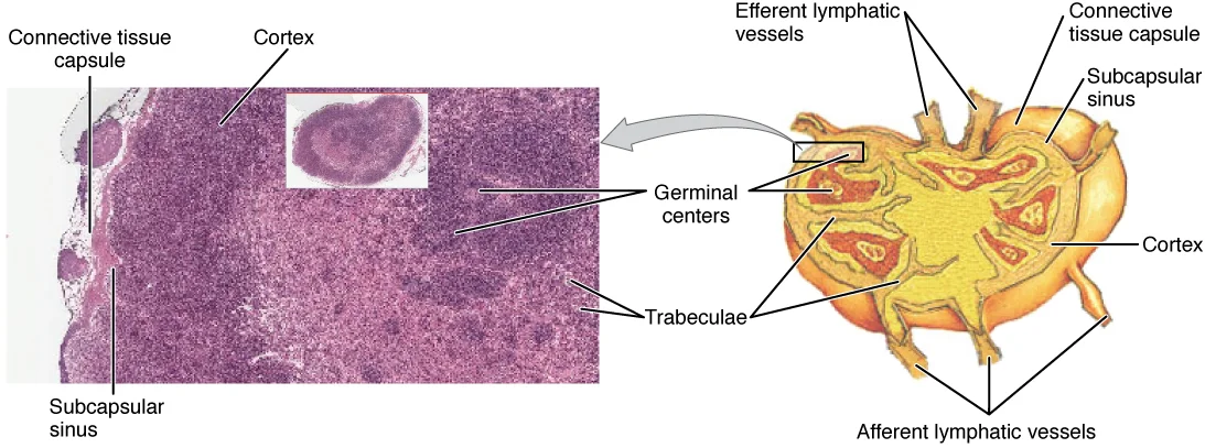
Lymph nodes are small, bean-shaped organs found throughout the lymphatic system, with humans having about 500–600 nodes[1]. They are commonly located near the groin, armpits, neck, chest, and abdomen[1]. Each node is surrounded by a tough connective tissue capsule with trabeculae extending inward to create compartments[1]. Reticular fibers laid down by fibroblasts provide additional structural support[1]. Lymph enters via afferent lymphatic vessels into the subcapsular sinus, which contains dendritic cells, macrophages, and reticular fibers[1]. The cortex contains lymphoid follicles with germinal centers of rapidly dividing B cells surrounded by T cells and accessory cells[1]. The medulla consists of medullary cords of B cells and plasma cells, and medullary sinuses where lymph collects before exiting via efferent lymphatic vessels[1].
The overall drainage system is asymmetrical[1]. The right lymphatic duct receives lymph from only the upper right side of the body[1]. The thoracic duct receives lymph from the rest of the body via all remaining lymphatic trunks[1]. Both ducts empty into the circulatory system at the junction of the jugular and subclavian veins in the neck[1]. Superficial lymphatics of the skin follow venous routes, while deep lymphatic vessels of the viscera generally follow arterial paths[1].
Primary lymphoid organs, the bone marrow and thymus gland, are the locations where lymphocytes of the adaptive immune system proliferate and mature[1]. Secondary lymphoid organs are site in which mature lymphocytes congregate to mount immune responses. Many immune system cells use the lymphatic and circulatory systems for transport throughout the body to search for and then protect against pathogens.
Lymph from the upper right body quadrant drains into the right lymphatic duct[1]. Lymph from the left side of the head, neck, thorax, left upper limb, and the entire body below the diaphragm drains into the thoracic duct[1]. Superficial vessels accompany veins; deep vessels accompany arteries[1].
Bacteria infecting interstitial fluid are taken up by lymphatic capillaries and transported to regional lymph nodes[1]. Dendritic cells and macrophages within nodes internalize and kill many pathogens, removing them from the body[1]. Nodes are also sites of adaptive immune responses mediated by T cells, B cells, and accessory cells[1]. This drainage pattern means infections and malignant cells can spread along lymphatic channels to regional nodes.
Lymphoedema results when lymphatic drainage is impaired, causing accumulation of protein-rich interstitial fluid. The system normally returns about 3 litres of fluid daily to the bloodstream[1]; obstruction leads to chronic swelling, tissue fibrosis, and increased infection risk.
The spleen is sometimes called the “filter of the blood” because of its extensive vascularization and the presence of macrophages and dendritic cells that remove microbes and other materials from the blood, including dying red blood cells. [1]
| Vessel type | Wall structure | Primary role | Example |
|---|---|---|---|
| Lymphatic capillary | Single overlapping endothelial cells; anchored by collagen filaments | Fluid entry from interstitium | Dermal capillary, lacteal [1] |
| Collecting vessel | Endothelium plus smooth muscle and valves | Active transport of lymph | Superficial and deep lymphatics |
| Lymphatic trunk | Large calibre, thick wall, multiple valves | Confluence of collecting vessels | Jugular, subclavian, bronchomediastinal, lumbar, intestinal |
| Lymphatic duct | Largest calibre, opens into venous system | Return of lymph to blood | Right lymphatic duct, thoracic duct |
Collecting vessels are grouped into superficial and deep sets. Superficial lymphatics run in the subcutaneous tissue alongside veins; deep lymphatics accompany arteries. Both sets converge into lymphatic trunks. The principal trunks are paired or single: the jugular trunks drain the head and neck; the subclavian trunks drain the upper limbs; the bronchomediastinal trunks drain the thorax; the lumbar trunks drain the lower abdomen, pelvis, and lower limbs; and the intestinal trunk drains the gut.
| Trunk | Laterality | Structures drained |
|---|---|---|
| Jugular trunk | Left and right | Head, neck |
| Subclavian trunk | Left and right | Upper limb |
| Bronchomediastinal trunk | Left and right | Thoracic viscera, thoracic wall |
| Lumbar trunk | Left and right | Lower abdomen, pelvis, lower limb |
| Intestinal trunk | Single (midline) | Gastrointestinal tract |
Finally, the trunks empty into two main ducts. On the right side, the right lymphatic duct receives lymph from the right side of the head, thorax, and right upper limb and opens into the right subclavian vein. On the left, the larger thoracic duct begins beneath the diaphragm in the cisterna chyli, a sac-like chamber that collects lymph from the left and right lumbar trunks and the intestinal trunk, then ascends to drain into the left subclavian vein. If the system is blocked or destroyed, protein-rich interstitial fluid accumulates in the tissues, producing lymphoedema. [1]
Clinical pearlClinical correlation
The cisterna chyli receives the lipid-rich chyle from the intestinal trunk; obstruction here produces chylous ascites.
Figure 32201 Anatomy of the Lymphatic System
Long description
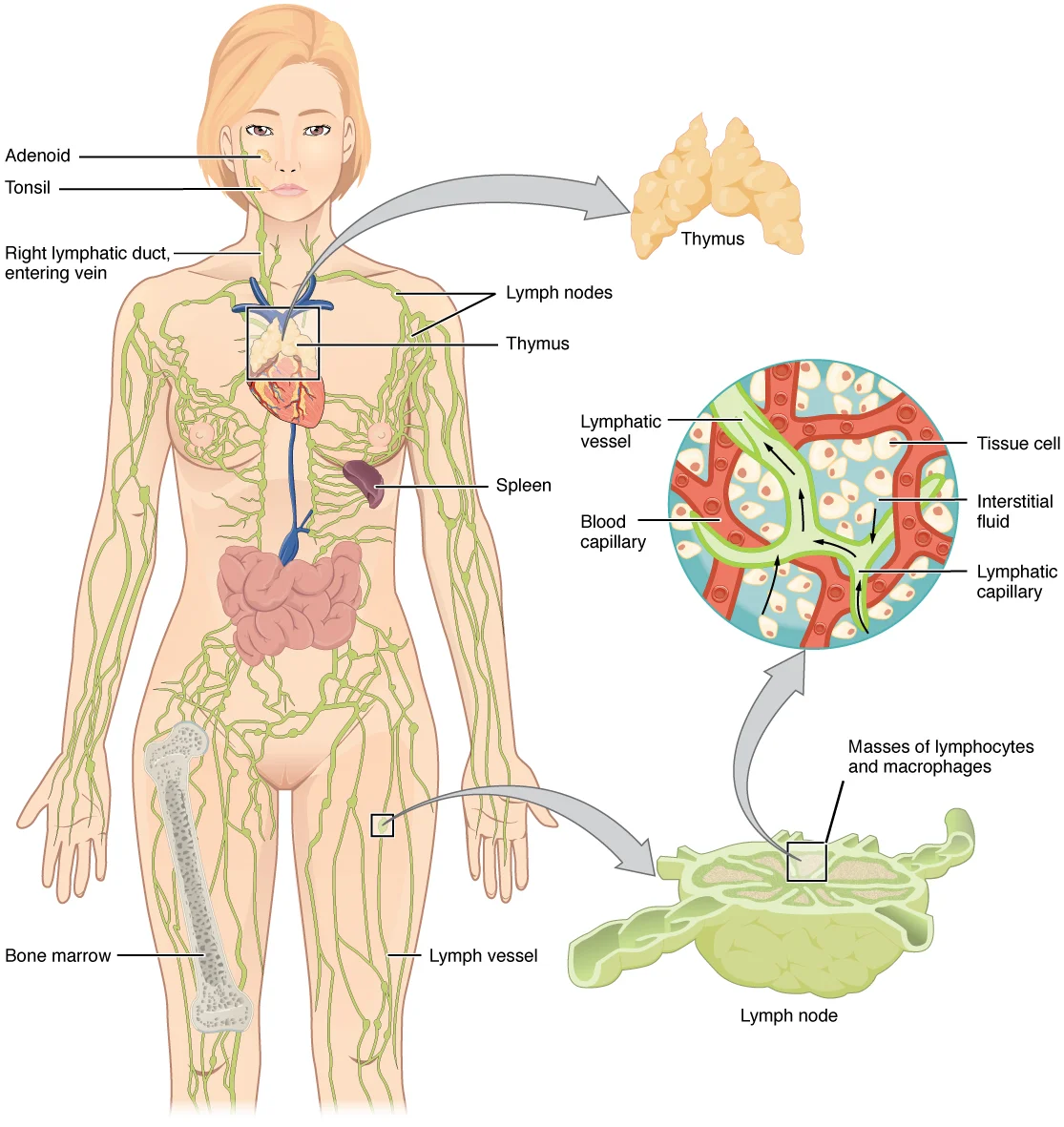
Lymphatic vessels in the arms and legs convey lymph to the larger lymphatic vessels in the torso.

Figure 3: Lymphatic vessels in the arms and legs convey lymph to the larger lymphatic vessels in the torso.
The lymphatic system is a network of vessels, tissues and organs that manages interstitial fluid return, lipid absorption and adaptive immunity. Its cellular agents are lymphocytes, which develop and mature in the primary lymphoid organs before entering the circulation as naïve cells. A naïve lymphocyte is defined as one that has left the primary organ and entered a secondary lymphoid organ; it is fully functional immunologically but has not yet encountered its specific antigen. This distinction underpins the anatomical separation between sites of lymphocyte genesis and sites of immune activation. [1]
In humans the primary lymphoid organs are the bone marrow and the thymus. Bone marrow is the site of haematopoiesis where all lymphoid lineages arise; B lymphocytes complete their maturation there. The thymus, located in the anterior superior mediastinum, receives bone‑marrow‑derived precursors and subjects them to positive and negative selection, yielding a self‑tolerant T‑cell repertoire. Both organs provide a specialised microenvironment of stromal cells, cytokines and cell‑contact signals that direct differentiation.
Secondary lymphoid organs are the anatomical sites where adaptive immune responses are mounted. They include the lymph nodes, the spleen, and lymphoid nodules. All these tissues share common features that support the interaction between antigens and lymphocytes. Lymphocytes recirculate continuously between the blood and the lymph, and they concentrate in secondary lymphoid organs to await antigen encounter. [1]
Lymph nodes are encapsulated, bean‑shaped organs positioned along lymphatic collecting vessels. Afferent lymphatic vessels pierce the convex capsule and drain into the subcapsular sinus. The hilum also admits the arterial supply and venous drainage. The cortex contains primary follicles (naïve B cells) and secondary follicles with germinal centres (activated B cells). The paracortex is rich in high endothelial venules through which naïve T cells enter from blood.
Figure 42207 Structure and Histology of a Lymph Node
Long description
Lymph nodes are masses of lymphatic tissue located along the larger lymph vessels. The micrograph of the lymph nodes shows a germinal center, which consists of rapidly dividing B cells surrounded by a layer of T cells and other accessory cells. LM × 128. (Micrograph provided by the Regents of the Un

Figure 4: Lymph nodes are masses of lymphatic tissue located along the larger lymph vessels.
Figure 5Gray621
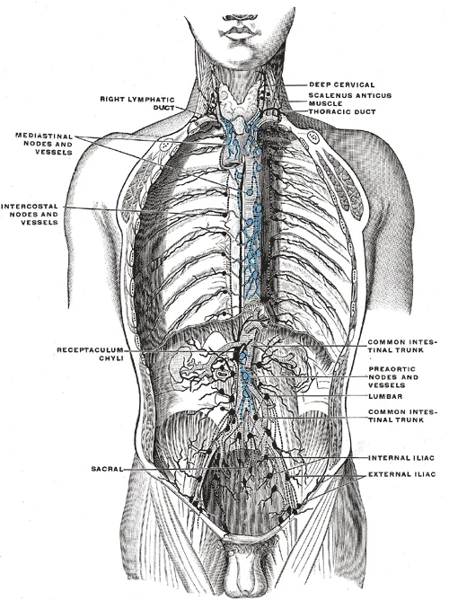
Long description
Deep lymph nodes and vessels of the thorax and abdomen (diagrammatic). Afferent vessels are represented by continuous lines, and efferent and internodular vessels by dotted lines. (Cunningham.)

Figure 5: Deep lymph nodes and vessels of the thorax and abdomen (diagrammatic).
The spleen is the largest secondary lymphoid organ, lying in the left upper quadrant beneath the diaphragm. It is surrounded by a fibrous capsule from which trabeculae extend into the parenchyma. The white pulp forms periarteriolar lymphoid sheaths (PALS) around central arterioles – T‑cell zones – with adjacent B‑cell follicles. The red pulp consists of splenic cords (Billroth’s cords) and venous sinuses that filter aged erythrocytes and blood‑borne antigens. Blood enters via the splenic artery, passes through the white pulp for immune surveillance, then traverses the red pulp before exiting via the splenic vein. [1]
Lymphoid nodules (also called mucosa‑associated lymphoid tissue, MALT) are unencapsulated aggregates of lymphocytes beneath epithelial surfaces. Examples include the palatine, tubal, pharyngeal and lingual tonsils (Waldeyer’s ring), Peyer’s patches in the ileum, and the vermiform appendix. They lack afferent lymphatics; instead they sample antigens directly from the lumen via specialised M cells in the overlying epithelium.
Lymphatic capillaries originate as blind‑ended tubes in tissue spaces, lined by overlapping endothelial cells that act as primary valves. They converge into collecting vessels with smooth muscle, valves and adventitia. Both ducts empty into the venous system at the junction of the internal jugular and subclavian veins.
The illustration depicts the primary lymphoid organs (bone marrow and thymus) at the top, with arrows indicating the egress of naïve lymphocytes into the bloodstream. The lymphatic vascular network is shown as a green mesh draining peripheral tissues, passing through regional lymph nodes (enlarged at neck, axilla, groin) and converging on the thoracic duct and right lymphatic duct. The secondary lymphoid organs – lymph nodes, spleen, and lymphoid nodules (tonsils, Peyer’s patches) – are highlighted in blue. The view is anterior, with the thoracic duct ascending along the vertebral column to drain into the left venous angle.
The capsule is the outermost dense connective tissue layer. Immediately deep lies the subcapsular sinus receiving lymph from afferent vessels (shown entering the convex surface). The cortex contains lymphoid follicles; some display pale germinal centres. The paracortex appears as a diffuse band between cortex and medulla. The medulla shows medullary cords and sinuses converging at the hilum, where the efferent lymphatic vessel exits alongside the artery and vein. The orientation is transverse, with the convex surface uppermost and the hilum at the lower medial aspect.
DefinitionNaïve lymphocyte
A lymphocyte that has matured in a primary lymphoid organ and entered a secondary lymphoid organ but has not yet encountered its specific antigen.
| Category | Examples | Key features |
|---|---|---|
| Primary lymphoid organs | Bone marrow, Thymus | Site of lymphocyte development and maturation; generate naïve lymphocytes |
| Secondary lymphoid organs | Lymph nodes, Spleen, Lymphoid nodules (tonsils, Peyer’s patches, appendix) | Mount adaptive immune responses; concentrate naïve lymphocytes and antigen-presenting cells |
Lymphocyte journey
- MaturationLymphocytes develop in primary lymphoid organs (bone marrow, thymus)
- CirculationNaïve lymphocytes travel in blood and lymph
- HomingCells accumulate in secondary lymphoid organs (lymph nodes, spleen, lymphoid nodules)
- ActivationAntigen encounter initiates adaptive immune response
The lymphatic system serves as the primary conduit for returning interstitial fluid and plasma proteins to the venous circulation, thereby maintaining tissue fluid homeostasis and preventing oedema. Specialised lymphatic capillaries called lacteals in the intestinal villi absorb dietary lipids as chyle and deliver it to the bloodstream via the thoracic duct. Lymph nodes stationed along the lymphatic pathways filter lymph, trap particulate matter, and provide the microenvironment where naïve lymphocytes encounter antigens. [1]
Lymph nodes are the first site of metastatic spread for most epithelial carcinomas; their clinical assessment by palpation, cross‑sectional imaging, and sentinel‑node biopsy underpins cancer staging and therapeutic decision‑making. Lymphadenopathy patterns — localised versus generalised, cervical versus supraclavicular, axillary versus inguinal — narrow the differential diagnosis among infectious, autoimmune, and neoplastic aetiologies. The thoracic duct and right lymphatic duct are vulnerable to iatrogenic injury during neck, thoracic, or upper abdominal surgery; leakage of chyle into the pleural or peritoneal cavity produces chylothorax or chylous ascites, which may require dietary modification, somatostatin analogues, or surgical ligation.
Knowledge of lymphatic watershed zones — anatomical boundaries between adjacent lymphatic territories — explains why lymphoedema after axillary clearance typically affects the arm but spares the forearm, and why inguinal node dissection spares the medial aspect of the leg. The valved, unidirectional flow of lymph dictates that manual lymphatic drainage and compression therapy must direct fluid toward functional nodal basins to be effective. In children, congenital lymphatic malformations such as lymphangioma or intestinal lymphangiectasia present with chylous effusions or protein‑losing enteropathy. During pregnancy, increased venous pressure and hormonal influences can exacerbate pre‑existing lymphatic insufficiency, particularly in the lower limbs. Older adults often have reduced lymphatic contractility and comorbid venous disease, accelerating the onset of chronic oedema. In low‑resource settings, advanced filarial lymphoedema (elephantiasis) remains a major cause of disability, and access to compression garments or specialised physiotherapy is limited, making community‑based hygiene and elevation strategies the cornerstone of management.
Clinical pearlSentinel node concept
The first node draining a tumour site predicts the status of the entire nodal basin; a negative sentinel node obviates complete lymphadenectomy.
| Cause | Typical distribution | Key feature |
|---|---|---|
| Axillary surgery ± radiotherapy | Ipsilateral upper limb | Forearm often spared (watershed) |
| Inguinal/femoral node dissection | Ipsilateral lower limb | Medial leg often spared |
| Filariasis (Wuchereria bancrofti) | Lower limbs, genitalia | Progressive, often bilateral |
| Malignant infiltration | Regional to primary tumour | Hard, fixed nodes |
Approach to peripheral lymphadenopathy
- Palpable node(s) detectedAssess size, consistency, mobility, tenderness, and distribution.
- Question: Localised or generalised?
Localised
- Examine drainage area for infection, inflammation, or malignancyConsider excisional biopsy if >1 cm, hard, fixed, or persistent >4 weeks.
Generalised
- Screen for systemic causesInfections (TB, HIV, EBV), autoimmune (SLE, RA), haematological malignancy.
- The spleen is the largest secondary lymphoid organ, measuring approximately 12 cm (5 in) in length and weighing 150–200 g in adults; it lies in the left upper quadrant beneath the 9th–11th ribs and is attached to the lateral border of the stomach by the gastrosplenic ligament and to the left kidney by the splenorenal ligament. [1]
- Because the spleen lacks a strong fibrous capsule and is highly vascular, it is the abdominal organ most vulnerable to blunt trauma; a fractured 10th or 11th rib often lacerates the splenic capsule, causing life-threatening haemorrhage that may present days after the initial injury. [1]
- Splenic red pulp contains numerous macrophages and dendritic cells that filter blood, removing encapsulated bacteria (especially Streptococcus pneumoniae, Neisseria meningitidis, Haemophilus influenzae), parasitised red cells, and senescent erythrocytes; this function explains the overwhelming post-splenectomy infection (OPSI) risk of 0.5–5 % in asplenic patients.
- The white pulp, organised around central arterioles as periarteriolar lymphoid sheaths (PALS) rich in T cells and follicular zones rich in B cells, is the principal site for mounting adaptive immune responses to blood-borne antigens.
- The oropharynx contains two distinct sets of tonsils, the palatine and lingual tonsils. A palatine tonsil is one of a pair of structures located laterally in the oropharynx in the area of the fauces, while the lingual tonsil is located at the base of the tongue. [2]
- Palatine and lingual tonsils are composed of lymphoid tissue that trap and destroy pathogens entering the body through the oral or nasal cavities. [2]
- Tonsils are often removed in those children who have recurring throat infections, especially those involving the palatine tonsils on either side of the throat, whose swelling may interfere with their breathing and/or swallowing. [1]
- Natural killer (NK) cells are circulating blood cells that contain cytotoxic (cell-killing) granules in their extensive cytoplasm. [1]
- NK cell activity is regulated by a balance of inhibitory receptors (recognising self-MHC class I) and activating receptors (recognising stress ligands); loss of MHC class I on transformed cells removes inhibition, triggering NK-mediated lysis—this “missing-self” recognition is a key innate surveillance mechanism.
- In neonates and young infants, passive maternal IgG provides temporary protection while tonsillar and splenic lymphoid tissue undergoes antigenic stimulation; consequently, the spleen and tonsils are relatively larger in proportion to body size during early childhood, regressing after puberty.
- Splenectomy or functional hyposplenism (e.g., sickle cell disease, coeliac disease) mandates lifelong pneumococcal, meningococcal, and H. influenzae type b vaccination plus daily penicillin prophylaxis in children under 5 years or for at least two years post-splenectomy to mitigate OPSI risk.
- A common exam trap: the palatine tonsils are often confused with the pharyngeal (adenoid) tonsil; the former are visible on oropharyngeal examination, whereas the latter lies in the nasopharynx and requires nasal endoscopy or lateral neck radiograph for assessment.
DefinitionPrimary lymphoid organs
The primary lymphoid organs are the bone marrow and thymus gland, where lymphocytes mature, proliferate, and are selected. [1]
Students often confuse primary and secondary lymphoid organs. The fact is that the primary lymphoid organs are specifically the bone marrow and thymus gland, and it is in these organs that lymphocytes mature, proliferate, and are selected to attack pathogens without harming the body’s own cells.
A second common mistake is to equate the pharyngeal tonsils with true lymph nodes. The pharyngeal tonsils are aggregates of lymphoid reticular tissue that are similar to a lymph node but are distinct structures located at the superior portion of the nasopharynx. [2]
Learners frequently assume that the pharyngeal tonsils remain prominent throughout life. In reality, the pharyngeal tonsils are large in children but tend to regress with age and may even disappear. [2]
Another pitfall is overlooking the functional role of the pharyngeal tonsil. The pharyngeal tonsil contains a rich supply of lymphocytes and is covered with ciliated epithelium that traps and destroys invading pathogens that enter during inhalation. [2]
Students sometimes forget that lymphoid organs are the sites where adaptive immunity is shaped. The lymphoid organs are where lymphocytes mature, proliferate, and are selected, which enables them to attack pathogens without harming the cells of the body.
A further error is mislocating the pharyngeal tonsil. It lies at the superior portion of the nasopharynx, flanked by the conchae of the nasal cavity, and serves only as an airway. [2]
Lymphocyte development in primary lymphoid organs
- Bone marrow and thymus (primary lymphoid organs)Site of lymphocyte maturation, proliferation, and selection
- Maturation and selectionLymphocytes learn to destroy only pathogens and spare self cells
- Mature lymphocytesEnter circulation to mediate adaptive immune response
Clinical pearlAdenoid hypertrophy in children
The pharyngeal tonsils are large in children and can cause nasal obstruction; they regress with age.
At the bedside, knowledge of the pharyngeal tonsils helps clinicians recognise adenoid hypertrophy as a cause of nasal obstruction, snoring, or recurrent otitis media in children. The pharyngeal tonsils are large in children but tend to regress with age and may even disappear, so the clinical picture changes over time.
In the operating theatre, understanding that the pharyngeal tonsil is an aggregate of lymphoid reticular tissue similar to a lymph node, located at the superior portion of the nasopharynx, guides adenoidectomy technique and awareness of surrounding structures such as the conchae of the nasal cavity. [2]
On imaging, the pharyngeal tonsil appears as a soft-tissue mass in the roof of the nasopharynx. Recognising its normal size variation — large in children, regressed in adults — prevents misinterpretation as a neoplasm. Its location at the superior portion of the nasopharynx, flanked by the conchae, is a key radiological landmark.
In examinations, students are tested on the distinction between primary and secondary lymphoid organs. The primary lymphoid organs are the bone marrow and thymus gland, where lymphocytes mature, proliferate, and are selected. They must also know that the pharyngeal tonsil (adenoid) is not a true lymph node but an aggregate of lymphoid reticular tissue covered by ciliated epithelium that traps and destroys inhaled pathogens.
| Structure | Type | Location | Key feature |
|---|---|---|---|
| Bone marrow | Primary lymphoid organ | — | Site of lymphocyte maturation, proliferation, selection |
| Thymus gland | Primary lymphoid organ | — | Site of lymphocyte maturation, proliferation, selection |
| Pharyngeal tonsil (adenoid) | Lymphoid aggregate (similar to lymph node) | Superior nasopharynx | Large in children, regresses with age; ciliated epithelium traps pathogens |
- The primary lymphoid organs are the bone marrow and thymus gland.
- Lymphoid organs are where lymphocytes mature, proliferate, and are selected.
- The pharyngeal tonsil (adenoid) is an aggregate of lymphoid reticular tissue similar to a lymph node.
- The pharyngeal tonsil lies at the superior portion of the nasopharynx.
- Pharyngeal tonsils are large in children but regress with age and may disappear.
- The pharyngeal tonsil contains lymphocytes and ciliated epithelium that traps and destroys inhaled pathogens.
- The nasopharynx serves only as an airway and is flanked by the conchae.
Answer each one in your head first, then open the answer.
What are the two primary lymphoid organs?
Show answerHide answer
Bone marrow and thymus glandWhere is the pharyngeal tonsil located?
Show answerHide answer
At the superior portion of the nasopharynxWhat happens to the pharyngeal tonsils with age?
Show answerHide answer
They tend to regress with age and may even disappearWhat is the function of the pharyngeal tonsil?
Show answerHide answer
Contains lymphocytes and ciliated epithelium that traps and destroys invading pathogens during inhalationWhat occurs in the primary lymphoid organs?
Show answerHide answer
Lymphocytes mature, proliferate, and are selected to attack pathogens without harming self cells
- Betts JG, Young KA, Wise JA, et al. Anatomy of the Lymphatic and Immune Systems. OpenStax, Anatomy and Physiology 2e (CC BY-NC-SA 4.0). Accessed .
- Betts JG, Young KA, Wise JA, et al. Organs and Structures of the Respiratory System. OpenStax, Anatomy and Physiology 2e (CC BY-NC-SA 4.0). Accessed .
Figure credits
- Figure 1: Termininja, via Wikimedia Commons, CC BY-SA 3.0, CC BY-SA 3.0.
- Figure 2: OpenStax College, via Wikimedia Commons, CC BY 3.0, CC BY 3.0.
- Figure 3: OpenStax College, via Wikimedia Commons, CC BY 3.0, CC BY 3.0.
- Figure 4: OpenStax College, via Wikimedia Commons, CC BY 3.0, CC BY 3.0.
- Figure 5: Henry Vandyke Carter, via Wikimedia Commons, Public domain, Public domain.
Educational content for medical students and doctors. It is not a substitute for clinical judgement or local guidelines.