Peripheral nerves: general anatomy
Overview of peripheral nerve anatomy including spinal nerves plexuses and coverings
A peripheral nerve is a cable-like bundle of axons in the peripheral nervous system that connects the central nervous system to the body. Nerves are classified by their attachment to the CNS: cranial nerves attach to the brain and spinal nerves attach to the spinal cord. Spinal nerves are all mixed nerves containing both sensory and motor fibres, whereas cranial nerves may be purely sensory, purely motor, or mixed. [1]
DefinitionSpinal nerve
A mixed nerve formed by the union of a dorsal sensory root and a ventral motor root that emerges from the spinal cord through an intervertebral foramen.
There are 31 pairs of spinal nerves named for the vertebral level at which they exit: eight cervical (C1–C8), twelve thoracic (T1–T12), five lumbar (L1–L5), five sacral (S1–S5), and one coccygeal pair. The first cervical nerve (C1) emerges between the occipital bone and the atlas; C2 exits between the atlas and axis; C3 to C7 exit above their correspondingly numbered vertebrae; C8 exits between the seventh cervical and first thoracic vertebrae. Each spinal nerve divides into two roots: the dorsal root carries sensory axons. [1]
Figure 1Gray802
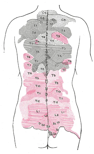
Long description
Areas of distribution of the cutaneous branches of the posterior divisions of the spinal nerves. The areas of the medial branches are in black, those of the lateral in red. (H. M. Johnston.)

Figure 1: Areas of distribution of the cutaneous branches of the posterior divisions of the spinal nerves.
Figure 2Gray801
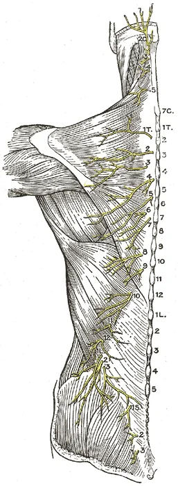
Long description
Diagram of the distribution of the cutaneous branches of the posterior divisions of the spinal nerves.

Figure 2: Diagram of the distribution of the cutaneous branches of the posterior divisions of the spinal nerves.
Figure 3Gray798
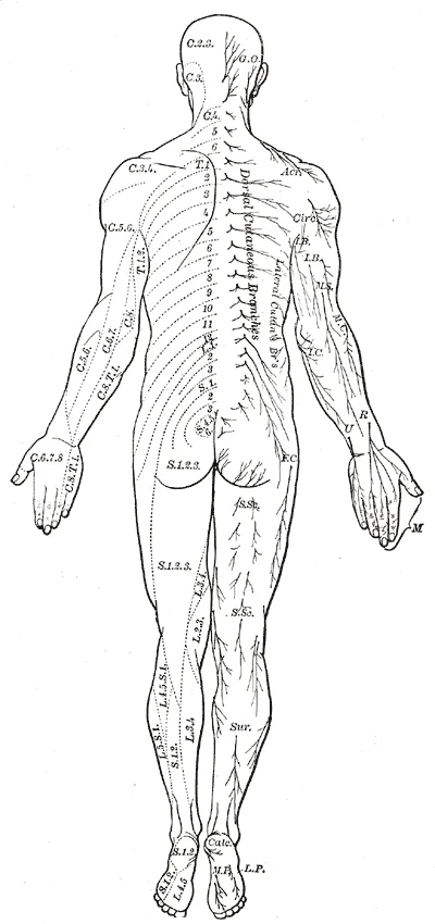
Long description
Distribution of cutaneous nerves. Dorsal aspect.

Figure 3: Distribution of cutaneous nerves (dorsal aspect).
Figure 4Gray797
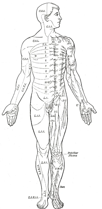
Long description
Distribution of cutaneous nerves. Ventral aspect.

Figure 4: Distribution of cutaneous nerves (ventral aspect).
Figure 5Gray796
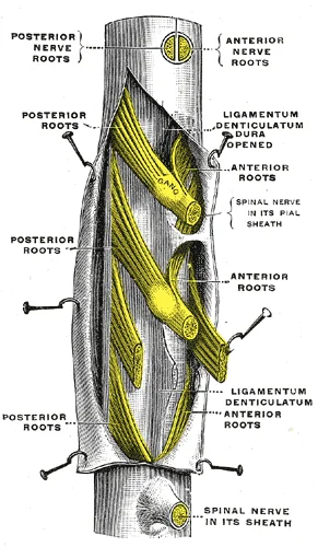
Long description
A portion of the spinal cord, showing its right lateral surface. The dura is opened and arranged to show the nerve roots. (Testut.)

Figure 5: A portion of the spinal cord, showing its right lateral surface.
Axons from different spinal nerves reorganise into four major plexuses — networks of nerve fibres without associated cell bodies. Two lie at the cervical level, one at the lumbar level, and one at the sacral level. Thoracic spinal nerves T2–T11 do not join plexuses but continue directly as intercostal nerves running between the ribs. [1] [3][4]
Figure 6Gray799
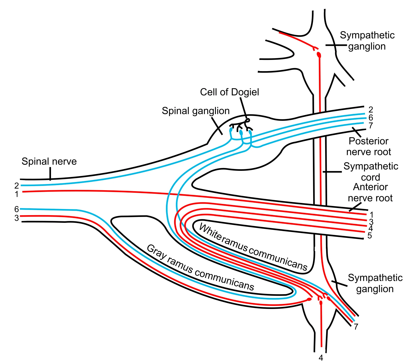
Long description
Scheme showing structure of a typical spinal nerve. 1. Somatic efferent. 2. Somatic afferent. 3,4,5. Sympathetic efferent. 6,7. Sympathetic afferent.

Figure 6: Scheme showing structure of a typical spinal nerve (1).
| Plexus | Spinal segments | Major branches |
|---|---|---|
| Cervical | C1–C5 | Phrenic nerve, cutaneous nerves of neck |
| Brachial | Radial, ulnar, median, axillary nerves [1] | |
| Lumbar | L1–L5 | Femoral nerve, saphenous nerve |
| Sacral | L4–S4 | Sciatic nerve (tibial + fibular) |
Autonomic motor fibres travel within the ventral roots alongside somatic motor fibres.
A dermatome is the area of skin supplied by a single spinal nerve; a myotome is the muscle group innervated by a single spinal nerve. Because plexuses redistribute axons, peripheral nerves contain fibres from multiple spinal segments, so cutaneous territories and muscle supplies do not follow simple segmental lines.
Cutaneous innervation derives from the sensory fibres that enter the dorsal root ganglia and travel through plexuses into named peripheral nerves. [4]
A peripheral nerve consists of axons bundled into fascicles. Each axon is surrounded by endoneurium; fascicles are wrapped by perineurium; the entire nerve is enclosed by epineurium. Blood vessels run in the epineurium. (Illustrative: a typical mixed nerve contains roughly equal numbers of sensory and motor axons.)
Spinal nerve formation and distribution
- Spinal cord segmentAnterior horn (motor), posterior horn (sensory)
- Root formationVentral root (motor) + Dorsal root (sensory) → Spinal nerve
- Question: Region?
Cervical, lumbar, sacral
- Plexus formationAxons reorganise into named peripheral nerves
Thoracic T2–T11
- Direct continuationIntercostal nerves
DefinitionCranial nerves
Twelve pairs of nerves that emerge directly from the brain or brainstem and serve the head, neck and thoraco‑abdominal viscera.
The twelve cranial nerves are numbered I through XII and can be grouped by the type of fibres they contain:
| Fibre type | Number of nerves | Examples |
|---|---|---|
| Though they are part of distinct sensory systems, the vestibular nuclei and the cochlear nuclei are close neighbors with adjacent inputs【P114】. [3] | ||
| Purely motor | 5 | Oculomotor (III), Trochlear (IV), Abducens (VI), Spinal accessory (XI), Hypoglossal (XII) |
| Mixed (sensory + motor) | 4 | Trigeminal (V), Facial (VII), Glossopharyngeal (IX), Vagus (X) |
These counts are taken directly from the anatomical description of the cranial nerves【P9】.
The cranial nerve examination is organised around four functional clusters that correspond to the fibre types:
- Special sensory – nerves that convey smell, vision, equilibrium and hearing (I, II, VIII)【P106】. [3]
- Eye‑movement (motor) – nerves that innervate the extra‑ocular muscles【P104】. [3]
- Oral cavity and superior pharynx – nerves that control facial expression, mastication, swallowing and speech【P104】. [3]
- Neck movement – the cranial nerve that assists head‑turning. [3]
These groups guide the sub‑tests of the cranial nerve exam, allowing the examiner to localise lesions to specific brain‑stem nuclei or peripheral segments. [3]
Two classic mnemonics summarise the functional role of each nerve:
- “Some Say Marry Money But My Brother Says Brains Beauty Matter More” – maps each nerve to sensory, motor or mixed status【P14】. [1]
- “On Old Olympus’ Towering Tops / A Finn And German Viewed Some Hops” – encodes the order of the twelve nerves for recall【P10】. [1]
These devices are routinely taught in introductory anatomy courses to help students retain the sequence and classification of the nerves【P10】.
| Functional group | Typical bedside test | What it assesses |
|---|---|---|
| Special sensory (I, II, VIII) | Smell identification, visual acuity, finger‑to‑nose (proprioception) | Integrity of pure sensory fibres |
| Eye‑movement | Pupillary light reflex, extra‑ocular movement tracking | Motor output to extra‑ocular muscles |
| Oral cavity & pharynx | Facial symmetry, taste on anterior 2/3 of tongue, gag reflex | Mixed sensory‑motor control of face and throat |
| Neck movement (XI) | Shoulder shrug against resistance | Pure motor function to sternocleidomastoid and trapezius |
These tests reflect the sub‑tests described for the cranial nerve exam and illustrate how the classification informs clinical assessment【P103】【P105】.
| Nerve | Fibre type | Functional group | Representative bedside test |
|---|---|---|---|
| I – Olfactory | Sensory | Special sensory | Smell identification |
| II – Optic | Sensory | Special sensory | Visual acuity |
| III – Oculomotor | Motor | Eye‑movement | Extra‑ocular movement |
| IV – Trochlear | Motor | Eye‑movement | Down‑and‑out gaze |
| V – Trigeminal | Mixed | Oral cavity & pharynx | Corneal reflex |
| VI – Abducens | Motor | Eye‑movement | Lateral gaze |
| VII – Facial | Mixed | Oral cavity & pharynx | Facial symmetry |
| VIII – Vestibulocochlear | Sensory | Special sensory | Hearing test |
| IX – Glossopharyngeal | Mixed | Oral cavity & pharynx | Taste posterior 1/3 |
| X – Vagus | Mixed | Oral cavity & pharynx | Gag reflex |
| XI – Spinal accessory | Motor | Neck movement | Shoulder shrug |
| XII – Hypoglossal | Motor | Neck movement | Tongue protrusion |
These classifications, mnemonics and bedside tests form the core knowledge that medical students must master for both written and practical examinations.
Figure 7Gray800
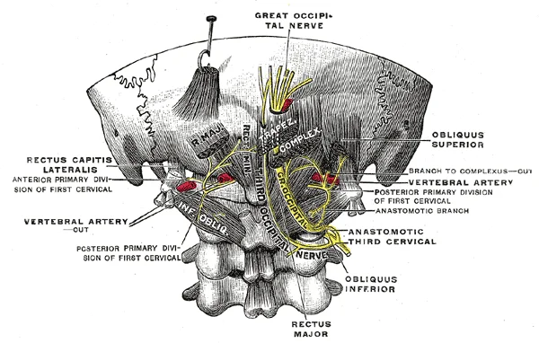
Long description
Posterior primary divisions of the upper three cervical nerves. (Testut.)

Figure 7: Posterior primary divisions of the upper three cervical nerves (Testut.).
Figure 8Gray803
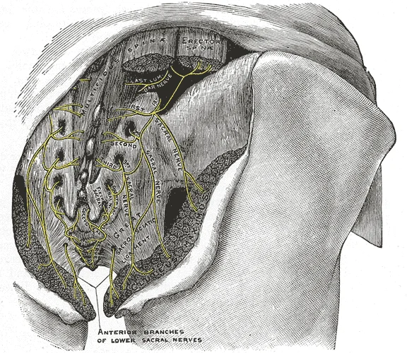
Long description
The posterior divisions of the sacral nerves.

Figure 8: The posterior divisions of the sacral nerves.
Figure 91319 Nerve StructureN
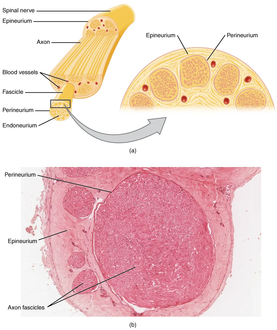
Long description
Illustration from Anatomy & Physiology, Connexions Web site. http://cnx.org/content/col11496/1.6/, Jun 19, 2013.

Figure 9: Illustration from Anatomy & Physiology, Connexions Web site (http://cnx.org/content/col11496/1.6/, jun 19, 2013).
The peripheral nervous system (PNS) is defined by exclusion: it comprises all neural tissue that is not the brain or spinal cord. Its two basic structural units are ganglia—collections of neuronal cell bodies—and nerves—bundles of axons running to and from the periphery. Ganglia are classified as sensory or autonomic. Sensory ganglia contain unipolar sensory neurons and sit on the dorsal root of every spinal nerve and on several cranial nerves. Autonomic ganglia lie in the sympathetic chain, in terminal ganglia, which are located near—or even within—the target effector. [1][4]
DefinitionGanglion types
Sensory ganglia hold unipolar afferent cell bodies on dorsal roots and selected cranial nerves; autonomic ganglia are motor relay stations in sympathetic chains, prevertebral plexuses, or terminal intramural sites. Terminal ganglia receiving cranial input are found in the head and neck, as well as the thoracic and upper abdominal cavities. [1][4]
Cranial nerves illustrate the functional range of peripheral nerves. The olfactory (I), optic (II), and vestibulocochlear (VIII) nerves are purely sensory for smell, vision, and balance/hearing. The trigeminal (V), facial (VII), and glossopharyngeal (IX) nerves are mixed: they convey facial somatosensation and split taste sensation between the anterior two‑thirds and posterior one‑third of the tongue respectively. [1][3]
| Nerve | Modality | Key targets / functions |
|---|---|---|
| I Olfactory | Special sensory | Smell |
| II Optic | Special sensory | Vision; fibres decussate at chiasm |
| III Oculomotor | Somatic motor + parasympathetic | Most extraocular muscles; levator palpebrae; pupil constriction [3] |
| IV Trochlear | Somatic motor | Superior oblique (intorsion) [3] |
| V Trigeminal | Mixed | Face somatosensation; anterior 2/3 taste |
| VI Abducens | Somatic motor | Lateral rectus (abduction) |
| VII Facial | Mixed | Face movement; anterior 2/3 taste; submandibular/sublingual secretion via pterygopalatine ganglion |
| VIII Vestibulocochlear | Special sensory | Balance and hearing |
| IX Glossopharyngeal | Mixed | Posterior 1/3 taste; parotid secretion via otic ganglion [3] |
| X Vagus | Mixed | Visceral afferents; gag reflex; thoracic/abdominal parasympathetic [1] |
Spinal nerves follow a segmental plan. Each forms from a dorsal (sensory) root bearing a dorsal root ganglion and a ventral (motor) root; the mixed nerve then divides into dorsal and ventral rami. General senses—touch, proprioception, kinesthesia, and visceral sensation—are distributed widely through mechanoreceptors in skin, muscle, and vessel walls. Special senses are confined to dedicated organs (eye, inner ear, tongue, nose).
Clinical pearlDorsal root ganglion
Every spinal nerve has a sensory ganglion on its dorsal root; cranial nerve sensory ganglia are homologous structures.
From outside to inside: the epineurium encloses the whole nerve; fascicles are wrapped by perineurium; individual axons with their endoneurium lie within fascicles. Blood vessels (vasa nervorum) run in the epineurium.
Exam trapOptic nerve is CNS
Despite its name, the optic nerve (II) is a CNS tract—myelinated by oligodendrocytes—and its fibres decussate at the chiasm.
Nerve injury and regeneration represent a critical area of clinical practice, particularly when managing trauma to the peripheral nervous system. Understanding the structural integrity of nerves is essential for predicting functional outcomes. The olfactory nerve and optic nerve are responsible for the sense of smell and vision, respectively. Damage to these special sensory nerves often results in permanent deficits because regeneration is limited. [1]
The facial nerve is responsible for the muscles involved in facial expressions, as well as part of the sense of taste and the production of saliva. Injury to this nerve, such as in Bell’s palsy, highlights the importance of distinguishing between motor and sensory components. Motor functions of the facial nerve are usually obvious if facial expressions are compromised, but can be tested by having the patient raise their eyebrows, smile, and frown. This clinical examination allows for rapid localisation of the lesion. [1] [3]
DefinitionNerve Regeneration
The process by which damaged axons regrow towards their target organs, guided by Schwann cells and basal lamina, is crucial for functional recovery in somatic nerves.
Lesions here affect swallowing and gag reflexes. The vagus nerve is responsible for contributing to homeostatic control of the organs of the thoracic and upper abdominal cavities. Its extensive parasympathetic distribution means that injury can have systemic effects on heart rate and digestion. [1]
| Nerve | Primary Function | Clinical Test |
|---|---|---|
| Oculomotor | Eye movements, eyelid lifting, pupillary constriction | Pupillary light reflex, eye movement |
| Trigeminal | Facial sensation, mastication | Corneal reflex, jaw strength |
| Facial | Facial expression, taste (anterior 2/3 tongue) | Raise eyebrows, smile, frown |
| Vagus | Visceral regulation, gag reflex | Gag reflex, voice quality |
| Accessory | Neck muscle control (sternocleidomastoid, trapezius) | Shoulder shrug, head rotation |
The spinal accessory nerve is responsible for controlling the muscles of the neck, along with cervical spinal nerves. The accessory nerve, also referred to as the spinal accessory nerve, innervates the sternocleidomastoid and trapezius muscles. When both the sternocleidomastoids contract, the head flexes forward; individually, they cause rotation to the opposite side. The trapezius can act as an antagonist, causing extension and hyperextension of the neck. These two superficial muscles are impor [1][3]
Exam trapAccessory Nerve
Do not confuse the cranial accessory component with the spinal accessory component; the latter is clinically tested via shoulder shrugging.
Movements of the tongue, soft palate, or superior pharynx can be observed directly while the patient swallows, while the gag reflex is elicited, or while the patient says repetitive consonant sounds. [3]
Clinical pearlVagal Tone
The vagus nerve's role in regulating heartbeat and digestion is a key indicator of autonomic nervous system health.
The terminal ganglia that receive input from cranial nerves are found in the head and neck, as well as the thoracic and upper abdominal cavities, whereas the terminal ganglia that receive sacral input are in the lower abdominal and pelvic cavities【P5】.
NoteIntramural Ganglia
Postganglionic vagal fibers are often located within the organ walls, allowing for precise local control of visceral functions.
The trigeminal nerve controls the muscles of chewing, which are tested for stretch reflexes. This reflex testing is vital for assessing the integrity of the trigeminal motor pathway. Understanding these anatomical relationships allows clinicians to pinpoint lesions based on specific motor or sensory deficits. [3]
- The first cervical nerve (C1) emerges between the occipital bone and the atlas, whereas the remaining cervical nerves generally exit above their correspondingly numbered vertebrae, with C8 exiting between the seventh cervical and first thoracic vertebrae.
- Thoracic spinal nerves T2 through T11 do not form plexuses, but instead continue directly as intercostal nerves running within the intercostal spaces.
- Sensory root ganglia are typically located within the intervertebral foramina, but exceptions exist, such as the sacral ganglia which lie inside the vertebral canal.
- A dermatome represents the area of skin supplied by a single spinal nerve, whereas a myotome represents the muscle group innervated by a single spinal nerve.
- Because nerve plexuses intermix axons from multiple spinal segments, peripheral nerve lesions result in cutaneous and muscular deficits that do not correspond to a single dermatome or myotome.
- The vestibulocochlear nerve (cranial nerve VIII) divides into the cochlear nerve for hearing, originating from spiral ganglion bipolar cells, and the vestibular nerve for equilibrium, originating from peripheral receptors in the semicircular canals, saccule, and utricle.
Exam trapMistaking peripheral nerve layers for muscle fibres
Students often label the epineurium as muscle tissue, confusing the protective connective tissue sheath with contractile fibres.
- Confusing the three connective‑tissue layers – many students list only the epineurium, omitting the perineurium and endoneurium. The correct hierarchy is: outer epineurium, intermediate perineurium surrounding each fascicle, and inner endoneurium encasing individual axons. [2]
- Reversing the direction of spinal nerve roots – the dorsal (posterior) root carries sensory afferents and the ventral (anterior) root carries motor efferents. Remember “Dorsal = sensory, Ventral = motor”. [1]
- Misidentifying the site of the brachial plexus – the plexus forms in the posterior triangle of the neck, not within the axilla. Its roots arise from C5–T1, then combine into trunks, divisions, cords and finally the five terminal branches.
- Overlooking the relationship of peripheral nerves to blood vessels – each nerve travels alongside an artery (e.g., the median nerve with the brachial artery). Ignoring this neurovascular bundle leads to errors in anatomy questions and clinical localisation.
Assuming all peripheral nerves are mixed – some nerves are purely sensory (e.g., the lateral femoral cutaneous nerve) or purely motor (e.g., the musculocutaneous nerve after it gives off its sensory branch). Recognising pure‑type nerves prevents mis‑labelled diagrams. - Neglecting the concept of proprioceptive fibres within peripheral nerves – proprioceptors are specialised endings (muscle spindles, Golgi tendon organs) that travel within the same peripheral nerve as motor fibres. Forgetting this leads to incomplete explanations of reflex arcs.
- Mixing up the terms “branch” and “division” in plexus anatomy – after the trunks split, the resulting anterior and posterior divisions recombine to form cords; cords then give rise to terminal branches. Using “branch” for a division is a common error.
- Ignoring the clinical significance of the endoneurial blood‑nerve barrier – this barrier protects individual axons; damage to it underlies neuropathic pain. Students who omit it lose a key point for exam scenarios on nerve injury.
NoteBedside relevance
Peripheral‑nerve anatomy underpins the neurological examination, localisation of lesions and safe regional anaesthesia.
- Sensory mapping – testing light touch, pin‑prick and vibration over the dermatomes supplied by the median, ulnar, radial and superficial peroneal nerves allows localisation of peripheral neuropathy.
- Motor testing – grading strength of muscles innervated by specific nerves (e.g., wrist extension by the radial nerve) confirms the level of a lesion.
- Reflex assessment – deep‑tendon reflexes depend on the afferent sensory fibre within a peripheral nerve (e.g., the biceps reflex uses the musculocutaneous nerve’s sensory component).
- Regional anaesthesia – ultrasound‑guided peripheral‑nerve blocks (e.g., interscalene block of the brachial plexus) require knowledge of the nerve’s course relative to the scalene muscles and the subclavian artery.
- Surgical exposure – orthopaedic procedures on the forearm demand identification of the median, ulnar and radial nerves to avoid iatrogenic injury.
- Nerve repair – microsurgical neurorrhaphy aligns fascicles within the epineurium and perineurium to restore continuity.
- High‑resolution ultrasound – visualises the hyperechoic epineurium and the fascicular pattern of the perineurium, aiding diagnosis of entrapment syndromes (e.g., carpal tunnel).
- MRI neurography – highlights the nerve’s signal intensity and its relationship to surrounding vessels, useful for tumour or inflammatory neuropathy assessment.
- Nerve conduction studies (NCS) – stimulate a peripheral nerve proximally and record the compound muscle action potential distally; the latency and amplitude reflect the integrity of the myelinated fibres within the endoneurium.
- Electromyography (EMG) – inserts a needle into muscles supplied by a specific nerve to detect denervation, confirming the level of a peripheral‑nerve lesion.
| Nerve | Origin (roots) | Course & Relations | Main Branches | Termination |
|---|---|---|---|---|
| Median | C5‑T1 (brachial plexus) | Travels down the arm medial to the brachial artery, enters the forearm between the two heads of pronator teres | Palmar cutaneous, thenar branch | Supplies most flexor muscles of the forearm and thenar eminence |
| Radial | C5‑T1 (posterior cord) | Runs in the radial groove of humerus, deep to brachioradialis, accompanied by the radial artery | Posterior cutaneous of arm, superficial radial | Extensor muscles of forearm and wrist, dorsal hand sensation |
| Ulnar | C8‑T1 (medial cord) | Passes posterior to medial epicondyle (funny bone), runs in the ulnar groove of ulna | Palmar cutaneous, dorsal cutaneous | Intrinsic hand muscles, skin of medial hand |
Peripheral‑nerve localisation in a patient with hand weakness
- History – onset, distribution of sensory lossIdentify dermatomal pattern
- Question: Is weakness isolated to thumb opposition?
Yes
- Test median nerve (thenar EMG)Look for median‑nerve lesion
No
- Assess ulnar and radial functionsBroaden differential
- Peripheral nerves consist of three connective‑tissue layers: epineurium, perineurium and endoneurium.
- Dorsal (posterior) roots are sensory; ventral (anterior) roots are motor.
- The brachial plexus forms in the posterior triangle of the neck from C5–T1.
- Each peripheral nerve travels with a corresponding artery, forming a neurovascular bundle.
- Pure sensory (e.g., lateral femoral cutaneous) and pure motor (e.g., musculocutaneous) nerves exist.
- Proprioceptive endings travel within mixed peripheral nerves.
- Knowledge of nerve anatomy guides regional blocks, surgical approaches and imaging interpretation.
- Nerve‑conduction studies assess the integrity of myelinated fibres within the endoneurium.
Answer each one in your head first, then open the answer.
Which connective‑tissue layer directly surrounds individual axons?
Show answerHide answer
EndoneuriumThe dorsal root of a spinal nerve carries which type of fibres?
Show answerHide answer
Sensory afferentsWhere does the brachial plexus form?
Show answerHide answer
Posterior triangle of the neckWhich artery accompanies the median nerve in the forearm?
Show answerHide answer
Brachial arteryName a pure sensory peripheral nerve of the lower limb.
Show answerHide answer
Lateral femoral cutaneous nerve
- Betts JG, Young KA, Wise JA, et al. The Peripheral Nervous System. OpenStax, Anatomy and Physiology 2e (CC BY-NC-SA 4.0). Accessed .
- Betts JG, Young KA, Wise JA, et al. Sensory Perception. OpenStax, Anatomy and Physiology 2e (CC BY-NC-SA 4.0). Accessed .
- Betts JG, Young KA, Wise JA, et al. The Cranial Nerve Exam. OpenStax, Anatomy and Physiology 2e (CC BY-NC-SA 4.0). Accessed .
- Betts JG, Young KA, Wise JA, et al. Divisions of the Autonomic Nervous System. OpenStax, Anatomy and Physiology 2e (CC BY-NC-SA 4.0). Accessed .
Figure credits
- Figure 1: Henry Vandyke Carter, via Wikimedia Commons, Public domain, Public domain.
- Figure 2: Henry Vandyke Carter, via Wikimedia Commons, Public domain, Public domain.
- Figure 3: Henry Vandyke Carter, via Wikimedia Commons, Public domain, Public domain.
- Figure 4: Henry Vandyke Carter, via Wikimedia Commons, Public domain, Public domain.
- Figure 5: Henry Vandyke Carter, via Wikimedia Commons, Public domain, Public domain.
- Figure 6: Henry Vandyke Carter, via Wikimedia Commons, Public domain, Public domain.
- Figure 7: Henry Vandyke Carter, via Wikimedia Commons, Public domain, Public domain.
- Figure 8: Henry Vandyke Carter, via Wikimedia Commons, Public domain, Public domain.
- Figure 9: OpenStax College, via Wikimedia Commons, CC BY 3.0, CC BY 3.0.
Educational content for medical students and doctors. It is not a substitute for clinical judgement or local guidelines.