Body cavities and the diaphragm: development
Embryological development of body cavities and diaphragm, including ventral wall defects.
The intraembryonic coelom forms the primary body cavities, which later differentiate into the pleural, pericardial, and peritoneal cavities. These spaces are initially continuous but become separated by the developing diaphragm and septum transversum. The diaphragm itself begins to develop at approximately 4 weeks of gestation and is fully formed by 12 weeks. This process involves the fusion of several structures, including the septum transversum, pleuroperitoneal membranes, dorsal mesentery of the oesophagus, and muscular ingrowths from the lateral body walls. [2]
The pleuroperitoneal canals, which normally close to separate the thoracic and abdominal cavities, are critical in this timeline. A defect in the pleuroperitoneal fold can result in a patent pleuroperitoneal canal, allowing abdominal contents to herniate into the thorax. This is the embryological basis for congenital diaphragmatic hernias (CDH). While the exact cause remains controversial, it was initially thought that the defect resulted from the failure of different parts of the diaphragm to fuse. However, alternative theories suggest that lung hypoplasia may be the primary causal factor in the pathophysiology of diaphragmatic hernia. If the development of the lung bud is disturbed, there is an impaired development of a post-hepatic mesenchymal plate (PHMP) that is closely related to lung development, resulting in a defective diaphragm. [2]
Ventral wall development involves the closure of the anterior abdominal wall. Defects in this process lead to conditions such as gastroschisis and omphalocele. These are distinct from diaphragmatic hernias but share the theme of incomplete closure of body cavities. Gastroschisis is typically a paraumbilical defect, whereas omphalocele involves herniation of abdominal contents into the base of the umbilical cord, covered by a membrane. The timing of these defects is crucial, with omphalocele resulting from failure of the intestines to return to the abdominal cavity around week 8, while gastroschisis occurs due to failure of the body wall folds to fuse properly.
The mesenteries also develop during this period, suspending the gut within the peritoneal cavity. The dorsal mesentery of the stomach becomes the greater omentum, while the ventral mesentery forms the lesser omentum and falciform ligament. Proper rotation and fixation of the gut are essential for normal anatomy. The diaphragm's development is particularly important as it separates the thoracic and abdominal cavities and plays a crucial role in respiration.
DefinitionIntraembryonic Coelom
The primitive body cavity that gives rise to the pleural, pericardial, and peritoneal cavities.
Clinical pearlDiaphragm Timeline
Development starts at 4 weeks and completes by 12 weeks of gestation.
Ventral wall defects arise from the failure of the lateral body folds to fuse properly at the midline. The two most significant clinical entities are omphalocele and gastroschisis. Omphalocele is characterised by the herniation of abdominal viscera into the base of the umbilical cord. The herniated contents are covered by a membrane consisting of peritoneum and amnion. This defect is associated with chromosomal abnormalities and other congenital anomalies in a significant proportion of cases. The size of the defect can vary, but the key feature is the membranous covering. Omphalocele occurs in approximately 1 in 5,000 live births and is often associated with other anomalies such as cardiac defects and neural tube defects.
Gastroschisis is a full-thickness defect of the abdominal wall, typically located to the right of the umbilicus. Unlike omphalocele, the herniated intestines are not covered by a membrane, exposing them directly to the amniotic fluid. This exposure can lead to inflammation and thickening of the bowel wall, which may compromise bowel function. Gastroschisis is usually an isolated defect, with fewer associated chromosomal abnormalities compared to omphalocele, occurring in approximately 1 in 3,000 live births. The incidence of gastroschisis has been increasing in recent years, particularly in younger mothers.
The timing of these defects is critical. Omphalocele results from the failure of the intestines to return to the abdominal cavity around week 8 of gestation, while gastroschisis occurs due to failure of the body wall folds to fuse properly during weeks 4-6. The location of gastroschisis is typically to the right of the umbilicus, as the right umbilical vein regresses earlier than the left, creating a weakness in the abdominal wall. Surgical management differs significantly between these two conditions, with omphalocele often requiring staged closure due to the size of the defect and the risk of abdominal compartment syndrome, while gastroschisis can often be managed with primary closure or silo placement.
| Feature | Omphalocele | Gastroschisis |
|---|---|---|
| Location | Base of umbilical cord | Paraumbilical, usually right |
| Covering | Membrane (peritoneum/amnion) | No membrane |
| Associated Anomalies | Common (chromosomal) | Rare |
| Pathophysiology | Failure of gut return | Failure of body wall closure |
| Incidence | 1 in 5,000 live births | 1 in 3,000 live births |
Exam trapOmphalocele vs Gastroschisis
Omphalocele has a membrane; gastroschisis does not. This distinction is crucial for surgical management and prognosis.
Congenital diaphragmatic hernias (CDH) occur when the diaphragm fails to close completely, allowing abdominal organs to enter the thoracic cavity. The most common type is the postero-lateral hernia, also known as a Bochdalek hernia. These account for 70–75% of cases. The majority of Bochdalek hernias occur on the left side (85%), with less frequent occurrence on the right side (13%) or bilaterally (2%). The defect may range from a small opening of the posterior muscle rim to complete absence of diaphragm. [2]
The embryological basis involves the failure of the pleuroperitoneal folds to fuse with the septum transversum and dorsal mesentery. This leaves a patent pleuroperitoneal canal. As the gut returns from the extraembryonic coelom, it can herniate through this defect into the thorax. This herniation can compress the developing lungs, leading to pulmonary hypoplasia, which is a major cause of morbidity and mortality. The severity of pulmonary hypoplasia correlates with the size of the hernia and the degree of mediastinal shift.
Anterior defects, known as Morgagni hernias, are less common, accounting for 23–28% of cases. These occur through the foramen of Morgagni, an area of weakness between the sternal and costal attachments of the diaphragm. Central hernias are rare, representing 2–7% of cases. The timing of diaphragmatic hernia development is critical, with the diaphragm beginning to develop at approximately 4 weeks of gestation and being fully formed by 12 weeks. Defects that occur earlier in development tend to be more severe. [2] [3]
The clinical presentation of CDH varies depending on the size and location of the defect. Small hernias may be asymptomatic and discovered incidentally, while large hernias can present with respiratory distress at birth due to pulmonary hypoplasia and mediastinal shift. The diagnosis is typically made prenatally with ultrasound, which can show abdominal contents in the chest and polyhydramnios due to impaired fetal swallowing. Postnatal management involves stabilization with mechanical ventilation and surgical repair, which is typically performed within the first week of life.
DefinitionBochdalek Hernia
A postero-lateral congenital diaphragmatic hernia, the most common type, usually on the left side.
NotePulmonary Hypoplasia
Lung hypoplasia may be a primary causal factor or a secondary consequence of herniation, impairing diaphragm development.
Figure 1Gray57
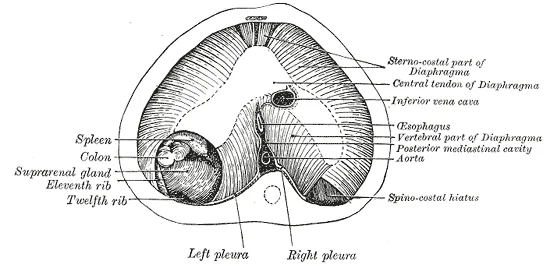
Long description
The thoracic aspect of the diaphragm of a newly born child in which the communication between the peritoneum and pleura has not been closed on the left side; the position of the opening is marked on the right side by the spinocostal hiatus. (After Keith.)

Figure 1: The thoracic aspect of the diaphragm of a newly born child in which the communication between the peritoneum and pleura has not been closed on the left side; the position of the opening is marked on the right side by the spinocostal hiatus (After Keith.).
The dorsal mesentery of the oesophagus contributes to the posterior crura. The lateral body wall muscles form the peripheral muscular portions.
The pre-embryonic stage covers the first two weeks of prenatal development, followed by the embryonic stage from weeks three to eight. By the end of the embryonic period, all organ systems are structured in rudimentary form. The diaphragm develops from four distinct components that converge to separate the thoracic and abdominal cavities. [1]
The septum transversum is the first component to appear. It is a thick plate of mesoderm that forms the central tendon of the diaphragm. It initially lies caudal to the developing heart and cranial to the yolk sac. As the embryo folds, it moves cranially to its final position.
The pleuroperitoneal membranes develop later to close the pericardioperitoneal canals. These canals connect the pericardial and peritoneal cavities. The membranes fuse with the septum transversum and the dorsal mesentery of the oesophagus. Defects in this closure can lead to congenital diaphragmatic hernia.
The dorsal mesentery of the oesophagus contributes to the posterior part of the diaphragm. It forms the crura of the diaphragm. These muscular pillars attach the diaphragm to the lumbar vertebrae.
The peripheral muscular portions of the diaphragm arise from ingrowths of the lateral body walls. These muscles interdigitate with the other components. They provide the contractile force for respiration.
A defect in the diaphragm leads to protrusion of abdominal contents into the thoracic cavity. This condition is known as congenital diaphragmatic hernia. The defect may range from a small aperture to complete absence of the diaphragm. The herniation interferes with normal lung development. Lung hypoplasia occurs on the ipsilateral side. The contralateral side is affected to a variable extent. The pathophysiology involves a combination of lung immaturity and hypoplasia. This can lead to persistent pulmonary hypertension of the newborn. [2]
Diaphragmatic eventration is a weakness in the diaphragm. It is more common on the right side. It is not associated with severe lung hypoplasia. It may be mistaken for a diaphragmatic hernia. Complete absence of the diaphragm results in diaphragmatic agenesis. This causes severe lung hypoplasia. [2]
The development of the body cavities and diaphragm is orchestrated by a complex interplay of molecular signals that guide the formation of the coelomic cavity and the separation of the thoracic and abdominal compartments. Understanding this process requires examining the spatial and temporal coordination of tissue interactions during early embryogenesis.
The diaphragm is represented as a developing muscular sheet inferior to the heart, separating the thoracic cavity above from the abdominal cavity below. The relationship between the heart and the developing diaphragm is critical, as the heart's descent into the thorax influences the final position and structure of the diaphragmatic musculature.
The heart begins its development as a tube-like structure, connected via capillaries to the chorionic villi. Cells of the primitive tube-shaped heart are capable of electrical conduction and contraction.
Defects in this molecular regulation can lead to congenital anomalies. Congenital Diaphragmatic hernia (CDH) is characterized by a defect in the diaphragm leading to the protrusion of abdominal contents into the thoracic cavity affecting the normal development of the lungs. The condition may present as an isolated lesion or as part of a syndrome. Evidence from electron microscopy in a rat model of CDH further supports the fact that when the development of the PHMP is impaired, a diaphragmatic de [2]
DefinitionCongenital Diaphragmatic Hernia
A defect in the diaphragm leading to the protrusion of abdominal contents into the thoracic cavity affecting the normal development of the lungs.
The central structure is the primitive gut tube, derived from the endoderm, which is suspended by the dorsal mesentery. Lateral to the gut tube lies the intraembryonic coelom, which will partition into the pleural, pericardial, and peritoneal cavities.
The development of the body cavities begins with the formation of the intraembryonic coelom. This cavity appears within the lateral plate mesoderm during the third week of development. As the embryo undergoes folding, the flat embryonic disc is converted into a cylindrical structure. The intraembryonic coelom is partitioned into three main cavities: the pericardial, pleural, and peritoneal cavities. The pericardial cavity forms the most cranial part, surrounding the developing heart. The pleural [1]
The diaphragm is a complex structure derived from four distinct embryonic components. The septum transversum is the most significant contributor. It forms as a thick plate of mesoderm that separates the pericardial cavity from the peritoneal cavity. Initially, the septum transversum is located at the level of the fourth cervical somite. As the embryo grows, the septum transversum moves caudally to its final position at the level of the fourth intercostal space. The body wall contributes the peri [1]
The yolk sac plays a crucial role in early development. It supplies nutrients absorbed from the trophoblast and provides primitive blood circulation to the developing embryo for the second and third week of development. When the placenta takes over nourishing the embryo at approximately week 4, the yolk sac has been greatly reduced in size and its main function is to serve as the source of blood cells and germ cells. During week 3, a finger-like outpocketing of the yolk sac develops into the allantois, a primitive excretory duct of the embryo that will become part of the urinary bladder. Together, the stalks of the yolk sac and allantois establish the outer structure of the umbilical cord. [1]
The following table summarises the key embryonic structures and their adult derivatives.
| Embryonic Structure | Adult Derivative |
|---|---|
| Septum transversum | Central tendon of the diaphragm |
| Pleuroperitoneal membranes | Crura of the diaphragm |
| Dorsal mesentery of oesophagus | Muscular components of diaphragm |
| Yolk sac | Primitive blood cells and germ cells |
| Allantois | Urinary bladder |
| Intraembryonic coelom | Pleural, pericardial, and peritoneal cavities |
The process of gastrulation establishes the three primary germ layers: the endoderm, mesoderm, and ectoderm. The endoderm forms the lining of the gut tube, which is suspended by the mesoderm-derived mesenteries. The mesoderm gives rise to the muscles and connective tissues of the diaphragm. The ectoderm forms the nervous system and skin. The coordination of these layers ensures the proper formation of the body cavities and the diaphragm. [1]
By the end of the embryonic period, the embryo is approximately 3 cm (1.2 in) from crown to rump and weighs approximately 8 g (0.25 oz). At this stage, the major organ systems are established, including the respiratory and digestive systems, which are intimately related to the diaphragm and body cavities. The diaphragm is fully formed and functional, allowing for the separation of the thoracic and abdominal contents. [1]
In the thoracic cavity, the lungs are compressed by abdominal viscera. The liver and intestines are displaced superiorly into the chest cavity, occupying the space normally reserved for lung development. This displacement is the hallmark of the diaphragmatic defect. The intestines protrude through this ring, covered only by a membrane, representing the umbilical anomaly.
Congenital diaphragmatic hernia is a defect in the diaphragm that allows abdominal organs to herniate into the thoracic cavity. The most common type is a Bochdalek hernia, which results from the failure of the pleuroperitoneal membranes to close the pleuroperitoneal canals. These canals are the embryological openings between the pleural and peritoneal cavities. Normally, these canals close during the eighth week of development. When closure fails, a hole remains, typically on the left side. The left side is more commonly affected because the right pleuroperitoneal canal closes earlier than the left. The herniation usually involves the stomach, intestines, and sometimes the liver. The presence of abdominal organs in the chest compresses the developing lungs. This compression leads to pulmonary hypoplasia, where the lung tissue is underdeveloped and has reduced surface area for gas exchange. The mediastinum is also shifted to the opposite side, further compromising respiratory function. The clinical presentation often includes severe respiratory distress immediately after birth. Diagnosis is confirmed by imaging showing bowel loops in the thorax.
Figure 2Gray55
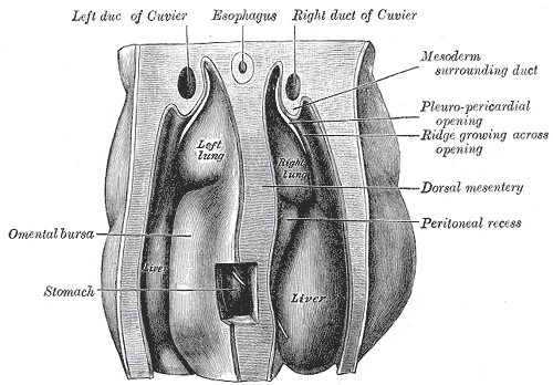
Long description
Upper part of celom of human embryo of 6.8 mm., seen from behind. (From model by Piper.)

Figure 2: Upper part of celom of human embryo of 6.8 mm., seen from behind (From model by Piper.).
Figure 3Gray54
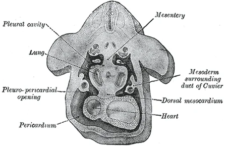
Long description
Figure obtained by combining several successive sections of a human embryo of about the fourth week (From Kollmann.) The upper arrow is in the pleuroperitoneal opening, the lower in the pleuropericardial.

Figure 3: Figure obtained by combining several successive sections of a human embryo of about the fourth week (From Kollmann.) The upper arrow is in the pleuroperitoneal opening, the lower in the pleuropericardial.
DefinitionBochdalek Hernia
A congenital defect caused by the failure of the pleuroperitoneal membrane to close the pleuroperitoneal canal, allowing abdominal viscera into the thorax.
Congenital umbilical hernia occurs when the umbilical ring fails to close completely after the physiological herniation of the midgut returns to the abdominal cavity. During early development, the midgut herniates into the umbilical cord due to limited space in the abdomen. It normally returns to the abdominal cavity by the tenth week. As it returns, the umbilical ring should narrow and close. If the ring remains patent, abdominal contents can protrude through it. This is visible as a soft bulge at the umbilicus. The hernia is covered by skin and peritoneum. It is common in infants and often resolves spontaneously as the abdominal muscles strengthen. However, if the defect is large, it may persist into adulthood. Unlike diaphragmatic hernias, umbilical hernias rarely cause immediate respiratory compromise. The primary concern is the potential for incarceration, although this is rare in infants.
NotePhysiological Herniation
The midgut normally herniates into the umbilical cord and returns by week 10; failure of the ring to close after this return causes umbilical hernia.
The umbilical region represents a critical anatomical junction where the abdominal wall is naturally weakest, formed by the passage of the umbilical cord during fetal life. The umbilical cord connects the conceptus to the placenta, carrying deoxygenated blood and wastes from the fetus through two umbilical arteries, while nutrients and oxygen are carried from the pregnant person to the fetus through the single umbilical vein. This structure is surrounded by the amnion, and the spaces within the cord around the blood vessels are filled with Wharton’s jelly, a mucous connective tissue that protects the vessels from compression. During the first several weeks of development, the cells of the endometrium—referred to as decidual cells—nourish the nascent embryo, but during prenatal weeks 4–12, the developing placenta gradually takes over the role of feeding the embryo, and the decidual cells are no longer needed.
In neonates, this is often a physiological finding due to the relative weakness of the abdominal muscles and the large size of the liver. Most small umbilical hernias in infants close spontaneously as the abdominal wall muscles strengthen, typically by the age of three to four years. However, larger defects are less likely to close and may require inte
In adults, the pathophysiology differs significantly. Umbilical hernias in adults are rarely congenital in the same sense; they are often acquired due to increased intra-abdominal pressure from obesity, pregnancy, or ascites. Unlike in children, adult umbilical hernias do not close spontaneously and carry a higher risk of complications such as strangulation. Consequently, surgical repair is the standard of care for symptomatic adult hernias.
However, prenatal care focuses on monitoring the integrity of the abdominal wall and the development of the umbilical cord structures. Any abnormalities in the cord insertion or associated anomalies may prompt further investigation. Postnatally, clinical examination is sufficient for diagnosis. The physical exam reveals a soft, reducible mass at the umbilicus that becomes more prominent with crying or straining. If the hernia is irreducible, tender, or discolored, it suggests incarceration or strangulation, which is a surgical emergency.
Management strategies must be tailored to the patient's age and defect size. For children, observation is preferred unless the defect is large or symptomatic. For adults, elective repair is recommended to prevent complications. The choice between open and laparoscopic repair depends on the surgeon's expertise and the patient's anatomy. In high-risk patients, such as those with obesity or previous abdominal surgeries, prophylactic mesh placement may be considered to reduce recurrence rates. The decision to use mesh involves weighing the benefits of reduced recurrence against the risks of chronic pain and infection.
Clinical pearlClinical Pearl
Umbilical hernias in children under 4 years old with defects less than 2 cm often close spontaneously; surgical intervention is reserved for larger defects or those persisting beyond this age.
Exam trapCommon Trap
Do not confuse umbilical hernia with epigastric hernia. Epigastric hernias occur along the linea alba between the umbilicus and xiphoid process and are less likely to contain bowel, often containing only preperitoneal fat.
| Feature | Children | Adults |
|---|---|---|
| Spontaneous Closure | Common in small defects | Rare |
| Primary Risk | Incarceration | Strangulation |
| Typical Repair | Suture repair | Mesh repair often preferred |
| Anesthesia | General | Local or General |
DefinitionFascial Closure
Closing the defect during laparoscopic repair reduces seroma and recurrence rates.
DefinitionCDH Syndromes
CDH is part of multiple syndromes including VACTERL and Pierre Robin sequence.
Students frequently conflate the embryological development of the body cavities with the clinical management of adult hernias. A primary error is assuming that all ventral hernias are equivalent in surgical planning. It is incorrect to group primary and incisional hernias together when studying outcomes, as their prognoses differ significantly. This distinction is vital because sub-populations of primary hernias may carry higher risks than some incisional variants, reflecting complex systemic factors rather than simple aetiology.
Another frequent mistake involves the diagnosis and measurement of diastasis recti. Students often fail to recognise that diastasis is defined by specific width thresholds at precise anatomical landmarks. A diagnosis is only valid if the width between the medial edges of the recti exceeds 22 mm at a point 3 cm superior to the umbilicus. Furthermore, the measurement at 2 cm inferior to the umbilicus must be greater than 16 mm to confirm the condition. Ignoring these specific criteria leads to misdiagnosis, as bulging alone is not diagnostic without quantitative measurement.
In surgical reconstruction, a critical error is attempting simultaneous anterior component separation (aCS) and transversus abdominis release (TAR) on the same side. This is strictly contraindicated; aCS and TAR should never be performed simultaneously at the same side. This rule exists to preserve vascular supply and tissue viability. Instead, TAR is indicated whenever midline closure of the linea alba is not possible in ventral hernia repair. The TAR technique can achieve midline closure in mo [4]
Students also overlook the specific indications for component separation. It is not a first-line option for all hernias but is indicated whenever it is not possible to close the linea alba in midline ventral hernia repair. Additionally, for recurrent hernias, reinforcement of the complete scar is recommended in both open and laparoscopic techniques. Failing to reinforce the entire scar increases the risk of further failure.
Finally, there is confusion regarding the reliability of imaging. While ultrasound is the gold standard for non-invasive measurement of inter-rectus distance (IRD) in parous women, it has limitations. The use of ultrasound is limited when IRD is large, and conventional imaging may not be sufficient. Students must know that extended field of view techniques, such as panoramic ultrasound or acoustic standoff pads, are required to visualise the linea alba completely in these cases. These techniques are highly correlated with conventional imaging, with correlation coefficients greater than 0.95.
This knowledge is applied at the bedside to assess abdominal wall integrity. Diastasis recti is a regular diagnostic challenge, typically presenting as bulging of the linea alba with increased intra-abdominal pressure. While it commonly develops in women during pregnancy, it usually regresses within a year after childbirth. However, in a third of those patients, the diastasis persists, requiring clinical intervention. Accurate measurement at the specified reference points allows for appropriate classification and management planning.
In the operating theatre, understanding these principles guides surgical technique. The European Hernia Society (EHS) classification is the most widely used system for categorising hernias. This classification is useful to identify patients at risk for complications, such as incarceration or the need for open surgery. Independent risk factors for postoperative complications include EHS width class, incarceration, open surgery, duration of surgery, Altemeier wound class, and therapeutic antibiotic treatment.
For large defects where primary closure is impossible, component separation techniques are employed. The transversus abdominis release (TAR) is a key technique for abdominal wall reconstruction, particularly in complex cases where anterior component separation has failed. When a recurrent hernia occurs after an anterior component separation, TAR has been shown to be an option for reconstruction. The ability to advance the abdominal wall by 8 to 12 cm per side makes TAR highly effective for achieving midline closure.
On imaging, extended field of view ultrasound is used when conventional methods fail. This is particularly relevant for patients with large inter-rectus distances where the linea alba cannot be fully visualised. The extended field of view technique allows complete visualization of the linea alba when the inter-rectus distance is large. This ensures accurate preoperative planning and assessment of the abdominal wall's functional capacity.
In examinations, students must distinguish between different hernia types and their management. It is inappropriate to combine primary and incisional hernias in study designs because their outcomes are different. Recognising that sub-populations of primary hernias may have worse outcomes than some incisional hernias demonstrates a nuanced understanding of surgical risk. Additionally, knowing that chronic groin pain may be related to mesh interaction, and that mesh removal can contribute to pain relief, is essential for managing postoperative complications.
- Primary and incisional hernias have different outcomes and should not be combined in studies. [3]
- Diastasis recti is diagnosed if the inter-rectus distance is >22 mm at 3 cm above the umbilicus. [4]
- The second diagnostic measurement for diastasis is >16 mm at 2 cm below the umbilicus. [4]
- Anterior component separation and transversus abdominis release should never be performed simultaneously on the same side.
- Transversus abdominis release allows an advancement of 8 to 12 cm per side. [4]
- The closure rate for transversus abdominis release is reported as 97.2%. [4]
- Extended field of view ultrasound is used when conventional imaging is insufficient for large inter-rectus distances. [3]
- Reinforcement of the complete scar is recommended for recurrent hernias in both open and laparoscopic repairs. [3]
Answer each one in your head first, then open the answer.
What is the inter-rectus distance threshold for diagnosing diastasis recti 3 cm superior to the umbilicus?
Show answerHide answer
Greater than 22 mmWhat is the inter-rectus distance threshold for diagnosing diastasis recti 2 cm inferior to the umbilicus?
Show answerHide answer
Greater than 16 mmWhich two component separation techniques should never be performed simultaneously on the same side?
Show answerHide answer
Anterior component separation and transversus abdominis releaseWhat is the reported closure rate for the transversus abdominis release technique?
Show answerHide answer
97.2%Why should primary and incisional hernias not be combined in outcome studies?
Show answerHide answer
Their outcomes are different
- Betts JG, Young KA, Wise JA, et al. Embryonic Development. OpenStax, Anatomy and Physiology 2e (CC BY-NC-SA 4.0). Accessed .
- Chandrasekharan PK, Rawat M, Madappa R, et al. Congenital Diaphragmatic hernia - a review. Maternal health, neonatology and perinatology 2017;3:6. doi:10.1186/s40748-017-0045-1. Accessed .
- Bittner R, Bain K, Bansal VK, et al. Update of Guidelines for laparoscopic treatment of ventral and incisional abdominal wall hernias (International Endohernia Society (IEHS))-Part A. Surgical endoscopy 2019;33(10):3069-3139. doi:10.1007/s00464-019-06907-7. Accessed .
- Bittner R, Bain K, Bansal VK, et al. Update of Guidelines for laparoscopic treatment of ventral and incisional abdominal wall hernias (International Endohernia Society (IEHS)): Part B. Surgical endoscopy 2019;33(11):3511-3549. doi:10.1007/s00464-019-06908-6. Accessed .
Figure credits
Educational content for medical students and doctors. It is not a substitute for clinical judgement or local guidelines.